
Investigation of a sensing approach based on a rapid reduction of azide to selectively measure bioavailability of H2S†
Gunwoo Kima,
Eunju Janga,
Alexis M. Pageb,
Ting Dinga,
Kimberly A. Carlson b and
Haishi Cao*a
b and
Haishi Cao*a
aDepartment of Chemistry, University of Nebraska-Kearney, Kearney, NE, USA 68849. E-mail: caoh1@unk.edu
bDepartment of Biology, University of Nebraska-Kearney, Kearney, NE, USA 68849
First published on 3rd October 2016
Abstract
A new reaction-based sensor (AHS) was synthesized for quantitative detection of H2S. AHS showed a high selectivity and sensitivity toward H2S over other thio-containing molecules, or reducing reagents with high abundance in living cells. In the presence of H2S, significant fluorescence enhancement (17-fold) was observed due to the reduction of the azide on AHS. The absorption (362 nm) and fluorescence emission (557 nm) of reduced AHS showed a highly linear correlation to H2S level, which were used to measure concentration of H2S in the range of 0–100 μM.
Introduction
Hydrogen sulfide (H2S) is an important gaseous signalling molecule, which widely exists in numerous physiological processes including anti-inflammation, redox regulation, and neuroprotection.1 In mammalian systems, endogenous H2S is mainly produced via cysteine metabolism catalysed by cystathionine-β-synthase (CBS), cystathionine-γ-lyase (CSE), and 3-mercatopyruvate sulfurtransferase (3MST).2 Cellular H2S is stored in the mitochondria and cytosol as both a free gas (20%) and a hydrosulfide anion (HS−, 80%) in pH 7.4 at 37 °C.3 The concentration of H2S in different tissues varies in the micromolar range.4 The increasing evidence indicates that H2S plays a critical role in many diseases such as stroke, Down's syndrome, and Parkinson's disease, in which the cellular level of H2S shows a significant change compared to normally physiological conditions.5 Therefore, accurate and reliable measurement of H2S in living cells becomes highly desired, which will afford important information to understand the functions of H2S under physiological and pathological conditions.6 In biological systems, multiple forms of H2S, large amounts of thiol-containing species, and reducing reagents with similar chemical structures and functions to H2S exist, thereby making quantitative detection of H2S extremely challenging.7 Many instrument based techniques including high pressure liquid chromatography (HPLC), gas chromatography (GC), and mass spectrometry (MS) have been applied to quantitatively analyse H2S, however, these approaches show limited capability for real-time measurement of H2S in biological samples.8Fluorescence sensing is a widely-used technique for detection of various molecules with low concentrations in biological samples due to high sensitivity, short response time, and non-destructivity.9 Reaction-based fluorescence sensors have particularly attracted great interest due to the improvement of selectivity.10 Currently, fluorescence sensors for detection of H2S are mainly designed on the basis of H2S-mediated reduction, nucleophilic reaction, and metal-sulfide precipitations.11 Although these reaction-based sensors provide high selectivity, the detection of H2S is still challenging because of the long response time due to organic reactions and low reactivity in the complicated biological environments.12
In this work, we report a novel fluorescent approach for detection of H2S on the basis of reduction of azide appended on 2,3-naphthalimide (2,3-NI). Compared to its isomer-1,8-naphthalimide (1,8-NI), 2,3-NI shows many unique photophysical properties (e.g., dual fluorescence and long emission) because of different steric environment and rotational dynamics imposed by the 5-membered imide ring of 2,3-NI at excited state.13 Therefore, 2,3-NI is used as the fluorophore to prepare the fluorescence sensor (AHS) for quantitative detection H2S. As a reaction-based fluorescence approach, AHS shows short reaction time, long emission wavelength, high sensitivity and selectivity to H2S, even in complicated living cellular environment. AHS provides a robust and reliable means for detection of H2S in biosamples.
Experimental
AHS was synthesized via a four-step reaction. 4-Nitro-α,α,α′,α′-tetrabromo-o-xylene was used as the starting material reacting with maleic anhydride to afford intermediate 1, which was coupled to 4-(2-aminoethyl)morpholine to yield NHS. NHS was reduced by palladium/carbon followed by nitroniumlization to yield AHS (Scheme 1).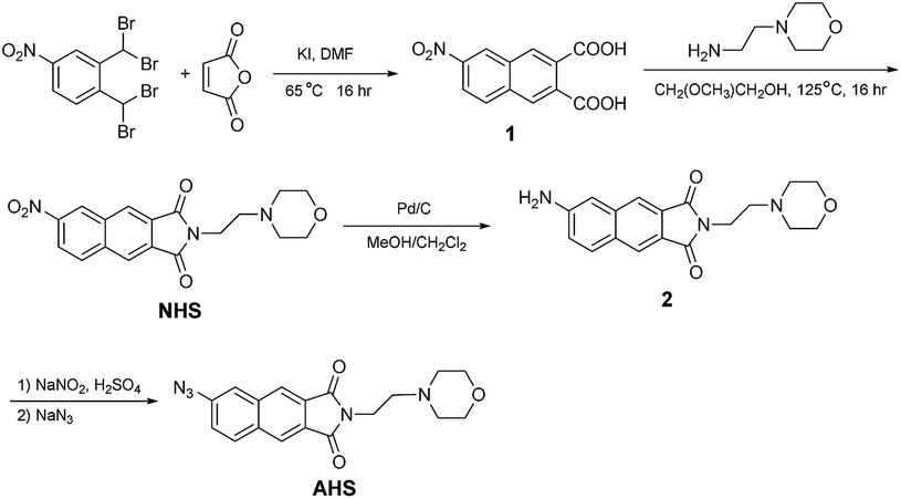 | ||
| Scheme 1 The synthetic route for AHS. | ||
Apparatus
Absorbance spectra were collected by Cary Series UV-vis Spectrophotometer (Agilent Technologies). Fluorescence measurements were all performed by using a FluoroMax-4 Spectrofluorometer (Horiba Jobin Yvon, USA). All of fluorescence spectra were recorded in a 1 cm quartz cuvette. The excitation and emission slits were set at 2 nm. 1H and 13C NMR spectra were recorded on (1H 300 MHz, 13C 75 MHz) Bruker 300 Ultra-Shield spectrometer at room temperature. TE2000-S inverted fluorescent microscope (Nikon, Melville, NY) was used for cell imaging. The HRMS data was collected in Nebraska Center of Mass Spectrometry at University of Nebraska-Lincoln by using GCT Mass Spectrometer (Waters, USA).Synthesis and characterization
![[thin space (1/6-em)]](https://www.rsc.org/images/entities/char_2009.gif) :CH2Cl2 = 2:1 to yield NHS as a light yellow solid (284 mg, 80%). 1H NMR (300 MHz, CDCl3) δ: 2.51–2.63 (m, 4H), 2.74 (t, J = 7.80 Hz, 2H), 3.61–3.72 (m, 4H), 3.96 (t, J = 6.90 Hz, 2H), 8.27 (d, J = 9.90 Hz, 1H), 8.46–8.53 (m, 2H), 8.58 (s, 1H), 9.03 (s, 1H). 13C NMR (75 MHz, CDCl3) δ: 29.7, 35.6, 56.0, 67.0, 122.4, 124.3, 126.1, 126.2, 129.8, 131.2, 131.8, 134.5, 138.0, 147.4, 167.0. TOF MS EI+: M+ m/z 355.1168 (calcd), 355.1155 (found).:1) for 45 min at room temperature. After filtration and rotoevaporation, 2 was obtained as a yellow solid (173 mg, 95%). 1H NMR (300 MHz, DMSO-d6) δ: 2.33–2.47 (m, 4H), 2.54 (t, J = 7.70 Hz, 2H), 3.43–3.59 (m, 4H), 3.72 (t, J = 6.70 Hz, 2H), 6.10 (s, 2H), 7.03–7.15 (m, 2H), 7.89 (d, J = 8.10 Hz, 1H), 8.02 (s, 1H), 8.18 (s, 1H). 13C NMR (75 MHz, CDCl3) δ: 34.9, 53.5, 56.1, 66.7, 108.6, 120.7, 121.6, 122.1, 124.8, 127.4, 128.4, 132.1, 138.2, 150.6, 168.2, 168.3.:CH2Cl2 (1:1) to give AHS as a light yellow solid (137 mg, 78%). AHS is stable at room temperature. No decomposition was found after storage at −20 °C for 60 days. 1H NMR (300 MHz, CDCl3) δ: 2.45–2.59 (m, 4H), 2.68 (t, J = 6.30 Hz, 2H), 3.58–3.68 (m, 4H), 3.89 (t, J = 7.80 Hz, 2H), 7.36 (d, J = 9.00 Hz, 1H), 7.64 (s, 1H), 8.04 (d, J = 9.00 Hz, 1H), 8.23 (s, 1H), 8.29 (s, 1H). 13C NMR (75 MHz, CDCl3) δ: 35.3, 53.6, 56.1, 67.0, 118.1, 121.8, 123.4, 124.5, 127.4, 129.2, 132.1, 132.8, 136.4, 141.3, 167.7, 167.8. TOF MS EI+: M+ m/z 351.1331 (calcd), 351.1318 (found).
:CH2Cl2 = 2:1 to yield NHS as a light yellow solid (284 mg, 80%). 1H NMR (300 MHz, CDCl3) δ: 2.51–2.63 (m, 4H), 2.74 (t, J = 7.80 Hz, 2H), 3.61–3.72 (m, 4H), 3.96 (t, J = 6.90 Hz, 2H), 8.27 (d, J = 9.90 Hz, 1H), 8.46–8.53 (m, 2H), 8.58 (s, 1H), 9.03 (s, 1H). 13C NMR (75 MHz, CDCl3) δ: 29.7, 35.6, 56.0, 67.0, 122.4, 124.3, 126.1, 126.2, 129.8, 131.2, 131.8, 134.5, 138.0, 147.4, 167.0. TOF MS EI+: M+ m/z 355.1168 (calcd), 355.1155 (found).:1) for 45 min at room temperature. After filtration and rotoevaporation, 2 was obtained as a yellow solid (173 mg, 95%). 1H NMR (300 MHz, DMSO-d6) δ: 2.33–2.47 (m, 4H), 2.54 (t, J = 7.70 Hz, 2H), 3.43–3.59 (m, 4H), 3.72 (t, J = 6.70 Hz, 2H), 6.10 (s, 2H), 7.03–7.15 (m, 2H), 7.89 (d, J = 8.10 Hz, 1H), 8.02 (s, 1H), 8.18 (s, 1H). 13C NMR (75 MHz, CDCl3) δ: 34.9, 53.5, 56.1, 66.7, 108.6, 120.7, 121.6, 122.1, 124.8, 127.4, 128.4, 132.1, 138.2, 150.6, 168.2, 168.3.:CH2Cl2 (1:1) to give AHS as a light yellow solid (137 mg, 78%). AHS is stable at room temperature. No decomposition was found after storage at −20 °C for 60 days. 1H NMR (300 MHz, CDCl3) δ: 2.45–2.59 (m, 4H), 2.68 (t, J = 6.30 Hz, 2H), 3.58–3.68 (m, 4H), 3.89 (t, J = 7.80 Hz, 2H), 7.36 (d, J = 9.00 Hz, 1H), 7.64 (s, 1H), 8.04 (d, J = 9.00 Hz, 1H), 8.23 (s, 1H), 8.29 (s, 1H). 13C NMR (75 MHz, CDCl3) δ: 35.3, 53.6, 56.1, 67.0, 118.1, 121.8, 123.4, 124.5, 127.4, 129.2, 132.1, 132.8, 136.4, 141.3, 167.7, 167.8. TOF MS EI+: M+ m/z 351.1331 (calcd), 351.1318 (found).Results and discussion
The photophysical properties of naphthalimides (1,8-NI and 2,3-NI) are very sensitive to substituent on naphthalene rings. The electronic characters of these substituents govern photophysical properties, particularly for absorption and emission spectra. Many fluorescence sensors have been developed based on 1,8-NI by using analyte triggered fluorescence change, but 2,3-NI was rarely investigated. In this research project, we designed a fluorescence approach for detection of H2S based on the absorption and fluorescence emission change of 2,3-NI conducted by H2S. Due to the electron withdrawing character, the azide group (–N3) on 2,3-NI functions as a quencher to have 2,3-NI as a non-fluorescent molecule. In the presence of H2S, the azide group is reduced into an amino group (–NH2) to turn on fluorescence (Scheme 2). | ||
| Scheme 2 The azide on 2,3-NI is reduced into an amino group by H2S, leading to fluorescence enhancement. | ||
Since the strong solvation effect for naphthalimides, six solvents were used for measurements of absorption and emission for 6-amino-2,3-NI (Table 1). In different solvents, no significant variation was found for absorption spectra within the range of 346–368 nm, but the emission spectra showed an obvious change from 469 nm to 557 nm. In protic solvent (i.e., MeOH), 6-amino-2,3-NI exhibited a long emission (561 nm) and low quantum yield (Φ = 0.02). On the contrary, a short emission and high quantum yield was observed in aprotic solvents. Based on the consideration of balancing long emission and high quantum yield, a mixture of DMSO/H2O (6:4, v/v) was chosen for detection of H2S via a N3 → NH2 reduction.
|
λab (nm) | λem (nm) | Φ |
|---|---|---|---|
| CH2Cl2 | 346 | 469 | 0.09 |
| EtOAc | 348 | 474 | 0.14 |
| MeCN | 348 | 508 | 0.15 |
| Acetone | 356 | 495 | 0.16 |
| MeOH | 361 | 561 | 0.02 |
| DMSO | 368 | 523 | 0.22 |
| DMSO/H2O (6:4) |
362 | 557 | 0.15 |

To investigate the response to H2S, the absorption and emission spectra of AHS were collected after incubating with different amounts of H2S in DMSO/PBS buffer (v/v = 6:4, pH = 7.4, 50 mM) for 20 min at 20 °C. As shown in Fig. 1a and b, the absorption peak at 332 nm given by AHS decreased and the absorption peak at 362 nm, which is formed by 6-amino-2,3-NI as a reduction product from AHS, significantly increased, indicating the reduction process was conducted by H2S. The reduction reaction was accomplished when the concentration of H2S reached 100 μM as shown in Fig. 1c. The absorption intensity at 362 nm showed a highly linear correlation with the amount of H2S in the range of 0–80 μM, which is the concentration range of H2S in the biological systems (Fig. 1d).
 | ||
| Fig. 1 (a)–(c) The absorption spectra change of AHS with addition of H2S in DMSO/H2O (6:4, v/v) at 20 °C; (d) the linear correlation between the absorption at 362 nm and concentration of H2S in the range of 0–80 μM (the relative standard deviation is less than 1.50%). | ||
The emission spectra of AHS were also collected with addition of H2S. Free AHS showed low fluorescence intensity at 514 nm due to the electron withdrawing feature of azide, which quenches the fluorescence of 2,3-NI. With the addition of H2S, the azide was reduced into an amino group (–NH2), leading to a strong emission with a significant red-shift and 17-fold fluorescence enhancement at 557 nm (Fig. 2a). The maximum emission was observed when the concentration of H2S reached 100 μM that is consistent to the absorption titration, indicating the accomplishment of reduction (Fig. 2b). During H2S titrations, H2S solution was used rather than NaHS or Na2S solutions, which allowed AHS to detect H2S rather than other sulphur-containing species.
 | ||
| Fig. 2 (a) The fluorescence spectra change of AHS with addition of H2S in DMSO/H2O (6:4, v/v) at 20 °C (λex = 362 nm); (b) the titration curve with a maximum fluorescence enhancement relative to H2S concentrations at 100 μM or higher (the relative standard deviation is less than 1.50%). | ||
For reaction-based fluorescence approaches, the reaction time is the essential factor determining their applications. In the aqueous media, the azide group on AHS showed a high reactivity with H2S to yield an amino group, which led to a significant fluorescence enhancement. The kinetics of a H2S-mediated reduction reaction was investigated at different temperatures, 20 °C, 25 °C, 30 °C, and 35 °C (Fig. 3). At 20 °C, the fluorescence gradually increased and achieved the maximum enhancement at 15 min. With increasing temperature, the titrations were saturated early, but the fluorescence enhancement was decreased. At pH 7.4, H2S can dissociate to H+, hydrosulphide anion (HS−) and sulphide anion (S2−). The dissociation rate increases with rising temperatures, which can explain the depletion of fluorescence enhancement for titration at higher temperatures. These titration results also suggested that reduction of azide was mainly conducted by H2S, rather than by other species. Since the pH may affect the fluorescence intensity, different buffer solutions with a pH between 4.0 and 9.0 were used to investigate the influence of pH change. As shown in Fig. 3b, the fluorescence intensity of 6-amino-2,3-NI was not significantly affected by pH in the range of 4.0–9.0, indicating AHS can work as a reliable sensor to detect H2S in physiological conditions.
 | ||
| Fig. 3 (a) The kinetic study of AHS reduction in the presence of H2S at different temperatures; (b) the influence of pH on fluorescence intensity of 6-amino-2,3-NI at 20 °C (the relative standard deviation is less than 3.00%). | ||
The affinity to analyte is another critical consideration for developing fluorescence sensors, particularly for detection of H2S because many thiol-containing species and reducing reagents may cause significant interference. To investigate the selectivity of AHS to H2S, several species with high abundance in biological systems, including HSO4−, HSO3−, S2O32−, L-cysteine (L-Cys), ascorbic acid, and glutathione (reduced GSH), have been examined as interferences. At 20 °C, the maximum fluorescence enhancement of AHS (10 μM) was observed after incubation with 100 μM H2S in DMSO/H2O (pH 7.4) in 15 min. Also, considering some species may reach 1 mM in living cells, the concentration of interfering species were used at 100 μM and 1000 μM. As shown in Fig. 4, no obvious fluorescence enhancement was detected after incubating AHS with HSO4−, S2O32−, L-Cys, and GSH (100 μM and 1000 μM). With the addition of HSO3− and ascorbic acid, slight fluorescence enhancement was observed with an intensity up to 11% caused by H2S. Also, the fluorescence enhancement was not altered after increasing the concentrations of HSO3− and ascorbic acid to 1000 μM. These results from interfering tests clearly indicate that AHS shows high selectivity to H2S over other thiol-containing anions or species with reducing ability, which allows AHS to work as a fluorescent approach for quantitative detection of H2S with high affinity.
 | ||
| Fig. 4 (a) The fluorescence spectra of AHS in the present of different reducing reagents; (b) the fluorescence enhancement of AHS with the addition of different reducing reagents (100 μM and 1000 μM) in DMSO/H2O (pH 7.4) for 15 min (the relative standard deviation is less than 10.00%). | ||
To evaluate the sensing ability of AHS to H2S in bio-samples, cell imaging was conducted by using the human monocytic cell line, U937. After incubating with AHS (10 mM) for 10 min, U937 cells were mixed with H2S solution at different concentrations (5 mM and 10 mM) for another 10 min. The supernatant was removed by aspiration, and the cells were visualized using a TE2000-S inverted fluorescent microscope. As shown in Fig. 5A and B, free AHS and H2S showed non-fluorescence in the U937 cells. However, with addition of H2S, a turn-on of fluorescence was observed, and the H2S at the higher concentration displayed a stronger fluorescence (Fig. 5C and D), which indicated that the AHS is able to function as sensor to measure H2S via reduction of azide in a complicated cellular environment. The experimental detection limit was measured up to 1 mM by using a TE2000-S inverted fluorescent microscope.
 | ||
| Fig. 5 Cell imaging in U937 monocytic cells (A) U937 cells with H2S (10 mM) (B) U937 cells with AHS (10 mM); (C) U937 cells with AHS (10 mM) and H2S (5 mM); (D) U937 cells with AHS (10 mM) and H2S (10 mM). UV filter was used with an excitation at 460 nm. | ||
Besides azide, nitro groups (–NO2) is able to be reduced by H2S, which could be used as a sensing method for detection H2S. Based on this consideration, the photophysical properties of NHS were investigated with addition of H2S under the same conditions used for AHS. As shown in Fig. 6a, the free NHS showed a weak emission at 471 nm. In the presence of H2S, a blue shift and fluorescence enhancement were observed. Since the emission of 6-amino-2,3-NI is at 557 nm, the enhancement shown here cannot be explained by a complete reduction of nitro group. The fluorescence enhancement could be caused by pH change or partial reduction of the nitro group. With addition of other reducing reagents, the fluorescence enhancement was also detected for HSO3−, L-Cys, and ascorbic acid, but the scale of enhancement was only approximately 2.5-fold (Fig. 6b). Also, higher concentration (i.e., 1000 μM) of HSO3−, L-Cys caused more fluorescence enhancement, indicating the less reactivity of the nitro group on NHS to reducing reagents. Compared to 4-nitro-1,8-NI, which has been reported for detection of H2S based on the NO2 → NH2 reduction reaction,16 the nitro group on 2,3-NI showed much less reactivity to H2S mediated reduction due to the different electronic properties. In 1,8-NI, the photophysical properties is highly sensitive to substitutes at position 3 or 4 (particularly for position 4) on naphthalene ring, which have been used to design various sensors.17 However, the high reactivity on position 3 or 4 may cause decrease of selectivity, which is also an important parameter for sensor. Therefore, the less reactivity of substituents on 2,3-NI could be beneficial for developing a strategy to increase selectivity of sensors, particularly for reaction-based sensors.
 | ||
| Fig. 6 The fluorescence change of NHS with addition of (a) different amounts of H2S; (b) different reducing reagents (100 μM and 1000 μM) in DMSO/H2O (pH 7.4) for 15 min (the relative standard deviation is less than 50.00%). | ||
Conclusions
In summary, a fluorescence approach based on 2,3-NI (AHS) was reported for quantitative detection of H2S via reduction of azide, which caused a 17-fold fluorescence enhancement. Although 1,8-NI has been widely used as a fluorophore for fluorescence sensing and cell imaging, 2,3-NI was rarely reported for other applications. Based on our results, the azide group on 2,3-NI showed high reactivity and affinity to H2S, even in living cells, which is able to be used as a reliable mechanism to measure H2S in complicated environment.Acknowledgements
Authors greatly appreciate the financial supports by the National Center for Research Resources (5P20RR016469), the National Institute for General Medical Science (8P20GM103427), and Nebraska Research Initiative.Notes and references
- L. Li and P. K. Moore, Trends Pharmacol. Sci., 2008, 29, 84–90 CrossRef CAS PubMed.
- G. K. Kolluru, X. Shen, S. C. Bir and C. G. Kevil, Nitric Oxide, 2013, 35, 5–20 CrossRef CAS PubMed.
- M. N. Hughes, M. N. Centelles and K. P. Moore, Free Radical Biol. Med., 2009, 47, 1346–1353 CrossRef CAS PubMed.
- M. Whiteman, S. L. Trionnaire, M. Chopra, B. Fox and J. Whatmore, Clin. Sci., 2011, 121, 459–488 CrossRef CAS PubMed.
- A. A. Varaksin and E. V. Puschina, Neurophysiology, 2011, 43, 62–70 CrossRef CAS.
- V. S. Lin and C. J. Chang, Curr. Opin. Chem. Biol., 2012, 16, 595–601 CrossRef CAS PubMed.
- X. Wang, J. Sun, W. Zhang, X. Ma, J. Lv and B. Tang, Chem. Sci., 2013, 4, 2551–2556 RSC.
- E. Galardon, A. Tomas, P. Roussel and I. Artaud, Dalton Trans., 2009, 9126–9130 RSC.
- L. P. Demchenko, Introduction to fluorescence sensing, Springer, 2009 Search PubMed.
- V. S. Lin, A. R. Lippert and C. J. Chang, Proc. Natl. Acad. Sci. U. S. A., 2013, 110, 7131–7135 CrossRef CAS PubMed.
- R. Wang, F. Yu, L. Chen, H. Chen, L. Wang and W. Zhang, Chem. Commun., 2012, 48, 11757–11759 RSC; T. Liu, Z. Xu, D. R. Spring and J. Cui, Org. Lett., 2013, 15, 2310–2313 CrossRef CAS PubMed; T. S. Bailey, L. N. Zakharov and M. D. Pluth, J. Am. Chem. Soc., 2014, 136, 10573–10576 CrossRef PubMed; T. Saha, D. Kand and P. Talukdar, Org. Biomol. Chem., 2013, 11, 8166–8170 Search PubMed; K. Sasakura, K. Hanaoka, N. Shibuya, Y. Mikami, Y. Kimura, T. Komatsu, T. Ueno, T. Terai, H. Kimura and T. Nagano, J. Am. Chem. Soc., 2011, 133, 18003–18005 CrossRef PubMed; B. Wang, P. Li, F. Yu, J. Chen, Z. Qu and K. Han, Chem. Commun., 2013, 49, 5790–5792 RSC; G. Zhou, H. Wang, Y. Ma and X. Chen, Tetrahedron, 2013, 69, 867–870 CrossRef.
- Y. Liu and G. Feng, Org. Biomol. Chem., 2014, 12, 438–445 CAS.
- P. Nandhikonda and M. D. Heagy, Org. Lett., 2010, 12, 4796–4799 CrossRef CAS PubMed.
- K. Baathulaa, Y. Xu and X. Qian, Nat. Protoc., 2011, 6, 1990 CrossRef CAS PubMed.
- H. Tian, J. Qian, Q. Sun, C. Jiang, R. Zhang and W. Zhang, Analyst, 2014, 139, 3373 RSC.
- L. A. Montoya and M. D. Pluth, Chem. Commun., 2012, 48, 4767–4769 RSC.
- H. Wang, H. Wu, L. Xue, Y. Shi and X. Li, Org. Biomol. Chem., 2011, 9, 5436–5444 Search PubMed; L. Song, Y. Yang, Q. Zhang, H. Tan and W. Zhu, J. Phys. Chem. B, 2011, 115, 14648–14658 CrossRef CAS PubMed; S. Banerjee, E. B. Veale, C. M. Phelan, S. A. Murphy, G. M. Tocci, L. J. Gillespie, D. O. Frimannsson, J. M. Kelly and T. Gunnlaugsson, Chem. Soc. Rev., 2013, 42, 1601–1618 RSC.
Footnote |
| † Electronic supplementary information (ESI) available. See DOI: 10.1039/c6ra20478c |
| This journal is © The Royal Society of Chemistry 2016 |