Biomimetic reconstruction of butterfly wing scale nanostructures for radiative cooling and structural coloration†
Jinwoo
Lee‡
a,
Yeongju
Jung‡
b,
MinJae
Lee
bc,
June Sik
Hwang
d,
Jiang
Guo
e,
Wooseop
Shin
b,
JinKi
Min
b,
Kyung Rok
Pyun
b,
Huseung
Lee
f,
Yaerim
Lee
e,
Junichiro
Shiomi
 e,
Young-Jin
Kim
 d,
Byung-Wook
Kim§
*c and
Seung Hwan
Ko
 *bg
*bg
aGeorge W. Woodruff School of Mechanical Engineering, Institute for Electronics and Nanotechnology, Georgia Institute of Technology, Atlanta, GA, 30332, USA
bApplied Nano and Thermal Science Lab, Department of Mechanical Engineering, Seoul National University, 1 Gwanak-ro, Gwanak-gu, Seoul, 08826, South Korea. E-mail: maxko@snu.ac.kr
cAdvanced Materials Research Team, Hyundai Motor Group, 37, Cheoldobangmulgwan-ro, Uiwang-si, Gyeonggi-do, 16082, South Korea. E-mail: byungwook@hyundai.com
dDepartment of Mechanical Engineering Korea Advanced Institute of Science and Technology (KAIST), 291 Daehak-ro, Yusung-gu, Daejeon 34141, South Korea
eDepartment of Mechanical Engineering, School of Engineering, The University of Tokyo, 7-3-1 Hongo, Bunkyo-ku, Tokyo 113-8656, Japan
fDepartment of Mechanical and Materials Engineering Education, Chungnam National University, 99 Daehak-ro, Yuseong-gu, Daejeon, 34134, South Korea
gInstitute of Advanced Machinery and Design (SNU-IAMD), Seoul National University, Gwanak-ro, Gwanak-gu, Seoul, 08826, South Korea
First published on 16th June 2022
Abstract
A great number of butterfly species in the warmer climate have evolved to exhibit fascinating optical properties on their wing scales which can both regulate the wing temperature and exhibit structural coloring in order to increase their chances of survival. In particular, the Archaeoprepona demophon dorsal wing demonstrates notable radiative cooling performance and iridescent colors based on the nanostructure of the wing scale that can be characterized by the nanoporous matrix with the periodic nanograting structure on the top matrix surface. Inspired by the natural species, we demonstrate a multifunctional biomimetic film that reconstructs the nanostructure of the Archaeoprepona demophon wing scales to replicate the radiative cooling and structural coloring functionalities. We resorted to the SiO2 sacrificial template-based solution process to mimic the random porous structure and laser-interference lithography to reproduce the nanograting architecture of the butterfly wing scale. As a result, the biomimetic structure of the nanograted surface on top of the porous film demonstrated desirable heat transfer and optical properties for outstanding radiative cooling performance and iridescent structural coloring. In this regard, the film is capable of inducing the maximum temperature drop of 8.45 °C, and the color gamut of the biomimetic film can cover 91.8% of the standardized color profile (sRGB).
New conceptsMany butterfly species in the warmer climate exhibit interesting optical properties on their wing scales which can cool down the wing temperature and exhibit structural coloring. In particular, the Archaeoprepona demophon dorsal wing demonstrates notable radiative cooling performance and iridescent colors based on the nanoporous and periodic grating nanostructure of the wing. Despite its practical functionalities, there has been no work that reconstructed the physical nanostructure of the butterfly species. In this work, we demonstrate a multifunctional biomimetic membrane that simulates the nanostructure of the Archaeoprepona demophon wing scales to replicate the radiative cooling and structural coloring performances. We resorted to the SiO2 sacrificial template-based solution process to mimic the random porous structure and laser-interference lithography to reproduce the nanograting architecture of the butterfly wing scale. As a result, the biomimetic structure of the nanograting with the porous matrix demonstrated iridescent structural coloring with a large color gamut and optical properties for substantial radiative cooling temperature reduction. We expect that our work on the biomimetic membrane would not only offer us clearer insights into the structure–function relationship of the butterfly wing scale but would also contribute to current state-of-the-art cooling technologies. |
Introduction
All species in nature evolved to adapt to the specific natural habitat in a way that increases the chance of survival because the climate is the most essential factor that affects the homeostasis and ultimately the survival of the species. For this reason, some terrestrial species have evolved over time so that they can regulate the body temperature in an arbitrary climate in a favorable manner. For example, the existence of hair, physical appendages, and the shift in the body size to regulate the surface-to-volume ratio substantiate the direct evidence of evolution to regulate the body temperature.1,2 Unlike simple heat dissipation strategies to regulate the body temperature, many species of butterflies that live in relatively warmer habitats possess distinctive scale nanostructures in their wings to induce radiative cooling by resorting to modulating the reflection and absorbance level of sunlight.3–9 In particular, the wing scale of Archaeoprepona demophon exhibits an outstanding radiative cooling functionality due to its high visible reflectance and infrared emittance.3 In addition to the radiative cooling capability, the scales of the butterfly wings exhibit iridescent colors which allow the species to camouflage into the environment and thereby protect themselves from predators,10,11 highlighting the significant functionalities of butterfly wing scales in survival.The structure of the butterfly wing scales consists of a highly porous matrix and periodically alternating ridge-like framework,3,4,8,12 and these nanostructures govern the performance of radiative cooling and coloration, thereby suggesting that understanding the physical properties of pores and ridges might allow further improvement of its thermal and optical functionalities. Several studies have looked to mimic the nanostructure of the butterfly wing scales, but much of the efforts focused on scrutinizing and replicating the structural coloring functionality,13–16 and other studies attempted to reproduce the wing scale structure of the specific species, the morphology of which substantially differs from that of our interest.17,18 Therefore, not much work has been devoted to reconstructing the radiative cooling functionality based on the biomimetic design, even though many of the butterfly species demonstrate the extraordinary cooling capability to cool down the localized temperature of the wing scale.3 A couple of reports predicted that the wing scale nanostructure of certain butterfly species possesses the radiative cooling capability based on heat transfer simulation, but they did not further proceed to fabricate the actual biomimetic materials that replicate the nanostructure of the wing scale.13,19
Thus, based on the biological inspirations, we realized that we could capture the novelty of this work by reproducing the dual functionalities of the butterfly species by mimicking the wing scale nanostructures and further enhancing the radiative cooling and structural cooling capabilities through experimental optimization. It was reported that the porous structure contributes to the radiative cooling enhancement,20–22 but these studies rely on phase separation methods to fabricate a porous film, which is not a precise method to control the pore size and thereby reproduce the porous structure of the butterfly wing scales. In contrast, the solution process to create an inverse opal structure offers a facile fabrication platform to precisely control the pore size and spatially distribute the pores because the size of the sacrificial template directly corresponds to the pore size of the inverse opal structure as much previous literature about inverse opal structures has demonstrated for other applications.23–27
The periodic ridge-like nanograting, on the other hand, induces the iridescent coloring of the butterfly wing scale due to Bragg diffraction.28 Unlike the clean-room-based photolithography and vacuum deposition processes29–31 that involve complicated multi-steps and large expense, laser interference lithography32–35 provides a scalable and precise patterning technique that is capable of reproducing the periodic grating structure of the butterfly wing scale. In this regard, we hypothesized that we will be able to reconstruct the butterfly wing scale nanostructure by combining the inverse opal fabrication process and laser interference lithography.
Here, we demonstrate a bio-inspired, multifunctional film that reconstructs the nanostructure of the Archaeoprepona demophon wing scales with radiative cooling and structural coloring functionalities based on the nanoporous and periodic grating nano-architecture. Polyvinylidene fluoride cohexafluoropropylene (PVDF-HFP) was selected as the radiative cooling material because it exhibits high emittance in the infrared atmospheric window where thermal radiation can be transmitted. Also, PVDF-HFP shows a chemically resistant property that enables our fabrication strategy. In order to make a hierarchical porous film, we dispersed PVDF-HFP and SiO2 nanospheres of different sizes in the solution and drop-coated it to fabricate the considerably thick film since the film needs to be free-standing and mechanically stable. SiO2 nanospheres were used as a sacrificial template to perforate the film since hydrofluoric acid (HF) can remove SiO2 nanospheres while leaving PVDF-HFP intact. On the other hand, the laser-interference lithography technique transformed the photoresist (PR) layer into a three-dimensional ridge-like nanograting, which was utilized as a mold for polydimethylsiloxane (PDMS) soft-imprinting. The integration of the ridge-like nanograting layer into a porous film not only emulates the physical nanostructure of the butterfly wing scale but also offers its biomimetic functionalities of radiative cooling and structural coloring. The biomimetic film exhibited an ![[R with combining macron]](https://www.rsc.org/images/entities/i_char_0052_0304.gif) visible value of 0.94 and an
visible value of 0.94 and an ![[small epsilon, Greek, macron]](https://www.rsc.org/images/entities/i_char_e0c6.gif) atmosphericwindow value of 0.96, thereby resulting in a maximum temperature reduction of 8.45 °C. Along with the radiative cooling capability, the biomimetic film produces the structural coloring of a large color gamut (91.8% of sRGB) based on the highly precise nature of the laser interference lithography technique, thereby capturing the essence of butterfly wing scale dual-functionalities.
atmosphericwindow value of 0.96, thereby resulting in a maximum temperature reduction of 8.45 °C. Along with the radiative cooling capability, the biomimetic film produces the structural coloring of a large color gamut (91.8% of sRGB) based on the highly precise nature of the laser interference lithography technique, thereby capturing the essence of butterfly wing scale dual-functionalities.
Results
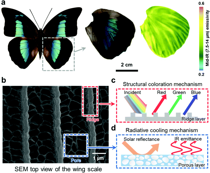
Fig. 1a includes the full snapshot of Archaeoprepona demophon and its dorsal wing along with mid-infrared mapping that exhibits high emissivity in most of its region. Fig. 1b illustrates the microscopic views of scanning electronic microscopy (SEM) for the dorsal wing scale of the butterfly, which comprises periodically spaced ridge-like structures and pores in between ridges as shown in the SEM image with the highest magnification. SEM images imply that pore size ranges from 200 nm to 800 nm in diameter, and several reports suggested that the porous structure serves to substantially increase the visible reflectance and infrared emittance by Mie scattering, thereby boosting the overall radiative cooling performance.20,36 The width of the ridge in the butterfly scale is approximately 500 nm, and the ridges are periodically interspaced by 2.3 μm, which contributes to the iridescent structural coloring of the butterfly wings.10,37 The underlying physical mechanisms for each structure of the butterflies that induce iridescent coloring and radiative cooling are depicted in Fig. 1c and d respectively. The spacing within the periodic grating determines the reflected color, as it induces Bragg diffraction of selective wavelength-band light.38 Thus, by precisely controlling the spacing of the periodic grating, we can generate structural colors, and therefore mimic the iridescent pattern of the butterfly wing scales. In addition to the ridge-like structure, the porous structure serves to reflect the light in the visible wavelength by Mie scattering (Fig. 1c),36 while radiative cooling polymers along with the porous structure serve to maximize light absorbance in the IR region, thereby enhancing the radiative cooling performance (Fig. 1d). | ||
| Fig. 1 The source of the biological inspiration and general overview of the study. (a) Digital photograph of the entire body of Archaeoprepona demophon (photograph by Didier Descouens, distributed under a CC-BY 3.0 license) and its dorsal wing with its corresponding mid-IR emissivity mapping (reproduced with permission from ref. 3. Copyright 2020 National Academy of Sciences). (b) SEM image of the Archaeoprepona demophon dorsal wing, which is characterized by periodic ridges and pores of different sizes. (c) Physical mechanisms to induce structural coloration and (d) radiative cooling based on ridge nanograting and porous structures, respectively. | ||
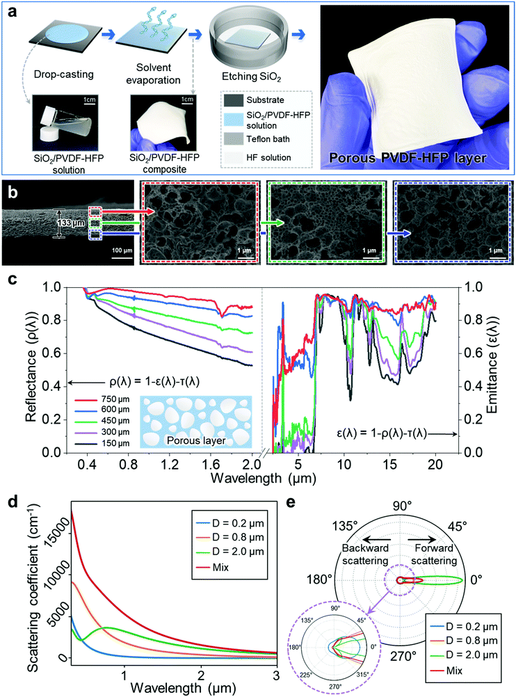
Inspired by the multifunctional structure of the butterfly wing scale that creates structural coloring and boosts the radiative cooling performance, we first devised a cost-effective solution-process fabrication method to artificially reconstruct the porous structure of the butterfly wing scale as is graphically illustrated in Fig. 2a. We synthesized SiO2 nanospheres of different sizes (200 nm, 800 nm, and 2 μm) based on the modified Stöber method and mixed them with PVDF-HFP powder in N-dimethylacetamide (DMAc) solution, which can disperse both SiO2 nanospheres and PVDF-HFP. We chose drop-casting as a solution depositing method since it offers a platform to fabricate a film with considerably high thickness for free-standing mechanical stability as well as for inducing radiative cooling. The subsequent heating process evaporates DMAc and results in a SiO2/PVDF-HFP composite film, which is then placed in an HF solution bath, producing an extremely porous film with random pore sizes. HF dissolves the SiO2 nanospheres into the water and SiF4 gas, while PVDF-HFP remains intact when in contact with HF due to high chemical resistant property of PVDF-HFP, resulting in a porous PVDF-HFP film as in the photograph on the right in Fig. 2a. Archaeoprepona demophon has pore sizes of 200 nm and 800 nm on its wing scale so we selected SiO2 sphere sizes of 200 nm and 800 nm to mimic the porous structure of its wing scale since the SiO2 sphere is directly translated into the pore size. We also included 2 μm pores in the porous film. Although pores of such a size are not present in the actual wing scale, it is reported that 2 μm pores serve to enhance the emittance in the middle wavelength infrared (MWIR) region.36
Cross-sectional SEM snapshots in Fig. 2b show the nanoporous PVDF-HFP film with three different pore sizes in the random configuration. It is also notable that the smaller pores perforated the surface of the larger pores, and this unique structure is likely to arise from the random hierarchical arrangement of the SiO2 nanospheres with different sizes in the composite film. The magnified SEM images indicate that HF completely permeates into the SiO2/PVDF-HFP composite and perforates the entire film without any SiO2 residue despite the hundreds-of-micrometer thickness of the porous film.
Notably, the porous PVDF-HFP structure serves to reflect the visible light due to light scattering20,36 and absorbs the infrared light as it is substantiated in the UV-Vis (ultraviolet-visible spectroscopy) and Fourier-transform infrared spectroscopy (FTIR) results of Fig. 2c, in which we measured visible reflectance and infrared emittance of porous films with varying thickness. The amount of the drop-cast SiO2/PVDF-HFP/DMAc solution has a directly proportional relationship with the thickness, so the larger the amount of drop-coated solution is, the thicker the porous PVDF-HFP film becomes. The UV-Vis result suggests that the visible reflectance increases as the porous film thickness becomes thicker because there would be more pores in the cross-sectional direction that would reflect the incident light on the film. Thus, the visible reflectance increases drastically from 0.78 to 0.99 for 150 μm and 750 μm samples, respectively, implying that the porous film with 750 μm thickness can reflect almost all the visible light that is incident on the film.
The FTIR result follows a similar pattern as the thicker film exhibits higher emittance as in Fig. 2c. The strong emission profile in the infrared region of the radiative cooling material originates both from the porous structure and vibration bands of the C–H and C–F bonding in PVDF-HFP itself.20,39 Thus, the synergistic effect of the structural and material properties increases as the film thickens because the thicker film would contain more C–H/C–F bonds and pores. A radiative cooling film of 750 μm thickness generates an overall ![[small epsilon, Greek, macron]](https://www.rsc.org/images/entities/i_char_e0c6.gif) 2–25μm value of 0.92 and
2–25μm value of 0.92 and ![[small epsilon, Greek, macron]](https://www.rsc.org/images/entities/i_char_e0c6.gif) atmosphericwindow value of 0.95, indicating that the film demonstrates high performance in absorbing both broadband and selective infrared lights. To further substantiate that the porous architecture contributes to the absorption increase in radiative cooling, we compared the infrared emittance of the porous PVDF-HFP film with that of a pristine one of equivalent weight so that we can neglect infrared emittance from material properties and only focus on the structural properties of PVDF-HFP films as shown in Fig. S1 (ESI†). The FTIR result demonstrates that the pores contribute to the substantial increase of emittance in the short infrared wavelength region (2 μm–8 μm) as the film thickness increases when compared to the pristine PVDF-HFP, corroborating the positive effect of pores on infrared emittance.
atmosphericwindow value of 0.95, indicating that the film demonstrates high performance in absorbing both broadband and selective infrared lights. To further substantiate that the porous architecture contributes to the absorption increase in radiative cooling, we compared the infrared emittance of the porous PVDF-HFP film with that of a pristine one of equivalent weight so that we can neglect infrared emittance from material properties and only focus on the structural properties of PVDF-HFP films as shown in Fig. S1 (ESI†). The FTIR result demonstrates that the pores contribute to the substantial increase of emittance in the short infrared wavelength region (2 μm–8 μm) as the film thickness increases when compared to the pristine PVDF-HFP, corroborating the positive effect of pores on infrared emittance.
To theoretically validate the experimentally acquired optical properties of the porous film, Fig. 2d delineates the plot for the scattering coefficient over the wavelength, which is directly related to the light scattering intensity. The result shows that 800 nm pores possess a strong scattering property in the visible light range, and 2 μm pores have a high scattering coefficient at longer wavelength, while 200 nm can be beneficial for UV solar light scattering or reflection. As a result, the mixed-pore condition that includes different sizes of pores demonstrates the summation of all the pore size scattering coefficients, encompassing all the highlighted advantages of different sized pores. The simulated result on the scattering mode in Fig. 2e implies that the forward scattering is dominant in the visible light reflection. The scattering phase function describes the angle probability that light may be scattered, and the result suggests that forward scattering tendency is apparent for larger size factor (diameter to wavelength) pores and small size factor pores exhibit larger backward scattering probability. The mixture of different sized pores can combine the scattering phase function probability and thus may be more adaptable to scatter different wavelength light.
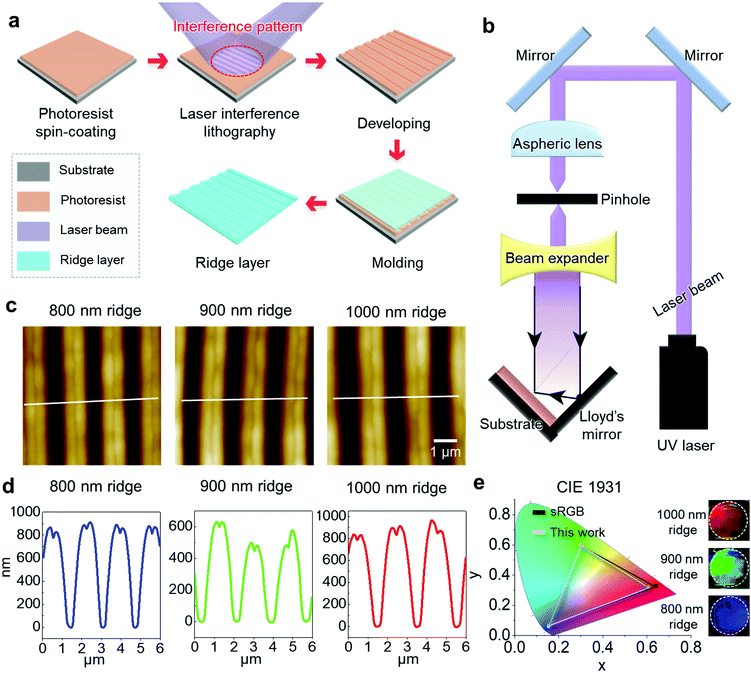
Along with the porous structure, we also aimed to reconstruct the nano-architecture of the ridge structure of the wing scale so that we can reproduce such multifunctional effects and manipulate its parameters for optimization. Laser interference lithography offers a cost-effective, rapid, and mask-less fabrication platform to generate a three-dimensional nano-grating pattern, unlike the conventional photolithography that involves complicated procedures and expensive cleanroom facilities.40Fig. 3a depicts the graphical representation of the laser interference lithography process in which the incident beam and reflected beam from the mirror were irradiated on the spin-coated PR film to create a nanograting pattern. Fig. 3b illustrates the optical setup of laser interference irradiation where the Lloyd mirror system enables the effective exposure of two interference beams with a single UV laser source. The angle between the incident beam and the mirror determines the distance between periodic gratings based on  , where d, λ, and θ correspond to the periodic distance, the wavelength of the incident light, and the angle between the incident beam and the mirror respectively. Fig. S2 (ESI†) shows the laser-interference patterned PR, and we were able to control the periodic grating of the PR mold from 400 nm to 900 nm as we manipulated θ. Thus, controlling the angle between the incident beam and the mirror allows us to fine-tune the distance between the ridges of the butterfly wing scale in the nanometer-region, as PDMS is imprinted and de-molded from the nano-grated PR film mold.
Fig. 3c shows the atomic force microscopy (AFM) image profiles of soft-imprinted PDMS films that have different nano-grating distances. Each film has interspacing distances of 800 nm, 900 nm, and 1000 nm, which generate structural coloring of blue, green, and red, respectively. Owing to the elaborate patterning nature of laser interference lithography, the nanograting pattern demonstrates precise periodicity as is also confirmed by the SEM images in Fig. S3 (ESI†). We utilized PR with a relatively high viscosity (∼8 cst) that results in a one to two micrometer thick PR film when spin-coated. For this reason, the AFM depth profile in Fig. 3d indicates that the height of the ridges reaches up to 800–900 nm, although the ridge height of the 900 nm ridge sample was not as high. Each periodic nanograting pattern created a clear red, green and blue color, and CIE 1931 in Fig. 3e shows the RGB color that each ridge grating produces. The simulated color gamut of the ridge grating structure covers 91.8% of sRGB, which represents the internationally approved color space that computer screens, printers, and web browsers utilize to express colors,41 suggesting that the ridge grating film can replicate most of the standardized color profiles.
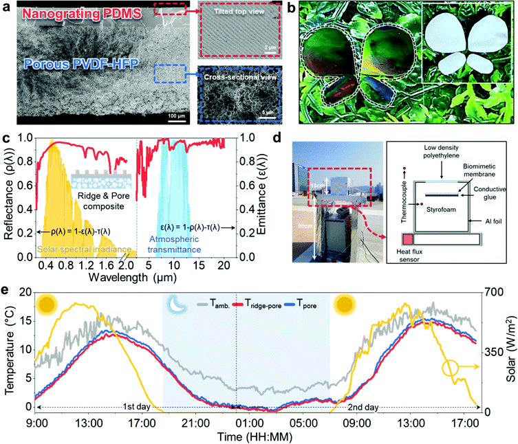
Fig. 4 shows the integrated structure of grated ridges with pores and further investigates effective radiative cooling and structural coloring of the biomimetic film. Fig. 4a shows the cross-sectional SEM image of the integrated porous and ridge structure that mimics the nanostructure of the butterfly wing scale. Just like the butterfly wing structure, the tilted top view of the biomimetic film shows a highly periodic nanograted surface whereas the magnified cross-sectional view of the porous region of the film exhibits densely populated and hierarchical pores of different sizes as in the insets of the figure. We acknowledge that the thickness of the biomimetic film herein is much thicker than that of the Archaeoprepona demophon wing scale. Rather than duplicating its exact microstructure with decreased radiative cooling performance, we aimed to develop a biomimetic film with improved radiative cooling capability based on biological inspirations since the biomimetic film thickness greatly affects the visible reflection and infrared emittance, which are key parameters for radiative cooling.
Fig. 4b shows the structural color functionality of the butterfly biomimetic film based on its nanograting structure. It exhibits a butterfly-resembling structure that imitates the iridescent coloring of butterfly wings including red, green, and blue. The structural color of the butterfly wings camouflages the species in its natural habitat, and we placed the butterfly biomimetic film on a grass background to corroborate this point. As can be observed from the figure, the nanograting structure of the biomimetic film enables it to camouflage into the background, while the sample without any nanograting texture on the top in the inset stands out, exemplifying the potential camouflage effect of the structural color functionality for butterflies. To demonstrate and quantify the radiative cooling performance along with structural coloring, we measured the optical properties of the integrated biomimetic structure as in Fig. 4c. The UV-Vis result indicates a ![[R with combining macron]](https://www.rsc.org/images/entities/i_char_0052_0304.gif) visible value of 0.944 for the integrated structure, which is, in fact, smaller than the
visible value of 0.944 for the integrated structure, which is, in fact, smaller than the ![[R with combining macron]](https://www.rsc.org/images/entities/i_char_0052_0304.gif) visible value of the porous film (=0.99) perhaps because the nanograted PDMS layer absorbs a portion of the incident light that should have been reflected in the porous film. On the other hand, the
visible value of the porous film (=0.99) perhaps because the nanograted PDMS layer absorbs a portion of the incident light that should have been reflected in the porous film. On the other hand, the ![[small epsilon, Greek, macron]](https://www.rsc.org/images/entities/i_char_e0c6.gif) 2–25μm value increased from 0.914 to 0.934 with an enhanced
2–25μm value increased from 0.914 to 0.934 with an enhanced ![[small epsilon, Greek, macron]](https://www.rsc.org/images/entities/i_char_e0c6.gif) atmosphericwindow value of 0.959 because the PDMS layer also possesses high emittance and also increases the effective thickness of the film as it contains Si–O, Si–C, and C–O bonds, the vibration bands of which serve to increase the emittance.42,43
atmosphericwindow value of 0.959 because the PDMS layer also possesses high emittance and also increases the effective thickness of the film as it contains Si–O, Si–C, and C–O bonds, the vibration bands of which serve to increase the emittance.42,43
To validate the radiative cooling performance of the biomimetic film, we examined the cooling temperature of the biomimetic film with the radiative cooling test setup that is made up of an acrylic box wrapped with aluminum foil to minimize the solar radiation transferred to the setup box as in Fig. 4d. As polystyrene foam (Styrofoam) possesses a great insulating property, we used polystyrene foam to fill the setup box to prevent external heat conduction from the outside system, and a transparent polyethylene film that was attached above the biomimetic film sample served to minimize the energy loss from external air heat convection. We examined the cooling performance of (i) porous film and (ii) the integrated film of porous and ridge structures to evaluate the radiative cooling capability of the biomimetic film and compare the cooling performance of the porous film and integrated film structure. Fig. 4e shows the recorded Tamb, Tridge-pore, and Tpore values over the approximately two-day time span with solar radiance plotted along with temperatures. As discussed previously in the previous section, the integrated film of the ridge-pore structure generated slightly higher ΔT than the porous film due to the covalent bonds in PDMS of the ridge-like structure that contributes to the radiative cooling capability and increases the effective thickness of the integrated film with a ridge-pore structure. The integrated film recorded an extremely large drop in maximum radiative cooling temperature of 8.45 °C, which primarily originates from the porous architecture since the porous film also generated a maximum radiative cooling temperature reduction of 8.27 °C and followed a similar radiative cooling pattern throughout the entire time-span of the experiment. This can suggest that the radiative cooling functionality of the actual wing scale of the butterfly species mainly stems from its porous structure as we also demonstrated the substantial radiative cooling with the reconstructed porous structure of the biomimetic film. Thus, we expect that the ridge-like structure of the butterfly wing scale is responsible for iridescent colors while the porous structure generates radiative cooling. Hence, we believe that our work might offer several valuable insights into the useful functionalities of natural species as we faithfully reconstructed their nano-microstructures by precise state-of-the-art nanoscale manufacturing technologies and replicated the dual functionalities of the natural species.
Conclusions
Inspired by the biological source, we developed a multifunctional biofilm that imitates the nanostructure of the Archaeoprepona demophon wing scale that exhibits radiative cooling and structural coloring functionalities by modulating the infrared absorbance and visible reflection level. We devised two different, cost-effective fabrication strategies for ridge and porous structures and integrated them to artificially reconstruct the butterfly wing scale. For the porous structure, we used a solution-based process that removes SiO2 nanospheres of different sizes to make an inverse opal structure. Coupled with the radiative cooling material property of PVDF-HFP, the random multi-porous structure plays a dominant role in maximizing the radiative cooling performance of butterfly species. On the other hand, for the ridge structure, we resorted to laser interference lithography to fabricate a periodic nanograting surface on which a soft elastomer can be imprinted to mimic the ridge structure of Archaeoprepona demophon. The ridge structure serves to generate structural coloring that mimics the visual appearance of the butterfly wing scale. Integration of porous and ridge structures resulted in radiative cooling and structural coloring functionalities of the butterfly wing scale, as it recorded the maximum radiative cooling temperature of 8.45 °C, while we imprinted the ridge structure with different grating distance so that each region with a different imprint generates distinctive color. Overall, we expect that the biomimetic film developed herein would not only offer us a clearer experimental insight into the structure–functionality relationship of the Archaeoprepona demophon wing scale, but it would also make substantial contributions to the state-of-the-art radiative cooling technologies that have the potential to address climate change.Experimental section
Materials
PVDF-HFP (Kynar Flex 2801, Arkema), PDMS (Sylgard 184, Dow), N-dimethylacetamide (Sigma Aldrich), tetraethyl orthosilicate (TEOS) (Sigma Aldrich), ammonium hydroxide (Sigma Aldrich), potassium chloride (Sigma Aldrich), photoresist (KL5310, KemLab), and Silc Pig™ black dye (Smooth-on) were used as received without purification.Methods
 | (1) |
The exposure was conducted following the typical process. The SiO2 substrate (SL.Sli1012, SciLab, Korea) with an area of 25.4 × 25.4 mm2 was ultrasonically cleaned in ethanol and deionized (DI) water for 5 min. The spin coating of positive PR (KL5310, KemLab, USA) was subsequently conducted at 3000 rpm for 60 s, and baking was conducted at 105 °C for 60 s. After 1 min of cooling, the collimated beam was irradiated on the substrate at an intensity of 2.55 μW cm−2 for 200 s. Finally, tetramethylammonium hydroxide (TMAH) developer solution was drop-cast onto the exposed substrate for 10 s. After puddle-type development, the substrate was rinsed with DI water, followed by N2 blowing. PDMS was prepared by mixing the base and cross-linker in a ratio of 10![[thin space (1/6-em)]](https://www.rsc.org/images/entities/char_2009.gif) :
:![[thin space (1/6-em)]](https://www.rsc.org/images/entities/char_2009.gif) 1, and then it was poured onto the laser-patterned template in a vacuum environment and cured subsequently to make a nano-grated elastomer. We made an additional black dyed PDMS layer by mixing the black dye with uncured PDMS and attached it beneath the nanograting layer so that it can reflect the selective light.
1, and then it was poured onto the laser-patterned template in a vacuum environment and cured subsequently to make a nano-grated elastomer. We made an additional black dyed PDMS layer by mixing the black dye with uncured PDMS and attached it beneath the nanograting layer so that it can reflect the selective light.
Synthesis of SiO2 nanospheres
Three different sizes of SiO2 nanospheres (200 nm, 800 nm, and 2 μm) were synthesized using different protocols based on the Stöber method. For 200 nm SiO2 nanospheres, 6 mL of TEOS, 300 mL of ethanol, 6 mL of deionized (DI) water, and 42 mL of NH4OH were poured into a quartz flask under vigorous stirring for 6 hours. For 800 nm nanospheres, Solution 1 was prepared by mixing NH4OH with 2.14 mL of TEOS and 33.3 mL of ethanol, and Solution 2 was made by mixing 12 mL of NH4OH, 65 mL of ethanol, 6.75 mL of DI water, and 0.017 g of KCl. Solution 1 was injected into Solution 2 at a rate of 0.02 mL min−1 under vigorous stirring. For 2 μm microspheres, 12.86 mL of TEOS and 33.3 mL of ethanol comprise Solution 1, while 16 mL of NH4OH, 65 mL of ethanol, 6.75 mL of DI water, and 0.017 g of KCl make up Solution 2. Just as in the 800 nm SiO2 nanosphere protocol, Solution 1 was injected into Solution 2 at 0.2 mL min−1 under vigorous stirring. After the synthesis process was finished, the obtained SiO2 nanoparticles were centrifuged in ethanol at 5000 rpm and the supernatant solution was discarded, leaving the precipitants only. The precipitant was dispersed in ethanol and tip-sonicated until it was completely dispersed. Such a cleaning process was repeated three times so that the resulting SiO2 nanospheres are free of undesired impurities.Fabrication of the porous PVDF-HFP structure and its integration with the nano-grated film
SiO2 nanospheres and PVDF-HFP (6![[thin space (1/6-em)]](https://www.rsc.org/images/entities/char_2009.gif) :
:![[thin space (1/6-em)]](https://www.rsc.org/images/entities/char_2009.gif) 4 wt. ratio) were dispersed in DMAc solution, and the solution was drop-cast on a plasma-treated glass slide and then heat-treated for solvent evaporation. After successive drop-casting and drying were conducted to achieve the target thickness of PVDF-HFP/SiO2, the film was placed in a diluted HF (10%) bath overnight to remove SiO2 nanospheres so that it results in a porous PVDF-HFP film. The porous film was rinsed several times to remove potentially remaining HF and then dried at room temperature. The nano-grated film was then integrated into the porous film by spin-coating PDMS at 2000 rpm for 30 seconds on the porous film and placing the nano-grated film on top of it under a vacuum environment. The integrated film was then cured at 60 degrees for an hour.
4 wt. ratio) were dispersed in DMAc solution, and the solution was drop-cast on a plasma-treated glass slide and then heat-treated for solvent evaporation. After successive drop-casting and drying were conducted to achieve the target thickness of PVDF-HFP/SiO2, the film was placed in a diluted HF (10%) bath overnight to remove SiO2 nanospheres so that it results in a porous PVDF-HFP film. The porous film was rinsed several times to remove potentially remaining HF and then dried at room temperature. The nano-grated film was then integrated into the porous film by spin-coating PDMS at 2000 rpm for 30 seconds on the porous film and placing the nano-grated film on top of it under a vacuum environment. The integrated film was then cured at 60 degrees for an hour.
Characterization of the biomimetic film and measurement of radiative cooling
To characterize the nanograting profile of the ridge structure, we resorted to AFM (NX-100, Park Systems) instead of SEM and measured with a 6 μm by 6 μm window because SEM cannot quantify its ridge depth information with a single view. UV-Vis (V-770, Jasco) and FTIR (Spectrum Paragon, PerkinElmer) measured the visible reflectance and infrared emissivity of the porous film and ridge-pore film to characterize and further compare the cooling performance of each film. To evaluate the cooling performance of the biomimetic film, we devised a homemade acrylic test box that is filled with styrofoam to minimize thermal conduction from the outside environment. To reduce the heat conduction from the outside environment as much as possible, we placed a box on the insulating table that is 90 cm high. The aluminum foil was utilized to wrap around the test box to reflect the sunlight for minimizing the solar radiation effect. We also attached the low-density polyethylene film right above the film sample to suppress the air heat convection into the system. The heat flux sensor was inserted inside the test box to measure the cooling temperature, and a separate thermocouple was set up outside the test system to measure the ambient temperature.Theoretical study for scattering phase functions
According to Mie scattering theory, the scattering or extinction efficiency is determined by the size factor and effective complex refractive index, which can be defined as m = (n − ki)/nm and x = 2 πr/λ, respectively, where r, λ, n, k and nm correspond to the pore radius, light wavelength, pore optical constant, and polymer matrix refractive index, respectively. The scattering Qsca and extinction efficiency Qext can be calculated as follows:where an and bn are the Mie scattering coefficients, which can be written as the following equations,
in which, ψn and ζn are the Riccati–Bessel functions. The phase function property is related to the scattering field pattern which can be derived as follows,
where θ is the scattering polar angle, πn and τn are related to Legendre polynomials Pn, which can be expressed as follows:
The volume-averaged scattering coefficient σs and extinction coefficient σe can be calculated using the following equations:
The volume-averaged phase function can be calculated as
where L is the light intensity,
![[s with combining right harpoon above (vector)]](https://www.rsc.org/images/entities/b_i_char_0073_20d1.gif) is the direction vector in the solid angle space, and r is the coordinate position. The first term on the right hand side in the equation describes the light attenuation by pores including scattering and absorption. The second term represents the addition of light intensity by scattered light intensity from the direction
is the direction vector in the solid angle space, and r is the coordinate position. The first term on the right hand side in the equation describes the light attenuation by pores including scattering and absorption. The second term represents the addition of light intensity by scattered light intensity from the direction ![[s with combining right harpoon above (vector)]](https://www.rsc.org/images/entities/b_i_char_0073_20d1.gif) ′ to the computing direction
′ to the computing direction ![[s with combining right harpoon above (vector)]](https://www.rsc.org/images/entities/b_i_char_0073_20d1.gif) . The integral-differential photon transport process needs numerical iterations to get convergent results due to complex scattering.
. The integral-differential photon transport process needs numerical iterations to get convergent results due to complex scattering.
Author contributions
J. L. and Y. J. contributed equally to this work. J. L., Y. J., and S.-H. K. conceived the work; M. L. built the measurement setup for radiative cooling temperature and characterized the radiative cooling performance; J.-S. H., H.-S. L., and Y.-J. K. fabricated the ridge-like structure of the butterfly wing scale based on laser-interference lithography; J. G., J. L., Y. J., Y. L, and J. S. discussed the theoretical background of optics/heat transfer for radiative cooling and J. G. computed the optical properties based on the simulation software; J. L., Y. J., and M. L. characterized the optical properties of the porous, ridge-like, and integrated films; Y. J., W. S., J. M. and K.-R. P. fabricated the test samples for characterization and radiative cooling performance; J. L., and S.-H. K. wrote the manuscript; J. L., Y. J., M. L., S.-H. K., and B.-W. K. discussed the overall result. S.-H. K. and B.-W. K. funded the project and led the work.Conflicts of interest
The authors declare no conflict of interest.Acknowledgements
This work was supported by the Hyundai Motor Company (HR-204429.0002) and the National Research Foundation of Korea (NRF) Grant (NRF-2021R1A2B5B03001691 and 2016R1A5A1938472).References
- S. Ryding, M. Klaassen, G. J. Tattersall, J. L. Gardner and M. R. Symonds, Trends Ecol. Evol., 2021, 36, 1036–1048 CrossRef PubMed.
- M. M. Hantak, B. S. McLean, D. Li and R. P. Guralnick, Commun. Biol., 2021, 4, 1–10 CrossRef PubMed.
- A. Krishna, X. Nie, A. D. Warren, J. E. Llorente-Bousquets, A. D. Briscoe and J. Lee, Proc. Natl. Acad. Sci. U. S. A., 2020, 117, 1566–1572 CrossRef CAS PubMed.
- R. H. Siddique, Y. J. Donie, G. Gomard, S. Yalamanchili, T. Merdzhanova, U. Lemmer and H. Hölscher, Sci. Adv., 2017, 3, e1700232 CrossRef PubMed.
- I. Medina, E. Newton, M. R. Kearney, R. A. Mulder, W. P. Porter and D. Stuart-Fox, Nat. Commun., 2018, 9, 1–7 CrossRef CAS PubMed.
- N. N. Shi, C.-C. Tsai, M. J. Carter, J. Mandal, A. C. Overvig, M. Y. Sfeir, M. Lu, C. L. Craig, G. D. Bernard and Y. Yang, Light: Sci. Appl., 2018, 7, 1–9 CrossRef CAS PubMed.
- N. N. Shi, C.-C. Tsai, F. Camino, G. D. Bernard, N. Yu and R. Wehner, Science, 2015, 349, 298–301 CrossRef CAS PubMed.
- C.-C. Tsai, R. A. Childers, N. N. Shi, C. Ren, J. N. Pelaez, G. D. Bernard, N. E. Pierce and N. Yu, Nat. Commun., 2020, 11, 1–14 CrossRef PubMed.
- H. Zhang, K. C. Ly, X. Liu, Z. Chen, M. Yan, Z. Wu, X. Wang, Y. Zheng, H. Zhou and T. Fan, Proc. Natl. Acad. Sci. U. S. A., 2020, 117, 14657–14666 CrossRef PubMed.
- B. D. Wilts, A. Matsushita, K. Arikawa and D. G. Stavenga, J. R. Soc., Interface, 2015, 12, 20150717 CrossRef PubMed.
- T. K. Suzuki, S. Tomita and H. Sezutsu, J. Morphol., 2019, 280, 149–166 CrossRef PubMed.
- A. McDougal, B. Miller, M. Singh and M. Kolle, J. Opt., 2019, 21, 073001 CrossRef CAS.
- X. Liu, D. Wang, Z. Yang, H. Zhou, Q. Zhao and T. Fan, Adv. Opt. Mater., 2019, 7, 1900687 CrossRef.
- S. Zhu, D. Zhang, Z. Chen, J. Gu, W. Li, H. Jiang and G. Zhou, Nanotechnology, 2009, 20, 315303 CrossRef PubMed.
- Z. Han, Z. Mu, B. Li, S. Niu, J. Zhang and L. Ren, Small, 2016, 12, 713–720 CrossRef CAS PubMed.
- H. Ding, D. Liu, B. Li, W. Ze, S. Niu, C. Xu, Z. Han and L. Ren, ACS Appl. Mater. Interfaces, 2021, 13, 19450–19459 CrossRef CAS PubMed.
- G. Zyla, A. Kovalev, E. L. Gurevich, C. Esen, Y. Liu, Y. Lu, S. Gorb and A. Ostendorf, Appl. Phys. A: Mater. Sci. Process., 2020, 126, 1–11 CrossRef.
- S. Zhang and Y. Chen, Sci. Rep., 2015, 5, 1–10 CrossRef.
- A. Didari and M. P. Mengüç, Sci. Rep., 2018, 8, 1–9 CAS.
- J. Mandal, Y. Fu, A. C. Overvig, M. Jia, K. Sun, N. N. Shi, H. Zhou, X. Xiao, N. Yu and Y. Yang, Science, 2018, 362, 315–319 CrossRef CAS PubMed.
- J. Wang, J. Sun, T. Guo, H. Zhang, M. Xie, J. Yang, X. Jiang, Z. Chu, D. Liu and S. Bai, Adv. Mater. Technol., 2021, 2100528 Search PubMed.
- B. Xiang, R. Zhang, Y. Luo, S. Zhang, L. Xu, H. Min, S. Tang and X. Meng, Nano Energy, 2021, 81, 105600 CrossRef CAS.
- B. Hatton, L. Mishchenko, S. Davis, K. H. Sandhage and J. Aizenberg, Proc. Natl. Acad. Sci. U. S. A., 2010, 107, 10354–10359 CrossRef CAS PubMed.
- L. Wang, Y. Wan, Y. Li, Z. Cai, H.-L. Li, X. Zhao and Q. Li, Langmuir, 2009, 25, 6753–6759 CrossRef CAS PubMed.
- S. W. Choi, Y. Zhang, M. R. MacEwan and Y. Xia, Adv. Healthcare Mater., 2013, 2, 145–154 CrossRef CAS PubMed.
- X. Chia and M. Pumera, ACS Appl. Mater. Interfaces, 2018, 10, 4937–4945 CrossRef CAS PubMed.
- G. H. Choi, D. K. Rhee, A. R. Park, M. J. Oh, S. Hong, J. J. Richardson, J. Guo, F. Caruso and P. J. Yoo, ACS Appl. Mater. Interfaces, 2016, 8, 3250–3257 CrossRef CAS PubMed.
- J. Sun, B. Bhushan and J. Tong, RSC Adv., 2013, 3, 14862–14889 RSC.
- K. T. Lee, C. Ji, D. Banerjee and L. J. Guo, Laser Photonics Rev., 2015, 9, 354–362 CrossRef CAS.
- I. Koirala, V. R. Shrestha, C.-S. Park, S.-S. Lee and D.-Y. Choi, Sci. Rep., 2017, 7, 1–8 CrossRef CAS PubMed.
- K. T. Lee, J. Y. Jang, S. J. Park, C. Ji, S. M. Yang, L. J. Guo and H. J. Park, Adv. Opt. Mater., 2016, 4, 1696–1702 CrossRef CAS.
- H. Jiang and B. Kaminska, ACS Nano, 2018, 12, 3112–3125 CrossRef CAS PubMed.
- H. Wu, Y. Jiao, C. Zhang, C. Chen, L. Yang, J. Li, J. Ni, Y. Zhang, C. Li and Y. Zhang, Nanoscale, 2019, 11, 4803–4810 RSC.
- J. S. Hwang, J.-E. Park, G. W. Kim, H. Lee and M. Yang, Appl. Surf. Sci., 2021, 547, 149178 CrossRef CAS.
- H. Kim, H. Jung, D.-H. Lee, K. B. Lee and H. Jeon, Appl. Opt., 2016, 55, 354–359 CrossRef PubMed.
- M. Chen, D. Pang, J. Mandal, X. Chen, H. Yan, Y. He, N. Yu and Y. Yang, Nano Lett., 2021, 21, 1412–1418 CrossRef CAS PubMed.
- S. Kinoshita, S. Yoshioka and J. Miyazaki, Rep. Prog. Phys., 2008, 71, 076401 CrossRef.
- I. Rashid, H. Butt, A. K. Yetisen, B. Dlubak, J. E. Davies, P. Seneor, A. Vechhiola, F. Bouamrane and S. Xavier, ACS Photonics, 2017, 4, 2402–2409 CrossRef CAS.
- A. Aili, Z. Wei, Y. Chen, D. Zhao, R. Yang and X. Yin, Mater. Today Phys., 2019, 10, 100127 CrossRef.
- C. Lu and R. Lipson, Laser Photonics Rev., 2010, 4, 568–580 CrossRef CAS.
- The Zen of sRGB Color Space, https://www.pixelz.com/blog/zen-srgb-color-space/, 2021.
- M. Zhou, H. Song, X. Xu, A. Shahsafi, Y. Qu, Z. Xia, Z. Ma, M. A. Kats, J. Zhu and B. S. Ooi, Proc. Natl. Acad. Sci. U. S. A., 2021, 118 Search PubMed.
- M. Chen, D. Pang, X. Chen and H. Yan, J. Phys. D: Appl. Phys., 2021, 54, 295501 CrossRef CAS.
Footnotes |
| † Electronic supplementary information (ESI) available. See DOI: https://doi.org/10.1039/d2nh00166g |
| ‡ J. Lee and Y. Jung equally contributed to this work. |
| § None of the material has been published or is under consideration elsewhere, including the Internet. |
| This journal is © The Royal Society of Chemistry 2022 |


