DOI:
10.1039/C6RA11453A
(Paper)
RSC Adv., 2016,
6, 76137-76141
Direct ultrasonic modification of a current collector with enhanced pseudocapacity†
Received
3rd May 2016
, Accepted 28th July 2016
First published on 28th July 2016
Abstract
In this study, we used a direct ultrasonic modified nickel foam in sulfuric acid, which achieved a simple and rapid binder-free electrode, improving the electronic transmission in the electrolyte. It produced a bed of electroactive nickel hydroxide and sulphide with a unique needle-like microstructure, which grew vertically on the surface of the Ni foam straight into the electrolyte, not only increasing the contact interface area between the active material and the electrolyte but also shortening the diffusion length of the ions. The obtained product possesses outstanding electrochemical performance, having a high specific capacitances of 6 F cm−2 at a current density of 5 mA cm−2, which can still retain 2.2 F cm−2, even at a current density as high as 40 mA cm−2. Furthermore, it has excellent cyclic stability with only 5% loss after 1000 cycles.
1. Introduction
Binder-free electrodes are a very important class of porous electrode, which simplify the preparation technique and effectively promote electronic and ionic transport properties.1 The formation processes always comprise the active materials growing on the current collectors directly by chemical methods (e.g., hydrothermal growth, chemical vapour deposition, atomic layer deposition, and electroplating). In contrast, the conventional binder electrode is a poor electronic conductor of the active material and cannot be effectively compensated for by the tight integration of the grown product on the current collector. For this reason, this design can neglect other auxiliary components of the configuration, such as conductive agents and binders, which are completely unnecessary in porous electrodes.2 It is effective to reduce the ionic diffusion resistance and improve the surface activity of the materials that will be exposed and soaked. Although it has been only a few years since this new concept came out, binder-free electrodes are being increasingly used in numerous types of energy storage devices, particularly supercapacitors.
Various metals, such as Al, Cu, Ni, and stainless steel, are used as current collectors with binder-free electrodes in the form of thin foils, meshes or foams. Among these, nickel foam has attracted great interest as a current collector in supercapacitors, due to its high corrosion resistance and three-dimensional structure, which provides a larger surface area and better active material/electrolyte contact. To date, both electric double layer capacitive materials3 (e.g., graphene and carbon nanotubes) and pseudocapacitive materials4 (e.g., Co3O4, MnO2, and ZnCo2O4) have been successfully grown on a nickel foam and are used as capacitor electrodes. Based on the reversible conversion of nickel oxidation states from bivalent to trivalent, nickel-containing compounds (Ni(OH)2, Ni3S2, NiO, Ni2P, NiCl2, and NiF2) have become one of the most significant categories for use as active materials in binder-free pseudocapacitor electrodes. Furthermore, the substantial deposits and affordable cost also make nickel-based active materials a promising candidate for commercial applications. One question that has been frequently asked: what happens to the pseudocapacitive properties of binder-free electrodes when there is an in situ modification on the current collectors? A more direct example of this broad question: can nickel elements from a nickel foam be transformed into nickel-containing compounds directly? Wang et al. reported a self-supported binder-free Ni/NiO electrode configuration constructed via a nickel foam-based pyrogenic oxidation process where the porous core–shell structured Ni/NiO delivered improved performance as an anode for a lithium ion battery.5 Analogously, Tang et al. proposed a simple two-step modification to activate a carbon cloth by wet chemical and thermal reduction treatments. The results of the modification showed excellent wettability and a high surface area, which greatly enhanced its capacitive performance.6
In this paper, a fast, practical and low cost electrode fabrication process is described. Nickel foam, used as the current collector, demonstrated a remarkably improved pseudocapacity via a simple ultrasonic modification in which a bed of electroactive nickel hydroxide and sulphide was formed on the surface of the foam directly through interfacial oxidation reactions. The enhanced electrochemical performance is primarily attributed to the surface formation of a unique needle-like microstructure, which minimized the contact resistance between the active materials and the Ni foam skeleton.
2. Experimental
2.1. Materials synthesis
A nickel foam (1 cm × 1 cm) was used as the current collector and cleaned with acetone and deionized water in an ultrasonic bath for 10 min. Subsequently, it was sonicated in a 6 M HCl solution for 20 min and washed with deionized water and absolute ethanol. Then, water and ethanol on the surface of the Ni foam were soaked using a filter paper. The dried Ni foam was placed into 95.5 wt% H2SO4 and subjected to ultrasonic processing for 5 min. The blackened Ni foam was soaked with a filter paper and placed into a 0.5 M KOH solution for another ultrasonic processing until no more floccule was generated on the surface. The obtained Ni foam was soaked with a filter paper and washed with deionized water and absolute ethanol three times. Finally, this modified Ni foam was soaked up once more with a filter paper and used as the working electrode for electrochemical tests.
2.2. Materials characterization
The as-prepared products were characterized with X-ray photo electron spectroscopy measurements (XPS; VG ESCALAB MARK II, Mg-Kα radiation), scanning electron microscopy (FESEM; JEOL, JSM-7600F), and transmission electron microscopy (TEM; JEOL, JEM-2100F) operated at 200 kV.
2.3. Electrochemical measurements
Cyclic voltammetry (CV) and electrochemical impedance spectroscopy (EIS) measurements were performed using a potentiostat/galvanostat (Ametek, PAR 2273). EIS tests were performed in a frequency range from 0.01 to 105 Hz with an amplitude of 5 mV as the open circuit potential. The galvanostatic charge–discharge measurements were conducted on an electrochemical analyser (Chenhua, CHI 660E). All the experiments were performed with a three-compartment cell in a 3 M KOH aqueous solution as the electrolyte. An Ag/AgCl electrode was used as the reference electrode, and a piece of platinum netting was employed as the counter electrode. To minimize any errors due to an iR drop in the electrolyte, the reference electrode was connected to the cell via a double salt bridge system and a Luggin capillary. After cell assembly, the working electrode was impregnated in the electrolyte for 2 h under vacuum to ensure that the electrode was thoroughly wetted. The specific capacitance (C) of the electrode was evaluated according to the following equation:
where C (F cm−2) is the specific capacitance of the electrode based on the area of electrode, I (mA cm−2) is the current density during the discharge process, Δt (s) is the discharge time, and ΔV (V) is the potential window (here ΔV = 0.45 V).
3. Results and discussion
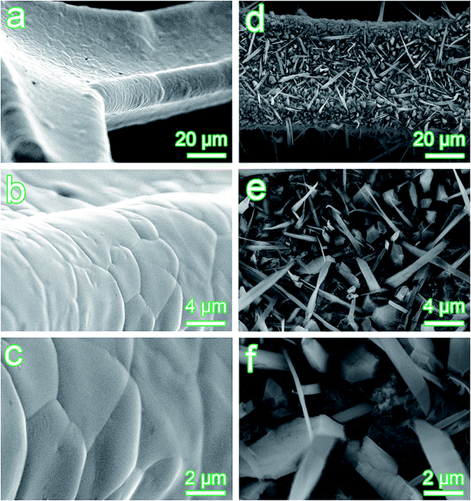
The morphologies of the obtained electrodes were characterized using scanning electron microscopy (SEM) and the results are shown in Fig. 1. Fig. 1a–c show the SEM images of the bare Ni foam, which exhibits a dense and smooth surface. These structural characteristics allow the electrolyte easy access to the entire surface of the generated materials, which facilitates charge transport and ion diffusion without the limitations associated about the binder. After ultrasonic modification (Fig. 1d–f), the surface of the entire Ni foam becomes chapped and rough, as revealed in the spaces between the needle-like microstructure. In addition, it was observed that the nickel substrate is clearly covered by the previous inexistence of materials. In addition, it was found that the materials had grown vertically on the entirety of the Ni foam to form a unique needle-like microstructure. The SEM images shown in Fig. S1† at other temperatures. The mesh structure is still maintained at 5 °C; at 30 °C, it exhibited a flake-like structure; the result of Fig. S1c† shows a globular structure and with the increase of temperature, the reunion completely covered the surface of nickel foam.
 |
| | Fig. 1 SEM images of the bare Ni foam and the modified nickel foam under different magnification. (a–c) The bare Ni foam; (d–f) the modified nickel foam. | |
Fig. 2 depicts the EDS pattern of generated materials on the surface of the Ni foam substrate and indicates the presence of Ni, O and S. The components of the generated materials are further verified by X-ray photoelectron spectroscopy (XPS) analysis. For the Ni 2p spectra (Fig. 3a), two Ni 2p core levels (2p1/2 and 2p3/2) and two satellite peaks are observed. The binding energy of the Ni 2p3/2 peak is distinctive from those found in the literature for NiO (853.7 eV) and Ni (852.6 eV),7,8 but standardized to Ni(OH)2 (856 eV).9 A spin-energy separation of 17.6 eV determined for 2p3/2 to 2p1/2 (873.5 eV) is also characteristic of the Ni(OH)2 phase and is in good agreement with the literature.10 Therefore, the results of this detailed analysis reveal that the components include Ni(OH)2 and NiO is absent. In terms of literature, the binding energy of NiS is located at 853.1 eV (ref. 11) and S 2p core is found at 162.8 eV;12 simultaneously, NiSO4 peak near 857.8 eV and S 2p core is found at 169.3 eV.12
 |
| | Fig. 2 EDS pattern of the target materials on the surface of Ni foam substrate. | |
 |
| | Fig. 3 The XPS spectra of the samples. (a) Ni 2p core; (b) O 1s and S 2p core. | |
Through Fig. 3a and b, we know that the smaller shoulder peak near 853.1 (±0.2) eV and S 2p core is found at 162.8 eV. The search reveals that the components contain NiS instead of NiSO4. The most likely source of the sulfide ion is the reduction of H2SO4 molecules.12 Furthermore, a more detailed verification of the formation of the electroactive nickel hydroxide and sulphide can be conducted using high-resolution transmission electron microscopy (HRTEM). In Fig. 4, lattice fringes of 0.2309 nm, 0.2331 nm, 0.1976 nm and 0.2588 nm, respectively, according to JCPDS card no., proved that the growth direction of Ni(OH)2 can be inferred as the crystal plane of (002) and (011), and NiS can be inferred as the crystal plane of (102) and (101). Fig. S2† represents the N2 adsorption and desorption isotherms of type IV accompanied by a H3 hysteresis loop. The pore size distribution curves are displayed in the inset, wherein the main presence was mesoporous (4 nm).
 |
| | Fig. 4 HRTEM images of the target materials on the surface of Ni foam substrate and analysis of the components based on the JCPDS card no., crystal plane and lattice fringes. | |
The electrochemical performances of the modified nickel foam when used as working electrodes for pseudocapacitors were investigated. The CV curves of the modified Ni foam (MNF) at different scan rates (1 to 20 mV s−1) within the potential window from 0 to 0.45 V in 3 M KOH solution are shown in Fig. 5a. The redox peaks are maintained at different scan rates, and the shapes of these curves are similar, meaning that the pseudocapacitive behaviour of the electrode is stable over this range. The anodic peak appeared indicates an oxidation process due to the oxidation of Ni(OH)2 to NiOOH and NiS to NiSOH, whereas the cathodic peak was observed owing to the reverse process. The corresponding equation can be expressed as follows:10,13
| Ni(OH)2 + OH− ↔ NiOOH + H2O + e− |
 |
| | Fig. 5 Electrochemical characterization of the MNF and BNF electrodes. (a) CV curves of the MNF electrode at scan rates of 1, 2, 5, 10, and 20 mV s−1; the inset shows the CV curves of the MNF and BNF at 10 mV s−1; (b) discharge curves of the MNF electrode at current densities of 5, 8, 10, 12, 15, 20, 25, 30, 35, and 40 mA cm−2; the inset shows the discharge curve of the MNF and BNF at 10 mA cm−2; (c) the corresponding specific capacitance (●) and capacity retention (○) as a function of different current densities; (d) expanded views of the high frequency region of the Nyquist plots with the imaginary part (Y-axis) vs. the real part (X-axis) of the impedance from the EIS studies; (e) electrical equivalent circuit used for fitting the impedance spectra and the calculated values of Rs, Rct, W and Cps determined via CNLS fitting of the experimental impedance spectra based on the equivalent circuit; (f) cycling performance of the MNF electrode measured at a current density of 10 mA cm−2. | |
The CV curves of the bare Ni foam (BNF) and the MNF measured at 10 mV s−1 are shown in the inset of Fig. 5a. The response current of BNF is much weaker than that of the MNF, which illustrates the enhanced pseudocapacity that can be obtained from the formation of electroactive nickel hydroxide and sulphide. The galvanostatic charge/discharge examination at 1 to 40 mA cm−2 is shown in Fig. 5b. The discharge range of BNF at 10 mA cm−2 is negligible compared with the MNF (see the inset in Fig. 5b). The relationships between the specific capacitance or capacity retention and current density are illustrated in Fig. 5c. The specific capacitance can achieve a maximum of 6 F cm−2 at a current density of 5 mA cm−2, which can still retain 2.2 F cm−2, even at a current density as high as 40 mA cm−2. For the BNF, the corresponding values at the same current densities are only 0.045 F cm−2 and 0.023 F cm−2. This result numerically demonstrates that a fast ultrasonic modification for binder-free electrodes at ambient temperatures and pressures can result in a hundredfold or greater enhancement to the capacitance. In contrast with the other reported materials, the area capacitance was 1.62 F cm−2 at a discharge current density of 30 mA cm−2 for Ni@Ni(OH)2 foams;14 Zhu acquired a specific capacitance of around 3.0 F cm−2 at a current density of 0.4 mA cm−2 for a NiO–TiO2 electrode.15 In addition, electrodes with different processing times are also studied to determine the optimal fabrication parameters (Fig. 6). An ultrasonic modification period of 5 min was found to result in the best enhancement. The electrochemical impedance spectrum (EIS) was further obtained to investigate the electrochemical behaviour of the BNF and MNF electrodes. The corresponding Nyquist plots in higher frequency areas are shown in Fig. 5d. The measured impedance spectra were fitted using an equivalent circuit (Fig. 5e), which consisted of a bulk solution resistance Rs, charge-transfer resistance Rct, constant phase angle element Q (or CPE), pseudocapacitive element Cps from the redox process, and W (Warburg impedance). The inconspicuous MNF loop agrees with the smaller Rct and may indicate a larger electroactive surface area.16 Thus, more faradic reactions will occur and enhance the capacitance, which is shown by the value of Cps (Fig. 5e). In addition, the long-term cyclic stability of the MNF electrodes was also investigated by repeating the galvanostatic charge/discharge tests at a very high current density of 10 mA cm−2 for ∼1000 cycles, as shown in Fig. 5f. The specific capacitance increases gradually over the first ∼300 cycles rather than decreases, which can be attributed to a complete activation of the modified electrodes.17 More significantly, the decrease in the specific capacitance based on the initial value after 1000 cycles is only ∼5%. The MNF electrode showed the excellent electrochemical performance.
 |
| | Fig. 6 The corresponding specific capacitance as a function of different current densities for the modified Ni foams with different processing times. | |
The abovementioned results are probably rooted to the following reasons. (i) The formation of nickel hydroxide and sulphide makes it possible for nickel in the Ni foam to take part in the faradic reactions. (ii) The one-dimensional needle-like microstructures grown vertically on the surface of the Ni foam go straight into the electrolyte, not only increasing the contact interface area between the active material and the electrolyte but also shortening the diffusion length of the ions.18 (iii) It demonstrates how a binder-free electrode can be composited by the in situ transformation from metallic nickel to electroactive nickel-containing compounds, which provides high conductivity transport of electrons in the entire electrode and facilitates electrical activation.
4. Conclusions
In summary, a nickel foam is modified via a fast and simple ultrasonic process at ambient temperatures and pressures. The generated active materials on the surface of the Ni substrate consisted of nickel hydroxide and sulphide, which exhibit a unique needle-like microstructure. During the examination of the capacitive performance of the product, the as-obtained electrodes demonstrate an ultra-high electrochemical activity (resulting in a hundredfold or more enhancement in the capacitance) and excellent cyclic stability (only ∼5% loss after 1000 cycles). The findings in this work demonstrate that an improved energy storage performance can be achievable quickly and practically using low cost fabrication methods.
Acknowledgements
This work was financially supported by the National Natural Science Foundation of China (No. 21266018), Science and technology projects of Science and Technology Department of Inner Mongolia Autonomous Region, P. R. China (No. 20110401 and No. 20130409), the Natural Science Foundation of Inner Mongolia, P. R. China (No. 2010MS0218), and the Program for Young Talents of Science and Technology in the Universities of Inner Mongolia Autonomous Region (No. NJYT-15-A04), the Ministry of Science and Technology China-South Africa Joint Research Program (No. CS08-L15) and the National Research Foundation (South Africa, CHN14033166025).
Notes and references
- G. Q. Zhang, H. B. Wu, H. E. Hoster, M. B. Chan-Park and X. W. Lou, Energy Environ. Sci., 2012, 5, 9453 CAS.
- J. Tian, Z. Xing, Q. Chu, Q. Liu, A. M. Asiri, A. H. Qusti, A. O. Al-Youbi and X. Sun, CrystEngComm, 2013, 15, 8300 RSC.
- Y. Zhai, Y. Dou, D. Zhao, P. F. Fulvio, R. T. Mayes and S. Dai, Adv. Mater., 2011, 23, 4828 CrossRef CAS PubMed.
- S. Liu, S. Sun and X.-Z. You, Nanoscale, 2014, 6, 2037 RSC.
- X. Li, A. Dhanabalan, K. Bechtold and C. Wang, Electrochem. Commun., 2010, 12, 1222 CrossRef CAS.
- S. Jiang, T. Shi, X. Zhan, H. Long, S. Xi, H. Hu and Z. Tang, J. Power Sources, 2014, 272, 16 CrossRef CAS.
- G. S. Gund, D. P. Dubal, S. B. Jambure, S. S. Shinde and C. D. Lokhande, J. Mater. Chem. A, 2013, 1, 4793 CAS.
- M. C. Biesinger, B. P. Payne, L. W. M. Lau, A. Gerson and R. S. C. Smart, Surf. Interface Anal., 2009, 41, 324 CrossRef CAS.
- D. L. Legrand, H. W. Nesbitt and G. M. Bancroft, Am. Mineral., 1998, 83, 1256 CrossRef CAS.
- J. Yan, W. Sun, T. Wei, Q. Zhang, Z. Fan and F. Wei, J. Mater. Chem., 2012, 22, 11494 RSC.
- H. W. Nesbitt, D. Legrand and G. M. Bancroft, Phys. Chem. Miner., 2000, 27, 357 CrossRef CAS.
- J. R. Kish, M. B. Ives and J. R. Rodda, J. Electrochem. Soc., 2000, 147, 3637 CrossRef CAS.
- C. C. Sun, M. Z. Ma, J. Yang, Y. F. Zhang, P. Chen, W. Huang and X. C. Dong, Sci. Rep., 2014, 4, 7054 CrossRef CAS PubMed.
- Q. Zhou, M. Cui, K. Tao, Y. Yang, X. Liu and L. Kang, Appl. Surf. Sci., 2016, 365, 125–130 CrossRef CAS.
- J. H. Kim, K. Zhu, Y. F. Yan, C. L. Perkins and A. J. Frank, Nano Lett., 2010, 10, 4099–4104 CrossRef CAS PubMed.
- Q. Zhou, J. Xing, Y. Gao, X. Lv, Y. He, Z. Guo and Y. Li, ACS Appl. Mater. Interfaces, 2014, 6, 11394 CAS.
- H. Jiang, J. Ma and C. Li, Chem. Commun., 2012, 48, 4465 RSC.
- X. Sun, C. Yan, Y. Chen, W. Si, J. Deng, S. Oswald, L. Liu and O. G. Schmidt, Adv. Energy Mater., 2014, 4, 1300912 CrossRef.
Footnote |
| † Electronic supplementary information (ESI) available. See DOI: 10.1039/c6ra11453a |
|
| This journal is © The Royal Society of Chemistry 2016 |
Click here to see how this site uses Cookies. View our privacy policy here. 
 ,
Yang Liu,
Yanfang Gao* and
Jinrong Liu
,
Yang Liu,
Yanfang Gao* and
Jinrong Liu