Zhaohui Dongab,
Fengping Xiaoac,
Ankang Zhaoa,
Lijia Liuade,
Tsun-Kong Shamae and
Yang Song*ae
aDepartment of Chemistry, The University of Western Ontario, London, Ontario N6A 5B7, Canada. E-mail: yang.song@uwo.ca; Fax: +1-519-661-3022; Tel: +1-519-661-2111 ext. 86310
bShanghai Synchrotron Radiation Facility (SSRF), Shanghai Institute of Applied Physics, CAS, Shanghai, 201204, PR China
cCollege of Chemistry and Chemical Engineering, Zhaoqing University, Zhaoqing, Guangdong 526061, PR China
dInstitute of Functional Nano and Soft Materials (FUNSOM), Soochow University, Suzhou, Jiangsu 215123, PR China
eSoochow University-Western University Centre for Synchrotron Radiation Research, The University of Western Ontario, London, Ontario N6A 5B7, Canada
First published on 3rd August 2016
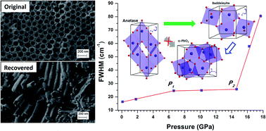
Anatase titanium dioxide (TiO2) nanotubes synthesized via electrochemical anodization were studied under high pressure up to 31 GPa. The structural transformations were characterized by in situ Raman spectroscopy and synchrotron X-ray diffraction. Raman measurements suggest that anatase TiO2 nanotubes transform to an amorphous phase upon compression to 17.7 GPa and remain mostly amorphous upon recovery but with minor α-PbO2 and anatase phases as a mixture. In contrast, direct anatase to baddeleyite phase transition was unambiguously observed at ∼14 GPa in the diffraction measurements. Structural refinement allows the quantitative analysis of the transition sequence and reveals that the recovered phase is mostly crystalline α-PbO2. This discrepancy can be attributed to the special surface and interfacial structure associated with the tube morphology. Moreover, the compression behavior such as the compressibility of both anatase and baddeleyite phases of TiO2 nanotubes was examined in parallel with other nanostructured TiO2 materials. Our analysis shows that morphology plays a more prominent role than size in affecting the high pressure behaviors of 1D TiO2 nanomaterials compared to nanoparticles, and that the interplay of multiple factors such as morphology, size, interfacial structures, as well as lattice defects can substantially influence the phase stability and thus the transformation sequence.
In addition to traditional synthetic routes, pressure provides an effective tool to produce new structures and to tune the properties of materials.19 As a result, extensive high-pressure studies have been carried out on TiO2 nanomaterials especially with anatase and rutile structures over the past a few years. For instance, a number of experimental and theoretical high-pressure studies indicate that both bulk and nanostructured TiO2 have a series of high-pressure phases, and the sequence of phase transitions is highly size and morphology dependent.20–43 At high pressure, both anatase and rutile bulk TiO2 attain phases that are isostructural with columbite (orthorhombic α-PbO2)22 and baddeleyite (monoclinic ZrO2),20 following a transition sequence from anatase phase to α-PbO2 phase and then to baddeleyite upon compression.21–25,27,43,44 However, this phase transition route is only applicable to bulk TiO2. For TiO2 nanomaterials, the transition sequences are different as their morphology and size vary. For instance, Wang and Saxena found that the anatase phase in TiO2 nanoparticles (with particle size ranging from 7 to 11 nm) was stable up to 24 GPa, and then turned to an amorphous phase upon further compression.45 The amorphous phase was found quenchable to ambient pressure. However, high-pressure study of anatase TiO2 nanoparticles with size of 30 nm showed that a baddeleyite phase formed at 16.4 GPa without pressure-induced amorphization.26 Such a large discrepancy was believed due to the variation in the grain size of nanoparticles. Systematic studies on TiO2 nanoparticles have revealed the correlation between their high-pressure behaviors and the particle size: (1) when the particle size is less than 10 nm, the nanoparticles underwent a pressure induced amorphization upon compression;31–33,36,43 (2) when the size of nanoparticle is between 12 and 50 nm, the anatase phase transformed into the baddeleyite phase directly;31,35,43 (3) when the particle size is larger than 50 nm, the phase transition sequence is from anatase to α-PbO2 and then to baddeleyite phase.23–25,28,31,43
However, such size-effects model is not applicable to 1D TiO2 nanomaterials. In addition to the factor of size, morphology was also found to play an important role in influencing the high pressure behaviors of 1D nanomaterials. For instance, the pressure-induced amorphization were found in TiO2-B nanoribbons with widths in the range of 50–200 nm and thickness of ∼20 nm, the size of which is far beyond the critical size of 10 nm for nanoparticles. Moreover, both our previous high-pressure study of anatase TiO2 nanowires with size of 50–100 nm or 150–200 nm and that by Li et al.37 suggest a phase transition sequence from anatase to baddeleyite phase without going through the α-PbO2 phase. Interestingly, nanostructured anatase TiO2 in another 1D morphology, i.e., nanotubes with a tube diameter of ∼8–10 nm, was found to irreversibly transform to amorphous phase with different densities upon compression and decompression, similar to nanoparticles.40 More recently, nanostructured anatase TiO2 with composition and morphology variations, such as Nb-doped TiO2 nanoparticles and TiO2 nanosheets show new interesting and unique high pressure behaviors.29
Despite the extensive high-pressure investigations and rationalizations of TiO2 nanoparticles, no systematic understanding of the high-pressure behaviors of 1D TiO2 nanomaterials, especially TiO2 nanotubes, is available due to the extremely sparse studies.37,39,40 Here we report the high-pressure study of electrochemically synthesized TiO2 nanotubes with tube diameter of ∼100 nm that allows the systematic study when compared with those with a significantly smaller tube diameter (i.e., ∼10 nm). Interesting and new high pressure behaviors of anatase TiO2 nanotubes that are substantially different from previous studies were observed and characterized by in situ Raman spectroscopy and synchrotron X-ray diffraction. By comparison with other 1D nanostructured anatase TiO2 particularly nanowires and nanotubes, the transformation mechanisms were examined and the morphology effect was addressed. This study contributes to the understanding of pressure tuning nanostructures involving the interplay of multiple influencing factors including morphology, dimensions as well as interfacial structures.
![]() | ||
Fig. 1 SEM images of TiO2 nanotubes collected before (a) and after (b) the compression and decompression cycle. |
High-pressure studies were carried out using a symmetric diamond anvil cell (DAC) with a pair of type I diamonds with a culet size of 400 μm. A hole with a diameter of 130 μm was drilled on a stainless steel gasket and used as the sample chamber. The pressure was determined by the well-established ruby fluorescence method. In situ Raman spectra were collected using a customized micro-Raman spectroscopy system with a diode pumped solid state laser (λ = 532 nm) as the excitation source. For Raman measurements, silicon oil was used as the pressure transmitting medium (PTM). In situ angle-dispersive XRD measurements were carried out at room temperature at the beamline 16ID-B of HPCAT at the Advanced Photon Source. The incident wavelength of the monochromatic X-ray beam was 0.3738 Å with a beam size of 4 μm × 5 μm. The diffraction data were recorded on a MAR 345 imaging plate. Neon gas was used as the PTM for XRD measurements. A motorized gear box was also employed to regulate the pressure with fine increments. The 2D Debye–Scherrer diffraction patterns were integrated by using Fit2D program for further analysis. The structural refinement was performed using GSAS software package.
The Raman spectra of TiO2 nanotube collected upon compression up to 17.7 GPa are shown in Fig. 2. Upon compression, all the Raman modes shifted to higher frequencies except for the Eg(2) mode, which exhibited a red shift until its disappearance at about 3 GPa. Upon compression, the intensity of all the peaks associated with the anatase phase gradually became weak. At 17.7 GPa, all the Raman modes were significantly suppressed leaving only one peak discernable at ∼183 cm−1, indicating that the sample was highly disordered at high pressure. Other than the profile broadening, no distinguishable new peak was observed, suggesting no phase transition below 17.7 GPa.
The reversibility of pressure effect on crystal structures provides important information on transformation mechanisms. Therefore, after sample was compressed to 17.7 GPa, Raman measurements of TiO2 nanotubes were also conducted upon decompression. In general, the intensity of all the Raman peaks increased gradually as pressure decreasing, and all the Raman modes shifted to lower frequencies. At 4.6 GPa, a new peak appeared at 161 cm−1 indicating a possible phase transition. This new peak shifts to 148 cm−1 at 0.1 GPa. According to the reference values, this peak can be associated with the α-PbO2 phase.39 The rest four peaks can be assigned to the recovered anatase phase although all the Raman peaks shift to slightly higher frequencies than before compression. Thus the retrieved phase can be interpreted as the mixture of the anatase phase and α-PbO2 phase. The large variation in the Raman profile can be attributed to the pressure induced structural modification, which is mostly irreversible upon compression.
Upon decompression, all the reflections shift to lower 2θ angle gradually, indicating the expansion of the unit cells. As shown in Fig. 3, when the pressure was decreased to 4.9 GPa, a new reflection appeared at 7.5595°, suggesting the onset of a phase transition. The new phase persisted down to ambient pressure, at which all reflections of the baddeleyite phase disappeared completely. The XRD pattern can be interpreted by the single α-PbO2 phase by structural refinement (Fig. 4c), suggesting that the sample is in almost pure α-PbO2 phase.
Starting TiO2 | Phase transition pressureb (GPa) | Bulk modulus (GPa) | Experimental method | ||
---|---|---|---|---|---|
Morphology | Sizea (nm) | Anatase | Baddeleyite | ||
a The column shows particle size, length, tube diameters for nanoparticles, nanowires and nanotubes, respectively. For nanosheets, l and t are side length and the sheet thickness.b The column shows phase transition pressures in TiO2. For the bulk row, values outside and inside the parentheses are the phase transition pressures for the anatase to α-PbO2 phase transition and α-PbO2 to baddeleyite phase transition, respectively. For the nanoparticles row, the italic values indicate transition pressures for the anatase to amorphous phase transition. The rest values are transition pressures for the direct anatase to baddeleyite phase transition.c This work. | |||||
Bulk | 4.3–4.6 | Raman28 | |||
∼5 (12–15) | Raman43 | ||||
5.4 (∼10) | 59 | XRD22 | |||
4.5–7 (13–17) | Raman23 | ||||
4.5 (∼13) | 179 | 290 | XRD24 | ||
Nano-particles | 4 | >24 | Raman31 | ||
8 | >21 | Raman31 | |||
7–11 | >24 | Raman26 | |||
12 | ∼18 | Raman43 | |||
20 | 15 and 16 | Raman31 | |||
32 | 11–15 | Raman & XRD31 | |||
30–34 | 18–20 | 243 | XRD42 | ||
Nanosheets | l: 20–40 | 14.6–22.8 | 317 | Raman & XRD29 | |
t: 5–8 | |||||
Nanowires | 50–100 | ∼14 | 266.5 | 127.8 | Raman & XRD39 |
150–250 | ∼9 | 188.3 | 114.8 | Raman & XRD39 | |
50–200 | ∼9 | 176 | Raman & XRD37 | ||
Nanotubes | 8–10 | ∼17.9 | 166 | XRD40 | |
∼100 | ∼14 | 164.2 | 182.8 | Raman & XRD c |
In the Raman measurements, contrastingly, no phase transition is found upon compression up to 17.7 GPa, a pressure substantially higher than the transition onset pressure (i.e., ∼14 GPa) found in the XRD measurements. In the case of TiO2 nanowires, Raman measurements clearly suggests the anatase-to-baddeleyite transition at similar pressures, consistent with the XRD measurements.39 We then carefully examined the difference between these different experiments. We noted that silicon oil was used as the PTM in Raman measurements while neon was used in the XRD experiments. Although neon provides a better hydrostatic condition above 10 GPa than silicon oil, the difference in the hydrostaticity is unlikely the primary factor for the different compression behavior of TiO2 nanotubes. Given the large tube diameter and open end morphology of the TiO2 nanotubes, as well as the chemical inertness of the PTM, the interaction between PTM with different molecular sizes and TiO2 nanotubes is expected to be similar. Indeed, in Li et al.’s similar study on TiO2 nanotubes where 4:
1 methanol–ethanol mixture as PTM was used, it was believed that PTM can penetrate into the tubes even with much a smaller diameter (i.e., ∼5 nm).40 Based on these analyses, we believe TiO2 nanotubes in the current study have substantially different and more complicated surface or interfacial structures than nanowires studied before or the nanotubes in Li's study. In particular, the large tube diameter (i.e., ∼100 nm) and the wall thickness (i.e., ∼10 nm) as well as the way the tubes are aligned upon production may result in a much more inhomogeneous pressure response along the radial direction upon compression. As a result, the inner part of the tube and tube surface layers may undergo different transition sequence. Considering that Raman spectroscopy is a surface sensitive probe while that bulk penetrating X-ray provides information involving long-range orderness, we may conclude that the bulk TiO2 nanotubes and especially the inner layers transform to baddeleyite phase whereas the surface layers become amorphous upon compression.
In order to further reconcile the “discrepancy” between Raman and XRD measurements as well as to probe the transition mechanism, the full width at half maximum (FWHM) of the most intense Eg(1) mode for TiO2 nanotubes is plotted in Fig. 5. Two tuning points (labeled as P1 and P2) are found at 6 GPa and 14 GPa, respectively, consistent with the observation in TiO2 nanowires reported before.39 For TiO2 nanowires, P2 coincides with the anatase to baddeleyite phase transition pressure. Interestingly, the P2 in the current study, which is 14 GPa, is also coincidental with the anatase to baddeleyite phase transition pressure identified in the XRD measurements. This coincidence may suggest that the anatase to baddeleyite phase did occur, although undetectable in the Raman measurements given the above discussion. Based on our earlier high-pressure study of TiO2 nanowires, these two turning points are consistent with the three-stage process proposed for the phase transition, which involves the competition between formations of the α-PbO2 phase and baddeleyite phase. In the first stage (0 GPa – P1), specifically, the anatase to α-PbO2 phase transition route may be favored. However, due to the enhanced surface energy, a high energy barrier for the formation of the α-PbO2 structure could substantially delay the transition to the α-PbO2 phase.45 Therefore, before the α-PbO2 phase is eventually formed, the baddeleyite phase became the more energetically favored structure in competition with the α-PbO2 phase at the second stage (P1 – P2), as seen in Fig. 5. At the third stage (>P2), the phase transformation followed anatase-to-baddeleyite route monotonically, accompanied by the significantly faster increase in the FWHM of the Eg(1) mode than the first two stages.
Furthermore, detailed analysis of the XRD results of TiO2 nanotubes allows the understanding of the pressure dependence of unit cell parameters as well as the compressibility in comparison with earlier studies of bulk and nanostructured TiO2 materials. The normalized unit cell lengths (i.e., a/a0 and c/c0) as a function of pressure for the TiO2 nanotubes are plotted in Fig. 6 in comparison with the corresponding bulk materials. First of all, the c-axis exhibits a linear compressibility that is three times larger than a-axis, consistent with earlies studies on bulk TiO224,47 as well as TiO2 nanowires.37,39 The different compressibilities are believed to be associated with the intrinsic anatase crystal structure. Specifically, there are four occupied TiO6 octahedra and four empty O6 octahedra per unit cell. The Ti atom inside the occupied octahedra (TiO6) makes the polyhedra much harder to compress than the empty ones (O6). Thus the higher compressibility of the c-axis than the a-axis can be interpreted in terms of the difference in the directional population of the hard occupied (TiO6) and soft empty (O6) oxygen octahedra. The specific c/a compression ratio further indicates a consequence of the alignment of the empty O6 octahedra along the c-axis and of the greater density of atoms along the a- and b-axes than along the c-axis. Moreover, compared to bulk TiO2, the c-axis of TiO2 nanotubes shows higher compressibility, while the compressibility along a-axis is similar. In general, the c-axis for all the nanostructured anatase TiO2 is more compressible than the corresponding bulk material, indicating that the morphology and the crystalline growth direction of TiO2 nanomaterials play an important role in the anisotropic behavior.34,37
![]() | ||
Fig. 6 Pressure dependences of cell parameters of anatase TiO2 nanotubes in comparison with those of bulk TiO2. Open and solid symbols denote the cell parameters of the TiO2 nanotubes and bulk TiO2 cited from ref. 24, respectively. Circles and squares represent a/a0 and c/c0 ratios, respectively. The solid lines are just for eye guidance. |
The pressure-volume data of TiO2 nanotubes are shown in the Fig. 7. By fitting the third-order Birch–Murnaghan equation of state (EOS), the bulk modulus (B0) of the anatase and baddeleyite phases for TiO2 nanotubes obtained are 164.2 GPa and 182.8 GPa, respectively, with the first derivative (B′0) fixed at 4. For comparison purposes, the bulk modulus of the anatase phase of corresponding bulk materials, nanoparticles and nanowires are plotted together with TiO2 nanotubes. Similar to Li et al.’s study on TiO2 nanotubes with a much smaller diameter (i.e., ∼10 nm) for which the bulk modulus was reported to be 158 GPa, the bulk modulus of TiO2 nanotubes in the current study (i.e., 164 GPa) is also slightly lower than that of TiO2 bulk materials (i.e., 179 GPa), indicating the dimension of the TiO2 nanotubes has a negligible influence in the compressibility. In contrast, other nanostructured TiO2 materials such as nanowires and nanoparticles showed substantially enhanced bulk modulus, e.g., 266.5 GPa and 243 GPa, respectively. In addition, morphology-induced alterations of bulk modulus have been reported in high-pressure studies of other TiO2 nanomaterials. For example, strongly contrasting compressibilities were observed for TiO2 nanoparticles with rod and rice shapes.34 The bulk modulus of rod-shaped particles was reduced, whereas that of the rice-shaped particles was enhanced by over 50% relative to the corresponding bulk materials.34 All these observations suggest that morphology plays a dominant role in the compressibility of different TiO2 nanostructures due to the different contribution of surface energy states.
![]() | ||
Fig. 7 Pressure dependence of the unit cell volume for the anatase phase of TiO2 nanotubes in comparison with that for bulk, nanowires, and nanocrystals. The inset figure shows the EOS of baddeleyite phase of TiO2 nanotubes. The solid squares are from this work. The solid lines represent fitting to the third order Birch–Murnaghan EOS. Dotted lines, dashed lines, and dash dotted lines are the EOS for bulk, nanowires and nanocrystals reported from ref. 24, 40 and 43, respectively. |
Another interesting observation is that the baddeleyite phase exhibited a much higher bulk modulus in TiO2 nanotubes than nanowires (182.8 GPa vs. 127.8 GPa).39 The SEM image of the recovered materials (Fig. 1b) shows that although the alignment of the nanotubes was substantially modified, the tube morphology is still clearly distinguishable. The preservation of the morphology on compressed materials has great implications for new applications and has been reported for TiO2 nanowires and nanotubes before.37,40 In addition to the open-end tube morphology and use of PTM as important factors, we believe the high stillness of baddeleyite lattice during the anatase-to-baddeleyite transition in TiO2 nanotube also contributes to the morphology stability upon compression. Indeed, in our earlier studies of TiO2 nanowires for which the baddeleyite phase has a lower stiffness, the wire morphologies were not preserved.39
Finally, the interplay of multiple factors must be considered collectively to interpret the different mechanical properties, phase stability as well as transition sequence of nanomaterials. Park et al. suggested that bulk modulus may be influenced by the crystal growth directions of TiO2 nanomaterials.34 For instance, the anatase TiO2 nanorods grown along the a-axis showed a lower bulk modulus (243 GPa) than TiO2 nanorices grown along the c-axis (319 GPa). Li et al. obtained the similar results in the study of anatase TiO2 nanowires.37 Their nanowires have the same growth direction as the nanorods, given a similar bulk modulus of 176 GPa as the bulk counterpart (i.e., 179 GPa). Therefore, we can infer that the primary growth direction of TiO2 nanotubes synthesized in this study is also along a-axis. However, our TiO2 nanotubes exhibit a “normal” transition sequence whereas those produced by Li et al. suggest that only amorphous TiO2 with different densities was observed upon compression.40 Crystalline defects and lattice impurities, which strongly depend on synthetic methods, can substantially influence the structural stabilities at the nanoscale.48,49 Indeed, plenty of crystal defects were identified in the hydrothermally synthesized TiO2 nanomaterials.40 Our Raman results on electrochemically synthesized TiO2 nanotubes suggest inhomogeneous interfacial structures, from which crystal defects can also be inferred, but likely with a different distribution along and across the nanotubes at nanoscale. Ultimately, it would be of great interest to fabricate energy devices based on TiO2 nanotubes produced via different routes both in the as-made form and retrieved from compression and to test the performance comparatively for practical applications.
This journal is © The Royal Society of Chemistry 2016 |