
Recovery of antioxidant and antiproliferative compounds from watercress using pressurized fluid extraction
Liliana Rodriguesa,
Inês Silvaa,
Joana Poejoa,
Ana Teresa Serraab,
Ana A. Matiasab,
Ana Luísa Simplícioab,
Maria R. Bronzeabc and
Catarina M. M. Duarte*ab
aiBET, Instituto de Biologia Experimental e Tecnológica, Apartado 12, 2780-901 Oeiras, Portugal. E-mail: cduarte@ibet.pt
bITQB, Instituto de Tecnologia Química e Biológica António Xavier, Universidade Nova de Lisboa, Av. da República, 2780-157 Oeiras, Portugal
cFaculdade de Farmácia, Universidade de Lisboa, Avenida das Forças Armadas, 1649-019 Lisboa, Portugal
First published on 11th March 2016
Abstract
In this work, pressurized fluid extraction was explored to recover isothiocyanates (ITCs) and phenolic compounds from watercress. Pretreatment of the raw material was studied, using different conditions of temperature (25 °C; 35 °C), pressure (Patm; 25 MPa), incubation time (0; 30; 60; 120 min) and moisture content (125; 250; 900% of water, dry basis), aiming at promoting the enzymatic hydrolysis of glucosinolates in ITCs. Extractions of ITCs were performed with supercritical CO2, and different mixtures of CO2![[thin space (1/6-em)]](https://www.rsc.org/images/entities/char_2009.gif) :ethanol (0–50, % w/w) were applied to obtain phenolic-enriched ITCs extracts. Extractions were performed at 35 °C and 25 MPa for 2 h and extracts analyzed concerning total ITCs, phenethyl isothiocyanate (PEITC) content, phenolic composition, antioxidant capacity (using ORAC, HORAC and HOSC assays) and antiproliferative effect in a human colorectal cancer cell line (HT-29). Results showed that supercritical CO2 was highly selective in isolating ITCs from watercress (up to 31.7 ± 1.6 μmol ITC per g). When mixtures of CO2:ethanol were used, extraction of phenolics (up to 10.1 ± 0.8 mg GAE per g) and antioxidants (up to 204.4 ± 21.5 μmol TE per g concerning ORAC, 70.8 ± 10.7 μmol CAE per g for HORAC and 189.5 ± 22.9 μmol TE per g for HOSC), was promoted. PEITC was the main compound responsible for the inhibition of cancer cell growth of all extracts (EC5024 h = 27.8 ± 1.9 μM PEITC). However, extracts obtained by supercritical CO2 extraction after a 30 minute incubation period with CO2 and with CO2:ethanol mixtures (80:20 and 60:40, % w/w) were revealed to be effective in isolating other bioactive compounds that enhanced the antiproliferative response of extracts (EC5024 h values of 23.1 ± 0.9; 20.7 ± 1.9 and 19.8 ± 0.7 μM PEITC, respectively).
:ethanol (0–50, % w/w) were applied to obtain phenolic-enriched ITCs extracts. Extractions were performed at 35 °C and 25 MPa for 2 h and extracts analyzed concerning total ITCs, phenethyl isothiocyanate (PEITC) content, phenolic composition, antioxidant capacity (using ORAC, HORAC and HOSC assays) and antiproliferative effect in a human colorectal cancer cell line (HT-29). Results showed that supercritical CO2 was highly selective in isolating ITCs from watercress (up to 31.7 ± 1.6 μmol ITC per g). When mixtures of CO2:ethanol were used, extraction of phenolics (up to 10.1 ± 0.8 mg GAE per g) and antioxidants (up to 204.4 ± 21.5 μmol TE per g concerning ORAC, 70.8 ± 10.7 μmol CAE per g for HORAC and 189.5 ± 22.9 μmol TE per g for HOSC), was promoted. PEITC was the main compound responsible for the inhibition of cancer cell growth of all extracts (EC5024 h = 27.8 ± 1.9 μM PEITC). However, extracts obtained by supercritical CO2 extraction after a 30 minute incubation period with CO2 and with CO2:ethanol mixtures (80:20 and 60:40, % w/w) were revealed to be effective in isolating other bioactive compounds that enhanced the antiproliferative response of extracts (EC5024 h values of 23.1 ± 0.9; 20.7 ± 1.9 and 19.8 ± 0.7 μM PEITC, respectively).
Introduction
Watercress (Nasturtium officinale) is a cruciferous vegetable of the Brassicaceae (or Cruciferae) family, frequently classed as a “superfood”. Being exceptionally concentrated in nutrients and phytochemicals, including β-carotene, lutein, quercetin, phenolic acids, and glucosinolates (phenethyl glucosinolate (gluconasturtiin) and lower amounts of 7-methylsulfinylheptyl, 8-methyl-sulfinyloctyl, 3-indolylmethyl and 4-methoxy-3-indolylmethyl glucosinolates), this vegetable has been known since ancient times for its health benefits.1 In fact, over the years, several epidemiological studies have established a positive correlation between a diet rich in watercress and a reduced risk of chronic diseases such as diabetes, cardiovascular diseases and cancer.2–4 In particular, the claimed role of cruciferous vegetables on cancer chemoprevention is consistently associated to glucosinolates (GLs) hydrolysis products. Typically glucosinolates are highly stable and biologically inactive. However, upon plant tissue disruption, these group of hydrophilic, sulphur-containing metabolites are rapidly degraded, by action of endogenous myrosinase (β-thioglucoside glucohydrolase, EC 3.2.3.147) or by myrosinase-positive human gut bacteria when ingested, in a wide variety of hydrolysis products, being isothiocyanates (ITCs) the most common.5,6 Watercress is the richest known source of gluconasturtiin (GLNT), the precursor of phenethyl isothiocyanate (PEITC), which has been widely studied in a large number of in vivo and in vitro models. As reviewed by Gupta et al. (2014), pure PEITC has been shown to act as chemopreventive agent in about 30 different targets present in tumor cells against several types of cancer, including breast, prostate, leukemia, lung, colon, liver, oral, multiple myeloma, ovary, and cervical cancer, operating through complementary and sometimes overlapping mechanisms of action. PEITC also revealed to promote inhibition of anti-apoptotic pathways in in vivo studies using preclinical mouse models and its anticancer effects on humans are currently under study in phase I and phase II clinical trials.7 Concerning watercress extracts, there are some studies in literature reporting the anticarcinogenic effect of crude extracts in human cancer cell lines. In particular, Rose and co-workers (2005) used a watercress extract to assess its effects on chemically induced cancer cell invasion in human breast cancer cells (MDA-MB-23). The extract revealed to be an inhibitor of metalloproteinase-9 action, which has been associated with increased invasive and metastatic potential, and suppressed the invasive potential of the breast cancer cells.8 In a later work, Boyd et al. (2006) showed for the first time that an extract of watercress juice could inhibit the three crucial stages of carcinogenesis pathway (initiation, proliferation of cancer cells and metastasis). In Boyd's work, the watercress extract significantly inhibited DNA damage induced by oxidative stress, delayed the cell cycle of human colon cancer cells (HT-29) and significantly blocked the cells' invasive or metastatic actions.9Regarding fruits and vegetables, protection against chronic diseases has often been, at least partly, attributed to antioxidant nutrients, such as vitamins and minerals. However, many phytochemicals such as phenolic compounds or some terpenoids, also possess powerful antioxidant activity and there are some evidence suggesting that they may work additively and synergistically.10,11 Watercress has already been recognized as a source of powerful antioxidants, which were tested against different oxidative systems in vitro. The antioxidant properties of this vegetable bioactive compounds revealed to be effective as scavengers of free radicals (with an ORAC value of 434.5 μmol TE per g dry watercress),12 as reductants, as metal chelating agents and as lipid peroxidation inhibitors.13–16
Nowadays, the health and safety of consumers, has led to a preference for natural products over synthetic. However, even though phytochemicals naturally occur in plants, their intake in daily diet might not be sufficient to promote beneficial effects on health. Consequently, the need to develop new processing techniques which enable a more selective and effective recover of these high-value components from natural sources has increased. In this field, high pressure extraction (HPE) using supercritical and pressurized fluids has revealed to be a particularly interesting technique to isolate and recover bioactive compounds from plant materials. In fact, HPE has been an important process in pharmaceutical, nutraceutical, food and cosmetic industries. Studies on the extraction of essential oils, phenolic compounds, carotenoids, fatty acids, alkaloids, among other classes phytochemicals, with different potential applications within these industries, have been published in the last years, as reviewed by Sovová and Stateva (2011).17 Traditionally, the extraction of natural products is commonly achieved by solvent boiling methods, such as hydrodistillation or Soxhlet extraction. However, most of conventional methods typically use destructive conditions or hazardous organic solvents, which cannot be used in the preparation of food, nutraceutical or pharmaceutical products.18 Consequently, high pressure processes have emerged as promising alternatives for the recovery of bioactive compounds from natural sources, as they do not involve the use of toxic solvents, increasing the selectivity toward valuable bioactive compounds, while preserving their bioactivity.19,20
Supercritical fluid extraction (SFE), using carbon dioxide as solvent was already applied for recovery of bioactive enriched fractions from several cruciferous vegetables. Recently, Solana et al. (2014) explored the potential of supercritical carbon dioxide using small percentages of water and ethanol as co-solvents to isolate glucosinolates, which are more stable although less lipophilic than ITCs, and phenols from rocket salad. The richest glucosinolate/phenol fraction was obtained when operating at 30 MPa and 75 °C using 8% (w/w) of water as co-solvent, with a total glucosinolate content of 1.96 mg g−1 dry matrix and a total phenolic content of 1.48 mg g−1 dry matrix.21 In a more recent work conducted by Ares and co-workers (2015), pressurized liquid extraction, using a mixture of ethanol and water, was optimized to isolate GLs from broccoli leaves, by applying a methodology for extraction, separation and quantification of intact glucosinolates.22 For isothiocyanates, the extraction of sulforaphane present in white cabbage23 and allyl isothiocyanate (AITC) present in horseradish24,25 and wasabi26 were already reported using supercritical fluid technology in ranges of pressure between 15 and 25 MPa and temperatures up to 60 °C. In particular, in the work of Li et al. (2010) the highest ITC yield (4.08 mg AITC per g dry matrix) was achieved when using supercritical CO2 at 25 MPa and 35 °C with a wasabi moisture content of 125% dry basis (d.b.) to induce glucosinolate hydrolysis.26 Additionally, concerning the extraction of phenolic compounds, ethanol has been widely used as co-solvent in SFE by several authors to extract claimed health-promoting ingredients, including flavonoids, from a broad group of natural sources.27
The purpose of this study is to exploit for the first time the use of high pressure technology to extract valuable bioactive ingredients from watercress, namely isothiocyanates and phenolic compounds. In a first stage and prior to extraction, different pretreatment conditions, including incubation time, moisture content, pressure and temperature were applied to freeze dried watercress in order to promote the enzymatic hydrolysis of glucosinolates to isothiocyanates. The addition of exogenous myrosinase to the raw material was also tested, aiming at maximizing the hydrolysis reaction and promoting the total conversion of GLNT in PEITC. Due to the lipophilic nature of isothiocyanates, after watercress pretreatment, extraction of PEITC was performed using supercritical CO2 as solvent, applying the pressure and temperature conditions optimized for the extraction of AITC from wasabi by Li and co-workers (2010) (25 MPa and 35 °C).26 In a second stage, gas-expanded liquid extractions were performed using different mixtures containing CO2 and ethanol (up to 50%), after applying the optimal pretreatment condition to both fresh and freeze dried watercress. The influence of CO2 and ethanol mixtures composition, as well as watercress moisture content, was studied through the characterization of total ITCs and PEITC yields, phenolic content and antioxidant activity. The bioactivity of watercress extracts was also evaluated by analyzing and comparing the antiproliferative effect on human colorectal adenocarcinoma cells (HT-29) with standard PEITC.
Experimental
Raw material
Watercress was kindly provided by Vitacress Portugal, after harvesting, in April 2014 and stored at 4 °C in the absence of light. Firstly, the aerial parts of the plants, including leaves and stalks, were dehydrated in a freeze dryer at −40 °C. After 72 hours watercress was milled in a domestic chopper (Moulinex, A320R1, Paris, France) and the particle size of ground material was determined using an AS 200 basic vertical vibratory sieve shaker (Retsch, Haan, Germany), with a measuring range between 250 μm and 710 μm. The processed plants were protected from light and stored at room temperature in a desiccator until the day of experiments. For extractions performed with fresh raw material, watercress from Vitacress was used within shelf life period and milled immediately before each experiment, in order to preserve plant properties and prevent degradation of the bioactive ingredients.Chemicals
Carbon dioxide pure grade (99.95%, Air Liquide, Lisbon, Portugal) and absolute ethanol anhydrous (99.9%, Carlo Erba Reagents, Val de Reuil, France) were used for high pressure extraction experiments.Solvents used for conventional solvent extractions and analyses of resulting extracts included n-hexane (95%, Carlo Erba Reagents, Val de Reuil, France); methanol (99.9%, Carlo Erba Reagents, Val de Reuil, France), dimethyl sulphoxide (99.9%, Carlo Erba Reagents, Val de Reuil, France), ethyl acetate (99.8%, Lab-Scan, Dublin, Ireland), acetonitrile (99.9%, Sigma-Aldrich, Zwijndrecht, Netherlands), formic acid (98%, Panreac, Barcelona, Spain) bi-distilled water and ultrapure water purified with a Milli-Q water purification system (Merck Millipore, Billerica, MA, USA). Chemicals used for 1,2-benzenedithiole-based cyclocondensation assay and HPLC-DAD analyses were: 1,2-benzenedithiole (96%) and N-(tert-butoxycarbonyl)-L-cysteine methyl ester (N-tBoc-Cys-ME), 97%, from Sigma-Aldrich (St. Quentin Fallavier, France). Potassium phosphate monobasic anhydrous from Amresco (Solon, OH, USA), potassium phosphate dibasic from Sigma-Aldrich (Madrid, Spain) were used for phosphate buffer solution (PBS) preparation. Phenethyl isothiocyanate standard (99%) was purchased from Sigma-Aldrich (St. Quentin Fallavier, France), as well as thioglucosidase from Sinapis alba (white mustard) seed, used as exogenous enzyme and for indirect determination of glucosinolates (Schnelldorf, Germany). For total phenolic content determination: sodium carbonate (Na2CO3) was purchased from Sigma-Aldrich (St. Quentin Fallavier, France), Folin–Ciocalteu reagent was acquired from Panreac (Barcelona, Spain) and gallic acid was purchased from Fluka (Germany).
Chemicals used for antioxidant activity assays were: 2′,2′-azobis(2-amidinopropane)dihydrochloride (AAPH), 6-hydroxy-2,5,7,8-tetramethylchroman-2-carboxylic acid (Trolox), caffeic acid (C9H8O4), disodium fluorescein (FL), cobalt(II) fluoride tetrahydrate (CoF2), picolinic acid (C6H5NO2), iron(III) chloride (FeCl3), hydrogen peroxide (H2O2) and acetone (≥99.5%) from Sigma-Aldrich (St. Quentin Fallavier, France). Sodium chloride (NaCl), potassium chloride (KCl), sodium phosphate dibasic dihydrate (Na2HPO4·2H2O) from Sigma-Aldrich (St. Quentin Fallavier, France) and potassium phosphate monobasic anhydrous (KH2PO4) from Amresco (Solon, OH, USA) were used for phosphate buffer solution (PBS) preparation. Sodium phosphate dibasic dihydrate (Na2HPO4·2H2O) and sodium phosphate monobasic monohydrate (NaH2PO4·H2O) from Sigma-Aldrich (St. Quentin Fallavier, France) were used in sodium phosphate buffer solution preparation (SPB).
All cell culture media and supplements, namely fetal bovine serum (FBS), glutamine and RPMI 1640, were obtained from Invitrogen (Gibco, Invitrogen Corporation, Paisley, UK). Moreover, cell viability kit assays used for cytotoxicity and antiproliferative experiments were PrestoBlue® viability reagent (Molecular Probes, Invitrogen, USA) and Cell Titter® aqueous one solution cell proliferation assay (MTS) (Wisconsin, USA). Chemicals used in these assays included dimethyl sulphoxide (99.9%, Carlo Erba Reagents, Val de Reuil, France).
Extraction procedures
| Sample | Static period (min) | % moisture content (d.b.) | Temperature (°C) | Pressure (MPa) |
|---|---|---|---|---|
| A | 30 | 125 | 35 | 25 |
| B | 60 | 125 | 35 | 25 |
| C | — | 125 | — | — |
| D | 30 | 125 | 25 | P atm |
| E | 60 | 125 | 25 | P atm |
| F | 120 | 125 | 25 | P atm |
| G | 60 | 125 | 35 | P atm |
| H | 60 | 250 | 35 | P atm |
| I | 60 | 900 | 35 | P atm |
| J | 60 | 125 (with 250 mU of myrosinase) | 35 | P atm |
| Sample | Solvent mixture | Total ITCs content (μmol ITC per g dry weight) | PEITC content (μmol PEITC per g dry weight) |
|---|---|---|---|
| Freeze-dried watercress | |||
| ITC SE | Hexane | 25.1 ± 4.2 | 20.5 ± 2.7 |
| G | CO2 | 31.7 ± 1.6 | 29.3 ± 2.6 |
| K | CO2:EtOH (90:10) |
22.2 ± 2.9 | 22.1 ± 3.9 |
| L | CO2:EtOH (80:20) |
22.5 ± 1.6 | 17.2 ± 2.7 |
| M | CO2:EtOH (60:40) |
23.3 ± 2.8 | 20.3 ± 1.7 |
| N | CO2:EtOH (50:50) |
18.5 ± 4.0 | 14.0 ± 2.8 |
|
|||
| Fresh watercress | |||
| O | CO2:EtOH (90:10) |
5.7 ± 0.7 | 4.5 ± 0.9 |
| P | CO2:EtOH (80:20) |
9.0 ± 1.8 | 6.6 ± 1.1 |
| Q | CO2:EtOH (60:40) |
20.5 ± 0.4 | 15.5 ± 3.3 |
| R | CO2:EtOH (50:50) |
23.4 ± 1.0 | 16.0 ± 3.5 |
Phytochemical characterization of watercress extracts
:30 (% v/v) was added to 1 g of dried plant material and heated at 75 °C for 10 minutes, under constant stirring. The resulting extract (GLNT SE) was separated from the solid residues by filtration and stored at 4 °C until quantification experiments. The experiment was replicated. GLNT present in the extract was indirectly quantified by analyzing the amount of phenethyl isothiocyanate (PEITC) formed after enzymatic hydrolysis of gluconasturtiin. The protocol suggested by Budnowski et al. (2013)30 and optimized in this study for the quantification of PEITC in watercress extracts was applied, being the samples incubated for 2 h at 25 °C and 300 rpm with 0.1 M phosphate buffer solution (pH 6.7), a 427 mM solution of N-tBoc-Cys-ME in 60% (v/v) methanol and ultrapure water containing 100 mU of myrosinase (thioglucosidase from Sinapis alba seed) in order to promote GLNT hydrolysis. The peak detection for PEITC was performed at 284 nm and results expressed as μmol of PEITC per gram of plant dry weight, as a mean of at least three replicates.:2). GC-MS analyses were carried out on a Shimadzu GCMS-QP2010 (Shimadzu Corporation, Kyoto, Japan) gas chromatograph with an electronic impact (EI) source and a quadrupole detector. The separation of sample components was achieved by using a VF-5MS capillary column (Varian, Inc. FactorFour), 30 m × 0.25 mm I.D. and 0.25 μm phase thickness. The column temperature program started at 35 °C for 5 minutes and went up to 230 °C with a heating rate of 5 °C min−1. Temperature was then kept at 230 °C for 5 minutes. Total flow of the carrier gas, helium, was set at 9.0 mL min−1. Injector temperature and both MS interface and ion source temperatures were 250 °C. Mass spectrometry detection was performed at 0.2 kV and samples were scanned from m/z 29 to 299. GCMSsolution software was used for data acquisition and the mass spectra of the components of the natural extracts were compared with the mass spectra from libraries NIST21, NIST27, NIST107, NIST147 and WILEY229.:acetonitrile:ultrapure water 1:180:19%(v/v/v) (eluent B), were applied with a gradient of 0–5.6% B (0–0.1 min), 5.6–16.7% B (0.1–15 min), 16.7–22.2% B (15–20 min), 22.2% B (20–30 min), 22.2–33.3% B (30–55 min), 33.3–55.6% B (55–80 min), 55.6–100% B (80–120 min), 100% B (120–135 min), 100–5.6% B (135–140 min), 5.6% B (140–160 min), at a flow rate of 0.3 mL min−1. Photodiode array detection was performed scanning between 192 and 798 nm at a speed of 1 Hz with a bandwidth of 5 nm. The detection was monitored using three individual channels, 280, 320 and 360 nm, at a speed of 10 Hz with a bandwidth of 11 nm.Antioxidant activity
Cell based assays
Statistical analysis
Statistical analysis of the results was performed using GraphPad Prism software (GraphPad Software, Inc., La Jolla, CA) by one-way analysis of variance (ANOVA) followed by the Tukey method. P < 0.05 was accepted as statistically significant in all cases. Data are expressed as mean ± SD values.Results and discussion
Isothiocyanates-rich extracts production
In order to obtain natural extracts rich in bioactive ingredients with potential health-promoting effects, high pressure extraction was explored and applied to watercress. Since isothiocyanates (ITCs) are obtained through enzymatic hydrolysis from glucosinolates (GLs), preliminary pretreatment experiments were performed, in an attempt to maximize the conversion of GLs in ITCs by action of endogenous myrosinase. The impact of pressure, temperature, incubation time and moisture content in hydrolysis reaction were studied, as well as the addition of exogenous myrosinase to the humidified raw material. Fig. 1 shows the effectiveness of each pretreatment evaluated in terms of ITCs extracted from watercress. Regardless the pretreatment conditions applied, results obtained by UV spectroscopy for total ITCs content were quite similar to those obtained by HPLC for phenethyl isothiocyanate (PEITC), suggesting that all extracts are mainly constituted by PEITC. Yields of PEITC ranged from 1.0 ± 0.2 μmol PEITC per g dry watercress to 29.3 ± 2.6 μmol PEITC per g dry watercress and, among all extracts, (G) contained the highest PEITC content (P < 0.05). Furthermore, GC-MS results (Fig. 2) showed that supercritical CO2 (using extract (G) as example) was much more selective in extracting phenethyl isothiocyanate from watercress than conventional hexane extraction (ITC SE) or CO2–expanded ethanol extractions (using extract (N) as an example). Even though ITCs are known for their high chemical reactivity, it is important to note that in the work reported herein, the concentration of ITCs in watercress extracts remained stable for at least 12 months (data not shown). | ||
| Fig. 1 Concentration of total ITCs and PEITC determined by UV spectroscopy after cyclocondensation assay (CC assay) and HPLC-DAD, respectively, in watercress supercritical CO2 (A–I) and hexane (ITC SE) extracts. For columns presenting the same color, different lowercase letters represent a significant difference. Legend: (A) 125% water (d.b.), 30 min incubation, 35 °C, 25 MPa; (B) 125% water (d.b.), 60 min incubation, 35 °C, 25 MPa; (C) 125% water (d.b.), no incubation; (D) 125% water (d.b.), 30 min incubation, 25 °C, Patm; (E) 125% water (d.b.), 60 min incubation, 25 °C, Patm; (F) 125% water (d.b.), 120 min incubation, 25 °C, Patm; (G) 125% water (d.b.), 60 min incubation, 35 °C, Patm; (H) 250% water (d.b.), 60 min incubation, 35 °C, Patm; (I) 900% water (d.b.), 60 min incubation, 35 °C, Patm; (ITC SE) 125% water (d.b.), 60 min incubation, 35 °C, Patm. | ||
 | ||
| Fig. 2 Example of the chromatographic GC-MS profiles of watercress extracts obtained after a period of incubation with 125% of water d.b. for 60 minutes, at 35 °C and atmospheric pressure: (G) was obtained with supercritical CO2 extraction, (N) was extracted with CO2:EtOH (50:50, % w/w) and (ITC SE) was obtained by conventional solvent extraction with n-hexane. Legend: (1) phenethyl isothiocyanate. | ||
Comparison of isothiocyanates (ITCs) extraction methods
In order to evaluate the supercritical CO2 extraction performance when compared to conventional solvent extraction, the same extraction parameters used to obtain extract (G) (incubation with 125% of water d.b. at 35 °C and atmospheric pressure for 60 minutes), including water content, temperature, solvent/matrix ratio and static and dynamic times, were applied to a conventional extraction using hexane as solvent. When comparing the results obtained for extract (G) and conventional solvent ITCs-rich extract (ITC SE), a significantly higher concentration in PEITC (P < 0.0001) can be achieved when supercritical CO2 is used as solvent (Fig. 1). The higher yield obtained for extract (G) might be caused by an increased efficiency and selectivity associated with supercritical fluid extraction. Furthermore, there is also the possibility of losses of ITCs during filtration and solvent evaporation, required after solvent extraction, which does not occur in SFE since extractions are performed in a closed system and the extracts are recovered in ethanol which is a biocompatible solvent.Comparison of isothiocyanates (ITCs) extraction and watercress glucosinolates (GLs) content
It was important to confirm if the results reached for the best extract obtained by supercritical fluid extraction (SFE) were in accordance with the initial concentration of gluconasturtiin (GLNT) in watercress, i.e. if the conversion of GLNT to PEITC was completely achieved during pretreatment and which was the extension of extraction efficiency. The total amount of GLNT in watercress was recovered, using a conventional solvent extraction with a mixture of methanol/water, 70:30 (% v/v)31 and submitted to complete enzymatic hydrolysis of gluconasturtiin.30 The quantity of resultant PEITC was determined by HPLC-DAD and was found to be 64.1 ± 7.4 μmol PEITC per g dry watercress, as shown in Fig. 3. A comparison between this result and the richest ITC extract obtained by SFE, extract (G), shows that only approximately half of the gluconasturtiin available in the freeze-dried plant material was converted in PEITC and/or extracted with supercritical CO2 (Fig. 3). Considering this fact, and aiming at further enhancing the concentration of phenethyl isothiocyanate in the extracts, an attempt was made to study the effect of the addition of exogenous myrosinase to watercress. The amount of myrosinase used was determined by monitoring the kinetics of the conversion of GLNT into PEITC in the hydro-methanolic extract, after addition of myrosinase in concentrations ranging from 0.0625 mU to 100 mU (Fig. 4). Based on the curve obtained, 10 g of freeze-dried watercress were treated with 125% of water d.b. containing 250 mU of white mustard seed myrosinase, an amount that should have been sufficient for complete hydrolyzation of gluconasturtiin present in watercress. A static period of 60 minutes at 35 °C and atmospheric pressure was applied, followed by supercritical CO2 extraction at 25 MPa and 35 °C for 2 hours (extract J). Results showed that, unlike what would be expected, there was no significant increase on the concentration of PEITC in extract (J) (Fig. 3). According to the work of Halkier and Gershenzon (2006), in some cruciferous vegetables myrosinase and glucosinolates can be found in separate but adjacent cells whereas in others, the glucosinolate–myrosinase system may be spatially separated at the subcellular, rather than cellular level.47 Nevertheless, in either case, cell disruption is a crucial factor without which the hydrolysis of glucosinolates cannot occur. Moreover, in this work, the absence of stirring in the extractor, where pretreatments were performed, may have contributed for the lack of contact between exogenous myrosinase and gluconasturtiin present in watercress.
 | ||
| Fig. 3 Concentration of phenethyl isothiocyanate (PEITC) in watercress extracts obtained by HPLC-DAD. Quantification of gluconasturtiin (GLNT) in GLNT-rich extract (GLNT SE) was performed by indirect determination of GLNT upon exogenous myrosinase addition and subsequent analysis of the amount of PEITC formed after enzymatic hydrolysis. ****P < 0.0001 (significant differences between PEITC content in freeze-dried watercress and PEITC content of watercress extracts: (G) supercritical CO2 extract obtained after incubation with 125% water (d.b.) for 60 min at 35 °C and Patm; (J) supercritical CO2 extract obtained after incubation with 125% water (d.b.) containing 250 mU of myrosinase for 60 min at 35 °C and Patm; (GLNT SE) GLNT-rich hydro-methanolic extract). | ||
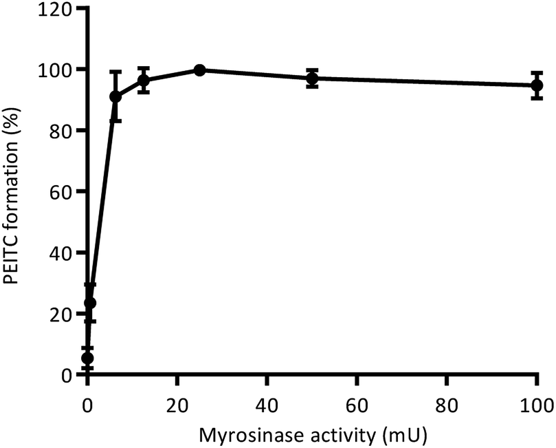 | ||
| Fig. 4 Formation of phenethyl isothiocyanate from gluconasturtiin-rich extract (GLNT SE) upon addition of different concentrations of exogenous myrosinase ranging from 0.0625 mU to 100 mU. | ||
Phenolic-enriched isothiocyanates (ITCs) extracts production
Although supercritical CO2 promoted a very selective extraction, resulting in an extract enriched in phenethyl isothiocyanate (PEITC), evidence is emerging that different mixtures of bioactive phytochemicals, rather than isolated compounds, may be far more effective in treating and protecting against cancer and other major chronic diseases.48,49 Therefore, in order to obtain an extract with a more complex composition consisting of isothiocyanates and phenolic compounds, different mixtures of CO2:ethanol were applied for extraction of both freeze-dried and fresh watercress. The optimal pretreatment and extraction conditions were applied, aiming at obtaining an extract with potential enhanced biological activity. Table 2 summarizes the solvent mixtures used and the respective total ITCs and PEITC contents obtained for each extract. Results showed that supercritical CO2 extraction (extract G) was significantly better in isolating phenethyl isothiocyanate relatively to conventional hexane extraction and that no significant increase in total ITCs or PEITC yields were achieved even when mixtures of CO2:ethanol were used. However, and as expected, when higher amounts of ethanol where used, a positive impact on the extraction of phenolic compounds was evident (Fig. 5(i)) as well as an enhanced antioxidant capacity of watercress extracts (Fig. 5(ii)–(iv)). This fact may be connected with covalent and dipole–dipole interactions which increased solubility of phenolic compounds.42 Furthermore, it was possible to obtain extracts with higher phenolic content and antioxidant capacity when extractions were performed on fresh watercress, which water content corresponds to approximately 1330% of water d.b. This is in accordance with several studies that have reported that hydro-alcoholic mixtures are more efficient than mono-solvent systems in the extraction of polyphenolic compounds.50,51 Fig. 5(v) shows the chromatographic profiles of CO2–expanded ethanol (CXE) extracts at 280 nm and, as it can be seen, residual phenolic compounds were detected in watercress supercritical CO2 extract (extract G). However, as the percentage of ethanol in the solvent mixture increased, compounds including hydroxycinnamic acids (caffeic and coumaric acids), flavonols (rutin) and hydroxycinnamic acid esters of malic acid (caffeoylmalate, coumaroylmalate, feruloylmalate and sinapoylmalate) were identified in watercress extracts. Among all extracts, (Q) and (R) obtained from fresh watercress with 40% and 50% of ethanol, respectively, presented the highest phenolic contents and antioxidant activities. However, the ratio between the antioxidant activity and TPC of watercress supercritical CO2 extract, considering in particular ORAC and HOSC values, was higher than for extracts obtained by CXE. Since PEITC presented very low antioxidant activity in the three assays performed (data not shown), this high ratio (above 90) may suggest the presence of non-phenolic antioxidants in extract (G) or eventually the existence of lipophilic phenolic antioxidants possessing a stronger radical scavenging activity than the phenolics present in CXE extracts. A statistically significant positive correlation was found between TPC of watercress extracts and ORAC values (R2 = 0.9770, P < 0.0001) and TPC and HOSC values (R2 = 0.9857, P < 0.0001), reflecting the scavenging capacity of the phenolic compounds extracted against peroxyl and hydroxyl radicals, being the correlation between ORAC values and total phenolic content of watercress extracts in accordance with results obtained by other authors.12 A marginally significant positive correlation was also found between TPC and HORAC values (R2 = 0.5387, P = 0.0244). From these observations, TPC seems to be a good indicator of antioxidant capacity of watercress extracts.
 | ||
| Fig. 5 Phenolic and antioxidant characterization of CO2–expanded ethanol extracts (K–R) and supercritical CO2 extract (G) obtained from watercress. Extracts (G) to (N) correspond to high pressure extractions of freeze-dried watercress, humidified with 125% water d.b., using CO2:EtOH ratios (% w/w) of 100:0 (G), 90:10 (K), 80:20 (L), 60:40 (M) and 50:50 (N); extracts (O) to (R) correspond to high pressure extractions of fresh watercress, using CO2:EtOH ratios of 90:10 (O), 80:20 (P), 60:40 (Q) and 50:50 (R). (i) Total phenolic content, (ii) oxygen radical absorbance capacity (ORAC), (iii) hydroxyl radical adverting capacity (HORAC) and (iv) hydroxyl radical scavenging capacity (HOSC) of watercress extracts. In each graph, different lowercase letters represent a significant difference. (v) Chromatographic profile of watercress extracts obtained by HPLC at 280 nm. Legend: (1) unidentified non-phenolic organic acid; (2) adenine; (3) caffeic acid; (4) tryptophan; (5) caffeoylmalate; (6) coumaric acid and rutin (co-eluting); (7) coumaroylmalate; (8) feruloylmalate; (9) sinapoylmalate. | ||
Evaluation of the antiproliferative effect of watercress extracts
Before antiproliferative assays, phenethyl isothiocyanate (PEITC) and watercress extracts were tested for their toxicity using confluent and undifferentiated Caco-2 cells. This cell model shares some characteristics with crypt enterocytes and thus it has been considered as an accepted intestinal model widely implemented to assess the effect of chemical and food compounds on the intestinal function.39–41 According to this, there are some published studies focusing on cytotoxicity and viability evaluation using undifferentiated Caco-2 phenotypes.40,41,52 In particular, Bony et al. (2006) reported that undifferentiated Caco-2 cells were found to be more sensitive to the tested compounds than differentiated cells, suggesting that this model can be considered as the most conservative approach for hazard characterization. IC50 values (inhibitory concentration that decreases 50% of cell viability) obtained after 24 h of incubation time are shown in Table 3. Results showed that, when compared with PEITC, supercritical fluid extracts presented similar or lower cytotoxicity than pure compound, with exception of extract (G) obtained from raw material pretreated for 60 min with 125% of water d.b. at 35 °C and atmospheric pressure. On the contrary, watercress extract obtained by conventional solvent extraction with hexane (ITC SE) showed higher toxicity in Caco-2 cells as well as CO2–expanded ethanol (CXE) extracts produced from freeze-dried watercress using 10, 20 and 50% w/w of ethanol. These results suggest that these extracts contain other compounds that probably increased the toxicity of samples. It is important to note that for CXE extracts derived from fresh watercress, no cytotoxicity was observed in Caco-2 cells for all the concentrations tested, indicating that the presence of water in raw material did not extract potential harmful compounds to intestinal cells. Based on the results obtained for cytotoxicity, non-cytotoxic concentrations were selected to be used on antiproliferative experiments.| Samples | IC50 (μM of PEITC) |
|---|---|
| PEITC | 79.1 ± 6.0 |
|
|
| Watercress extracts | |
| Supercritical CO2 extracts | |
| A | >100 |
| B | 73.4 ± 6.4 |
| C | >100 |
| D | >100 |
| E | 97.8 ± 5.1 |
| F | 81.6 ± 5.1 |
| G | 67.0 ± 4.1 |
| Conventional extract | |
| ITC SE | 57.8 ± 5.4 |
| CO2–expanded ethanol extractions | |
| K | 57.7 ± 8.9 |
| L | 48.3 ± 2.4 |
| M | >78 |
| N | 34.6 ± 0.8 |
| O | >100 |
| P | >100 |
| Q | >100 |
| R | >100 |
In order to evaluate the antiproliferative effect of watercress extracts, a human colorectal adenocarcinoma cell line (HT-29) was subjected to treatment with non-cytotoxic concentrations of extracts for 24 h. The percentage of viable cells was determined, dose–response curves were generated and EC50 values were calculated. For all extracts, results were expressed in terms of PEITC concentration in cellular media and compared with the standard compound, aiming at evaluating if the bioactive effect was mainly related with this ITC or if there are other compounds present in the extracts that interfered with the bioactive response. Results showed that all extracts inhibited HT-29 cell proliferation (Fig. 6). Fig. 6(i) compares the EC50 values of all watercress extracts derived from dry material with the standard compound, and demonstrates that the majority of samples, including conventional hexane extract, presented a similar effect to PEITC standard (EC5024 h = 27.8 ± 1.9 μM), indicating that their bioactivity is mainly related with the presence of this ITC, which has already been reported to inhibit HT-29 cells growth.53 However, it is important to highlight that for the sample obtained with supercritical CO2 after a 30 minutes incubation period with 125% of water d.b. at 35 °C and 25 MPa (extract A) as well as for the samples developed with CO2-expanded with 20% and 40% w/w of ethanol (extracts (L) and (M), respectively) a significant increase in the antiproliferative response was observed when compared with PEITC, i.e. the EC50 values of these watercress extracts were significantly lower (23.1 ± 0.9 μM PEITC, 20.7 ± 1.9 μM PEITC and 19.8 ± 0.7 μM PEITC, respectively) than the one obtained for PEITC alone. These results suggest that these extracts contain other compounds with antiproliferative activity and/or compounds that can act synergistically with PEITC reinforcing the bioactive response.
 | ||
| Fig. 6 Antiproliferative activity of (i) supercritical CO2 (A–G), hexane (ITC SE) and CO2–expanded ethanol extracts using freeze-dried watercress (K–N) and (ii) CO2–expanded ethanol extracts using freeze-dried watercress (K–N) and fresh watercress (O–R) in human colorectal cancer cells after 24 h incubation. *P < 0.05, **P < 0.01, ***P < 0.001, ****P < 0.0001 (significant differences between the antiproliferative effect of (i) watercress extracts and PEITC standard and (ii) dry watercress extracts and fresh watercress extracts). | ||
When comparing the high pressure extracts resulting from freeze-dried and fresh watercress (Fig. 6(ii)) a significant decrease of the activity of extracts as cancer cell-growth inhibitors was observed when fresh raw material was used. This effect could be related with the presence of water in watercress (approximately 1330% d.b.) that could promote the extraction of compounds that have an antagonistic effect on the antiproliferative response of PEITC. In fact, in our previous studies with Opuntia ficus indica fruits we observed that the presence of water in the pressurized solvent composition (CO2:ethanol) decreased the antiproliferative effect of the obtained extracts.54 Future studies should be performed in order to identify which compounds are responsible for the synergistic and antagonistic effects on PEITC in these watercress extracts.
Conclusions
In the study reported herein, pressurized fluid extraction was explored for the first time to recover directly isothiocyanates from watercress. Results showed that supercritical CO2 extraction selectively isolated phenethyl isothiocyanate (PEITC), whereas extracts obtained by CO2–expanded ethanol extractions (CXE) revealed to have in their composition not only PEITC, but also different groups of phenolic compounds, including hydroxycinnamic acids, flavonols and hydroxycinnamic acid esters of malic acid. Pretreatment of the raw material revealed to be necessary in order to maximize the enzymatic hydrolysis of gluconasturtiin in PEITC. In particular, an incubation period of watercress humidified with 125% of water (dry basis), during 60 minutes at 35 °C and atmospheric pressure, showed to be the best pretreatment condition to improve the extraction yield of PEITC. Among all extraction processes, including conventional hexane extraction and CO2–expanded ethanol extractions, supercritical CO2 at 25 MPa and 35 °C, showed to be the elected conditions to reach a higher recovery yield of isothiocyanates and in particular phenethyl isothiocyanate from freeze-dried watercress (31.7 ± 1.6 μmol ITC per g and 29.3 ± 2.6 μmol PEITC per g, respectively). By performing CO2–expanded ethanol extractions from both freeze-dried and fresh watercress, it was possible to obtain phenolic-enriched ITCs extracts, with intensified antioxidant capacity against peroxyl and hydroxyl radicals. Furthermore, PEITC revealed to be the main responsible for extracts' ability to inhibit colorectal cancer cell-growth when using HT-29 cell line. However, extractions performed with (i) supercritical CO2 after a 30 minutes incubation period with 125% of water d.b. at 35 °C and 25 MPa and (ii) CXEs with 20% and 40% w/w of ethanol, revealed to be able to isolate other bioactive compounds that significantly enhanced the antiproliferative response of PEITC in HT-29 cells. From the results obtained it can be concluded that pressurized fluid extraction technology can be considered as a good alternative to traditional solvent extraction methods for development of high-value products from watercress with potential application in cancer prevention and therapy.Acknowledgements
The authors acknowledge the financial support received from Portuguese Fundação para a Ciência e Tecnologia (FCT) through the PTDC/AGR-TEC/3790/2012 project and PEst-OE/EQB/LA0004/2011 grant. iNOVA4Health – UID/Multi/04462/2013, a program financially supported by Fundação para a Ciência e Tecnologia (FCT)/Ministério da Educação e Ciência, through national funds and co-funded by FEDER under the PT2020 Partnership Agreement is acknowledged. To João Ferreira for LC-MS/MS analyses at Pharmacy Faculty, University of Lisbon. The authors are grateful to Jason de Sain from Vitacress Portugal for providing the raw material.References
- L. Costain, Watercress: The original superfood, 2007 Search PubMed.
- J. V. Higdon, B. Delage, D. E. Williams and R. H. Dashwood, Pharmacol. Res., 2007, 55, 224–236 CrossRef CAS PubMed.
- S. Bahramikia and R. Yazdanparast, J. Ethnopharmacol., 2008, 115, 116–121 CrossRef PubMed.
- H. F. Hoseini, A. R. Gohari, S. Saeidnia, N. S. Majd and A. Hadjiakhoondi, Pharmacologyonline, 2009, 3, 866–871 Search PubMed.
- I. T. Johnson, Phytochem. Rev., 2002, 1, 183–188 CrossRef CAS.
- T. Oliviero, R. Verkerk, M. A. J. S. Van Boekel and M. Dekker, Food Chem., 2014, 163, 197–201 CrossRef CAS PubMed.
- P. Gupta, B. Kim, S.-H. Kim and S. K. Srivastava, Mol. Nutr. Food Res., 2014, 58, 1685–1707 CAS.
- P. Rose, Q. Huang, C. N. Ong and M. Whiteman, Toxicol. Appl. Pharmacol., 2005, 209, 105–113 CrossRef CAS PubMed.
- L. A. Boyd, M. J. Mccann, Y. Hashim, R. N. Bennett, C. I. R. Gill and I. R. Rowland, Nutr. Cancer, 2006, 55, 232–241 CrossRef CAS PubMed.
- R. H. Liu, J. Nutr., 2004, 134, 3479S–3485S CAS.
- B. L. Halvorsen, K. Holte, M. C. W. Myhrstad, I. Barikmo, E. Hvattum, S. F. Remberg, A.-B. Wold, K. Haffner, H. Baugerød, L. F. Andersen, J. Ø. Moskaug, D. R. Jacobs and R. Blomhoff, J. Nutr., 2002, 132, 461–471 CAS.
- M. Isabelle, B. L. Lee, M. T. Lim, W. P. Koh, D. Huang and C. N. Ong, Food Chem., 2010, 120, 993–1003 CrossRef CAS.
- A. Martínez-Sánchez, A. Gil-Izquierdo, M. I. Gil and F. Ferreres, J. Agric. Food Chem., 2008, 56, 2330–2340 CrossRef PubMed.
- R. Yazdanparast, S. Bahramikia and A. Ardestani, Chem.-Biol. Interact., 2008, 172, 176–184 CrossRef CAS PubMed.
- S. Bahramikia and R. Yazdanparast, J. Acupunct. Meridian Stud., 2010, 3, 283–290 CrossRef PubMed.
- A. Aires, R. Carvalho, E. a. S. Rosa and M. J. Saavedra, CyTA--J. Food, 2013, 11, 343–351 CrossRef CAS.
- H. Sovová and R. P. Stateva, Rev. Chem. Eng., 2011, 27, 79–156 Search PubMed.
- M. E. Lucchesi, F. Chemat and J. Smadja, J. Chromatogr. A, 2004, 1043, 323–327 CrossRef CAS PubMed.
- M. Herrero, J. A. Mendiola, A. Cifuentes and E. Ibáñez, J. Chromatogr. A, 2010, 1217, 2495–2511 CrossRef CAS PubMed.
- M. Herrero, M. Castro-Puyana, J. A. Mendiola and E. Ibañez, Trends Anal. Chem., 2013, 43, 67–83 CrossRef CAS.
- M. Solana, I. Boschiero, S. Dall'Acqua and A. Bertucco, J. Supercrit. Fluids, 2014, 94, 245–251 CrossRef CAS.
- A. M. Ares, J. Bernal, M. J. Nozal, C. Turner and M. Plaza, Food Res. Int., 2015, 76, 498–505 CrossRef CAS.
- V. Dal Prá, C. B. Dolwitsch, G. D. da Silveira, L. Porte, C. Frizzo, M. V. Tres, V. Mossi, M. A. Mazutti, P. C. do Nascimento, D. Bohrer, L. M. de Carvalho, C. Viana and M. B. da Rosa, Food Chem., 2013, 141, 3954–3959 CrossRef PubMed.
- M. Tanaguchi, R. Nomura, M. Kamihira, I. Kijima and T. Kobayashi, J. Ferment. Technol., 1988, 66, 347–353 CrossRef.
- H. Wu, G.-A. Zhang, S. Zeng and K. Lin, Pest Manage. Sci., 2009, 65, 1003–1008 CrossRef CAS PubMed.
- L. Li, W. Lee, W. J. Lee, J. H. Auh, S. S. Kim and J. Yoon, Food Sci. Biotechnol., 2010, 19, 405–410 CrossRef CAS.
- C. G. Pereira and M. A. A. Meireles, Food Bioprocess Technol., 2010, 3, 340–372 CrossRef CAS.
- A. N. Nunes, C. Saldanha do Carmo and C. M. M. Duarte, RSC Adv., 2015, 5, 83106–83114 RSC.
- Y. Zhang, C. G. Cho, G. H. Posner and P. Talalay, Anal. Biochem., 1992, 205, 100–107 CrossRef CAS PubMed.
- J. Budnowski, F. S. Hanschen, C. Lehmann, M. Haack, R. Brigelius-Flohé, L. W. Kroh, M. Blaut, S. Rohn and L. Hanske, Anal. Biochem., 2013, 441, 199–207 CrossRef CAS PubMed.
- A. Śmiechowska, A. Bartoszek and J. Namieśnik, Crit. Rev. Anal. Chem., 2010, 40, 202–216 CrossRef.
- V. L. Singleton and J. A. J. Rossi, Am. J. Enol. Vitic., 1965, 16, 144–158 CAS.
- A. T. Serra, A. A. Matias, A. V. M. Nunes, M. C. Leitão, D. Brito, R. Bronze, S. Silva, A. Pires, M. T. Crespo, M. V. San Romão and C. M. Duarte, Innovative Food Sci. Emerging Technol., 2008, 9, 311–319 CrossRef CAS.
- D. Huang, B. Ou, M. Hampsch-Woodill, J. A. Flanagan and R. L. Prior, J. Agric. Food Chem., 2002, 50, 4437–4444 CrossRef CAS PubMed.
- R. P. Feliciano, M. N. Bravo, M. M. Pires, A. T. Serra, C. M. Duarte, L. V. Boas and M. R. Bronze, Food Analytical Methods, 2008, 2, 149–161 CrossRef.
- B. Ou, M. Hampsch-Woodill, J. Flanagan, E. K. Deemer, R. L. Prior and D. Huang, J. Agric. Food Chem., 2002, 50, 2772–2777 CrossRef CAS PubMed.
- A. T. Serra, A. A. Matias, R. F. M. Frade, R. O. Duarte, R. P. Feliciano, M. R. Bronze, M. E. Figueira, A. de Carvalho and C. M. M. Duarte, J. Funct. Foods, 2010, 2, 46–53 CrossRef.
- J. Moore, J.-J. Yin and L. L. Yu, J. Agric. Food Chem., 2006, 54, 617–626 CrossRef CAS PubMed.
- Y. Sambruy, S. Ferruzza, G. Ranaldi and I. De Angelis, Cell Biol. Toxicol., 2001, 17, 301–317 CrossRef CAS PubMed.
- G. Manda, M. A. Mocanu, D. E. Marin and I. Taranu, Toxins, 2015, 7, 593–603 CrossRef CAS PubMed.
- G. Cano-Sancho, C. A. Gonzalez-Arias, A. J. Ramos, V. Sanchis and M. L. Fernandez-Cruz, Toxicol. In Vitro, 2015, 29, 1639–1646 CrossRef CAS PubMed.
- A. T. Serra, I. J. Seabra, M. E. M. Braga, M. R. Bronze, H. C. de Sousa and C. M. M. Duarte, J. Supercrit. Fluids, 2010, 55, 184–191 CrossRef CAS.
- Y. Yang, X. Liao, X. Hu, Y. Zhang, F. Chen and J. Wu, Int. J. Food Sci. Technol., 2011, 46, 1545–1553 CrossRef CAS.
- M. Ohtsuru and H. Kawatani, Agric. Biol. Chem., 1979, 43, 2249–2255 CrossRef CAS.
- Y. Ge, H. Yan, B. Hui, Y. Ni, S. Wang and T. Cai, J. Agric. Food Chem., 2002, 50, 685–689 CrossRef CAS PubMed.
- B. P. Nobre, A. F. Palavra, F. L. P. Pessoa and R. L. Mendes, Food Chem., 2009, 116, 680–685 CrossRef CAS.
- B. A. Halkier and J. Gershenzon, Annu. Rev. Plant Biol., 2006, 57, 303–333 CrossRef CAS PubMed.
- S. HemaIswarya and M. Doble, Phytother. Res., 2006, 20, 239–249 CrossRef CAS PubMed.
- T. M. De Kok, S. G. Van Breda and M. M. Manson, Eur. J. Nutr., 2008, 47, 51–59 CrossRef CAS PubMed.
- D. Franco, J. Sineiro, M. Rubilar, M. Sánchez, M. Jerez, M. Pinelo, N. Costoya and M. J. Núñez, Electron. J. Environ., Agric. Food Chem., 2008, 7, 3210–3216 CAS.
- M. Dent, V. Dragović-Uzelac, M. Penić, M. Brñić, T. Bosiljkov and B. Levaj, Food Technol. Biotechnol., 2013, 51, 84–91 CAS.
- S. Bony, M. Carcelen, L. Olivier and A. Devaux, Toxicol. Lett., 2006, 166, 67–76 CrossRef CAS PubMed.
- K. C. Lai, S. C. Hsu, C. L. Kuo, S. W. Ip, J. S. Yang, Y. M. Hsu, H. Y. Huang, S. H. Wu and J. G. Chung, J. Agric. Food Chem., 2010, 58, 11148–11155 CrossRef CAS PubMed.
- J. Poejo, MSc thesis, Universidade Nova de Lisboa, 2011.
| This journal is © The Royal Society of Chemistry 2016 |