 Open Access Article
Creative Commons Attribution 3.0 Unported Licence
Correction: TAK1 inhibition by natural cyclopeptide RA-V promotes apoptosis and inhibits protective autophagy in Kras-dependent non-small-cell lung carcinoma cells
Jianhong Yanga,
Tao Yanga,
Wei Yana,
Dan Lia,
Fang Wanga,
Zhe Wangb,
Yingjie Guoc,
Peng Baia,
Ninghua Tan*b and
Lijuan Chen*a
aCancer Center, West China Hospital, Sichuan University, Collaborative Innovation Center for Biotherapy, Chengdu, China. E-mail: chenlijuan125@163.com
bSchool of Traditional Chinese Pharmacy, State Key Laboratory of Natural Medicines, China Pharmaceutical University, Nanjing, China. E-mail: nhtan@cpu.edu.cn
cDepartment of Cardiology, The Second Affiliated Hospital of Guangdong Medical University, Zhanjiang, Guangdong, China
First published on 14th July 2025
Abstract
Correction for ‘TAK1 inhibition by natural cyclopeptide RA-V promotes apoptosis and inhibits protective autophagy in Kras-dependent non-small-cell lung carcinoma cells’ by Jianhong Yang et al., RSC Adv., 2018, 8, 23451–23458, https://doi.org/10.1039/C8RA04241A.
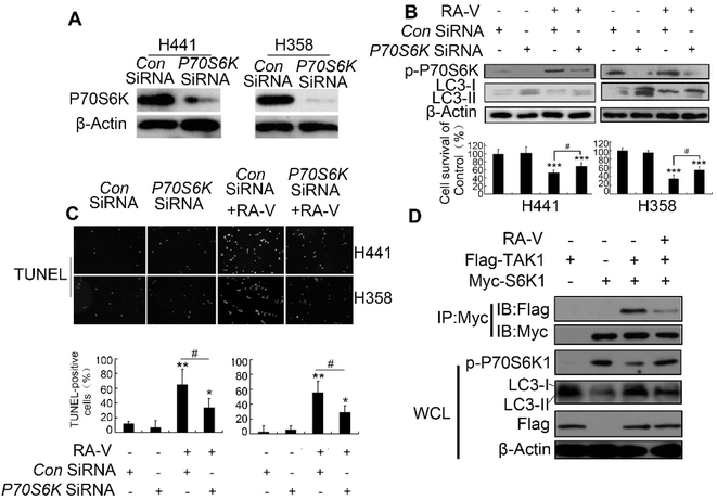
The authors regret an error in Fig. 4b where an incorrect β-actin band was used for the H441 cells. The corrected Fig. 4 is shown below.
The authors confirm that this correction does not affect the original conclusions of the study.
The Royal Society of Chemistry apologises for these errors and any consequent inconvenience to authors and readers.
| This journal is © The Royal Society of Chemistry 2025 |

