Facile preparation and bifunctional imaging of Eu-doped GdPO4 nanorods with MRI and cellular luminescence†
Abstract
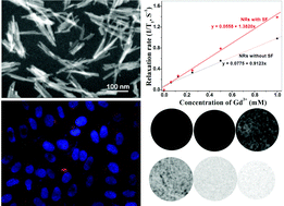
The biocompatibility of multifunctional nanomaterials is very important for their clinical applications. Herein, the hexagonal crystal Eu-doped GdPO4 nanorods (NRs) in the template of silk fibroin (SF) peptides are successfully synthesized via a mineralization process. The sizes of the Eu-doped GdPO4 NRs with SF peptides (SF-NRs) are ∼150 nm in length and ∼10 nm in diameter. The Eu-doped SF-NRs have strong pink luminescence and a mass magnetic susceptibility value of 1.27 emu g−1 in 20 000 G of magnetic field due to Eu ion doping. The cell test indicates that the Eu-doped SF-NRs obviously promote the viability of cells at an NR concentration of 25–200 μg mL−1. A growth mechanism of Eu-doped GdPO4 SF-NRs is proposed to explain their strong cellular luminescence, magnetic resonance (MR) imaging and good cyto-compatibility. Compared to NRs without SF, the Eu-doped SF-NRs not only exhibit a higher effective positive signal-enhancement ability (the longitudinal relaxivity r1 value is 1.38 (Gd mM s)−1) and in vivo T1 weighted MR imaging enhancement under a 7.0 T MRI system, but also show the better luminescence imaging of living cells under the fluorescence microscope. This indicates that the Eu-doped SF-NRs have potential as T1 MRI contrast agents and optical imaging probes.


 Please wait while we load your content...