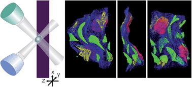
The combination of several micro-XRF analysis modes is presented for the investigation of an illuminated parchment manuscript. With a commercial instrument, conventional micro-XRF spot analysis (0D) and mapping (2D) are performed, yielding detailed lateral elemental information. Depth resolution becomes accessible by mounting an additional polycapillary lens in front of an SDD detector. Quantitative confocal depth profiles (1D) are presented as well as the full separation of the front and the backside decorations with the help of fast 3D mappings of specific areas. Only through the use of these multi-dimensional modes can elemental information be assigned both to lateral and depth positions, making the analysis of such heterogeneous samples feasible.
