
In situ photodeposition of loaded Co–MoSx for promoting visible-light g-C3N4 photocatalytic hydrogen production performance†
Yonggang
Lei
 abc,
Kim Hoong
Ng
d,
Chenyu
Zou
b,
Lejun
Chen
b,
Yuekun
Lai
*bc and
Jianying
Huang
*bc
abc,
Kim Hoong
Ng
d,
Chenyu
Zou
b,
Lejun
Chen
b,
Yuekun
Lai
*bc and
Jianying
Huang
*bc
aCollege of Chemistry and Chemical Engineering, Hexi University, Zhangye 734000, PR China
bNational Engineering Research Center of Chemical Fertilizer Catalyst (NERC-CFC), College of Chemical Engineering, Fuzhou University, Fuzhou 350116, PR China
cQingyuan Innovation Laboratory, Quanzhou 362801, PR China
dDepartment of Chemical Engineering, Ming Chi University of Technology, New Taipei City 243303, Taiwan, China. E-mail: jyhuang@fzu.edu.cn; yklai@fzu.edu.cn
First published on 12th January 2024
Abstract
Herein, Co–MoSx/CN photocatalysts were prepared by photodeposition using urea and ammonium tetrathiomolybdate as starting materials. The doping of the cocatalyst MoSx changed the electronic band structure of CN, shortened the band gap width, and exhibited excellent visible light response. Furthermore, the introduction of the transition metal Co not only forms a Co–Mo–S bond but also makes better use of the excess S2− generated by the decomposition of ammonium tetrathiomolybdate, resulting in more active sites and improving the utilization of raw materials.
Energy and environmental issues are the major hurdles that limit the rapid development of the modern economy and society. This could be a result of the overreliance of humans on traditional fossil fuels that come along with polluting emissions during energy generation.1 In response to this, photocatalytic technology that realizes hydrogen production from water enables a one-stop solution to both energy crisis and environmental protection.2–6 Significantly, graphite phase carbon nitride (g-C3N4, abbreviated as CN) with a two-dimensional lamellar structure is widely investigated as a photocatalytic material due to its suitable band gap structure, good physical and chemical stability, and high availability.7 One major drawback of such material is associated with its relatively large band gap (2.7 eV) for very limited solar utilization. Furthermore, CN normally possesses rather low crystallinity, a small specific surface area, and lacks active sites, and these features promote high charge recombination during photoreactions, leading to under-par kinetics for H2 generation. Various strategies have been incorporated to overcome the abovementioned limitations, with the coupling of a cocatalyst and sacrificial reagents being a few that have been approved by most researchers due to their proven efficiencies.
Significantly, cocatalyst loading is a simple and effective approach to improve charge separation, thereby enhancing the overall photocatalytic activity of CN.8–10 Pt, as an excellent cocatalyst, is widely used in CN-based catalysts, whereby a simple in situ photodeposition is sufficient to confer robust photocatalytic hydrogen evolution performance even with a meager loading of Pt.11 While being more economically commensurable, MoS2, with its well-known layered structure and rich exposed edges, could also permit outstanding co-catalytic features for better photo-activity of CN. Despite the improved activity with MoS2, past research indicates the possibility of further augmenting the co-catalytic features of MoS2 by introducing transition metal atoms.12–14 This effort enables better conductivity while anchoring active sites to the MoS2 co-catalyst, thus promising better photo-activity in photocatalytic HER. For instance, Chen et al. synthesized cobalt-doped graphite carbon nitride by one-step thermal polycondensation (Co CN), where the optimum photocatalyst consisting of 0.46 wt% Co showed a high hydrogen evolution rate (28.0%), tripling that of pure CN under standard evaluation conditions.15 This can be ascribed to the thermal condensation protocol for the co-existence of all precursors, resulting in tightly intercalated Co CN with a larger specific surface area and more Co–Nx active sites for HER under light. Wang et al. successfully loaded MoSx nanoparticles onto CN through sonochemical methods, where the following Ni-introduction was found to alleviate photo-corrosion of photocatalysts.16 The composite catalyst, therefore, exhibits Pt-like hydrogen production performance (5968 μmol g−1 h−1) in an extremely sustainable manner. Yu et al. successfully modified amorphous CoMoSx on the surface of CdS by a facile photoinduced deposition process, the resultant a-CoMoSx/CdS sample displayed a markedly higher photocatalytic H2-production performance than the blank CdS, a-MoSx/CdS, and Co(II)/CdS samples.17 Similar results were also attained by Attanayake et al., where their Co and Ni-doped MoS2 realized a low starting potential of 75 mV and an overvoltage of 145 mV, which are equivalent to those of Pt.18–20
While MoS2 is recognized for its remarkable co-catalytic features, its under-coordinated counterpart of MoSx is believed to serve as a better modifier. Significantly, MoSx consists of more edge unsaturated sites, which in theory, permits better co-catalytic properties than MoS2. Deducing from the case of MoS2, an in situ photo-deposition approach should be adequate to tightly anchor MoSx onto CN at room conditions, thereby providing a similarly greener pathway for photocatalyst preparation.21 Meanwhile, the works described above also illustrated the potential of using transition metals such as Fe, Co, and Ni to further augment the MoSx co-catalyst. It is speculated that, upon modifying, a facilitated charge dynamic could be imparted to the composite photocatalyst. Co is used as a modifier, and an active site with a “Co–Mo–S” structure would be established, synergistically resulting in even better hydrogen production performance under visible light irradiation.22–24
In this study, Co–MoSx particles were uniformly loaded on CN by photodeposition to construct a photocatalyst material with a close contact interface and achieve efficient catalytic decomposition of water for hydrogen production. Without using any precious metals, the prepared Co–MoSx/CN photocatalyst has excellent hydrogen production ability under visible light. Significantly, the presence of Co confers not only a “Co–Mo–S” active center but also makes full use of surplus S2− while evading raw material wastage during the catalyst preparation. Activity-check confirms the enhanced photocatalytic activity, mainly attributed to the close contact between Co–MoSx and CN, an increase of active sites, and accelerated charge separation in the catalyst. In this regard, this study provides a simple and green photo-deposition method for the modification of CN-based catalysts using non-noble metal cocatalysts.
The surface morphology of the selected photocatalyst was characterized by SEM and TEM, as shown in Fig. S1† and 1, respectively. Pure CN manifests a stacked lamellar structure with a high degree of irregularity in shape. Meanwhile, MCN resembles spherical particles of several micrometers in size, along with a poriferous surface generated by the decomposition of (NH4)2MoS4 during photodeposition. The microscopic morphology and element distribution of Co–MCN are shown in Fig. 2.
 | ||
| Fig. 1 The schematic diagram of the preparation process of the composite Co–MoSx/CN. | ||
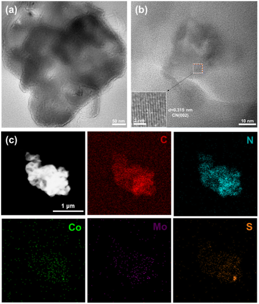 | ||
| Fig. 2 (a) TEM image of Co–MCN; (b) HRTEM image of Co–MCN; (c) HAADF image of Co–MCN and corresponding element mappings (C, N, Co, Mo, and S). | ||
Notably, the layered structure of CN is well-preserved upon incorporating with Co, as evidenced from the TEM image of Co–MCN shown in Fig. 2a. A clear lattice stripe of about 0.315 nm, which belongs to the (002) crystal plane of CN, is also seen in Fig. 2b. While shifting to the other location of the same sample using a high angle annular dark field scanning transmission electron microscope (HAADF-STEM, Fig. 2c), the element mapping analysis indicate the presence of five different elements, namely C, N, Co, Mo, and S, in this region. The abundance of CN in Co–MCN can be evidenced by the denser red and robin-egg blue colour tones in Fig. 2c. Meanwhile, the co-existing Co, Mo, and S are overlapping and well-distributed on C and N, demonstrating the successful incorporation of Co–MoSx onto CN.
Fig. 3a depicts the N2 adsorption–desorption isotherms of pure CN and MCN, alongside their pore size distribution, as shown in Fig. 3b. It can be seen that both the doped and bare CN resemble Type IV isotherms with H3 type hysteresis loops, thereby confirming their mesoporous structures with opening ranging from 0 to 120 nm. Table S1† shows the BET-specific surface area, pore volume, and pore size of bare CN, MCN, and Co–MCN. It can be seen that the specific surface area and average pore size of CN were increased from 35.9 m2 g−1 and 5.5835 nm to 41.2 m2 g−1 and 5.9694 nm, respectively, upon incorporation of MoSx. These can be due to the increased voids in MCN with the introduction of MoSx to CN. Further anchoring Co to MCN will, however, cover these voids, therefore contributing to the small surface area, pore volume, and pore size (30.8 m2 g−1, 0.0451 cm3 g−1, 5.8569 nm) of Co–MCN in Table S1.† Generally, larger surface area and porosity are more conducive to the hydrogen production reaction, but this is not the only factor that determines the rate of hydrogen production, it should be noted that, in the realm of photocatalysis, the presence of suitable and ample active sites is more important. This explains the ascendancy of Co–MCN in generating H2 from water against bare CN and MCN (discussed in the following section), despite its much smaller surface area, pore volume, and pore size.
 | ||
| Fig. 3 (a) N2 adsorption–desorption curves and (b) pore size distribution of pure CN, MCN, and Co–MCN. | ||
The crystallinity and crystal structure of the catalyst were analyzed by X-ray diffraction patterns (XRD; Fig. 4a). Pure CN exhibits a graphite-like lamellar structure with peaks emerging at 2θ = 13.3° and 27.6°, which can be associated with (100) and (002) phase of CN, respectively. The (100) plane absorption peak at 13.3° arises from the periodic planar arrangement of the tris homotriazine structure of CN, while the (002) plane absorption peak at 27.6° indicates the presence of stacked CN layers, which agrees with the SEM in Fig. S1.† While manifesting the characteristic peaks of CN at the same positions, no other peaks were seen in all the doped samples, regardless of the extents of MoSx- or Co–MoSx-modification (Fig. 3a and S2–S4†). These, therefore, imply the formation of tiny dopant particles and good dispersion on CN, which is consistent with TEM elemental mapping results in Fig. 2c.
 | ||
| Fig. 4 (a) XRD and (b) FT-IR of CN, MCN, and Co–MCN. | ||
Similarly, the FT-IR spectra shown in Fig. 4b for all three samples closely resemble each other. Broad peaks that centered at 3184 cm−1 indicate the adoption of water on the surface of all photocatalyst. Meanwhile, the respiratory pattern of heptazine ring in CN was also well-preserved too, regardless of MoSx- or Co-MoSx-modification, judging from the absorption bands peaked at 807 cm−1 for all analyzed samples.25 Meanwhile, the continuous absorption bands extending from 1200 to 1600 cm−1 are attributed to the tensile vibration modes of aromatic C–N and C![[double bond, length as m-dash]](https://www.rsc.org/images/entities/char_e001.gif) N heterocycles. Also, the absorptions at 1320 and 1640 cm−1 could also be associated with the vibrations of carbon and nitro single and double bonds, respectively.26 Overall, both XRD and FTIR data indicate the unaltered crystal attributes and surface functional group of CN upon loading of MoSx or Co–MoSx.27 Clearly, the chemical structure of the CN surface was characterized by Raman spectroscopy, as shown in Fig. S5,† which revealed that after CN modification by MoSx and Co–MoSx, the CN peaks at 703, 749, 977, and 1236 cm−1 were weaker. It is shown that MoSx and Co–MoSx have an effect on the chemical structure of their CN, which is recent proof that the synthesis of the materials is effective. The surface chemical state and elemental composition of the catalyst were studied by XPS. Similar to the EDX results, the signals for the same five species (Co, Mo, S, C, and N) were captured in the full XPS spectrum shown in Fig. 5a. Notably, the graphitic carbon peak at 284.8 eV was used as the calibration standard for the spectrum. The CN features of all analyzed samples can be confirmed from the high-resolution C 1s and N 1s peaks shown in Fig. 5b and c, respectively. The C 1s peak spectrum of the bare CN can be deconvoluted into three species, viz. C–C skeleton, sp3 hybrid carbon (C–O), and sp2 hybrid carbon (N–C
N heterocycles. Also, the absorptions at 1320 and 1640 cm−1 could also be associated with the vibrations of carbon and nitro single and double bonds, respectively.26 Overall, both XRD and FTIR data indicate the unaltered crystal attributes and surface functional group of CN upon loading of MoSx or Co–MoSx.27 Clearly, the chemical structure of the CN surface was characterized by Raman spectroscopy, as shown in Fig. S5,† which revealed that after CN modification by MoSx and Co–MoSx, the CN peaks at 703, 749, 977, and 1236 cm−1 were weaker. It is shown that MoSx and Co–MoSx have an effect on the chemical structure of their CN, which is recent proof that the synthesis of the materials is effective. The surface chemical state and elemental composition of the catalyst were studied by XPS. Similar to the EDX results, the signals for the same five species (Co, Mo, S, C, and N) were captured in the full XPS spectrum shown in Fig. 5a. Notably, the graphitic carbon peak at 284.8 eV was used as the calibration standard for the spectrum. The CN features of all analyzed samples can be confirmed from the high-resolution C 1s and N 1s peaks shown in Fig. 5b and c, respectively. The C 1s peak spectrum of the bare CN can be deconvoluted into three species, viz. C–C skeleton, sp3 hybrid carbon (C–O), and sp2 hybrid carbon (N–C![[triple bond, length as m-dash]](https://www.rsc.org/images/entities/char_e002.gif) N) in the s-triazine ring, with peaks at 284.8, 286.2, and 288.0 eV, respectively. Upon modification, the incorporated MoS2 or Co–MoSx would withdraw electrons from CN, leading to the reduced electron density in CN and thus the increased N–CN energy of 288.3 eV and 288.4 eV, respectively, as indicated in Fig. 5b. Meanwhile, four photoelectron peaks were observed for N 1s (Fig. 5c) of bare CN, with that centered at 398.5, 399.8, 401.1, and 404.0 eV corresponds to sp2 hybrid N (CN–C) in the triazine ring, the bridge N in the N–(C)3, the N (C–N–H) in the amino group, and the π excitation, respectively. Similar positive shiftings was also observed upon MoS2 or Co–MoSx-modifications, therefore, confirming their roles in facilitating electron migration during photoreaction.
N) in the s-triazine ring, with peaks at 284.8, 286.2, and 288.0 eV, respectively. Upon modification, the incorporated MoS2 or Co–MoSx would withdraw electrons from CN, leading to the reduced electron density in CN and thus the increased N–CN energy of 288.3 eV and 288.4 eV, respectively, as indicated in Fig. 5b. Meanwhile, four photoelectron peaks were observed for N 1s (Fig. 5c) of bare CN, with that centered at 398.5, 399.8, 401.1, and 404.0 eV corresponds to sp2 hybrid N (CN–C) in the triazine ring, the bridge N in the N–(C)3, the N (C–N–H) in the amino group, and the π excitation, respectively. Similar positive shiftings was also observed upon MoS2 or Co–MoSx-modifications, therefore, confirming their roles in facilitating electron migration during photoreaction.
 | ||
| Fig. 5 XPS spectra of pure CN, MCN, and Co–MCN: (a) survey (b) C 1s (c) N 1s (d) Mo 3d and (e) S 2p, and (f) Co 2p. | ||
The bimodal bands of Mo 3d in Fig. 5d indicate the co-existence of Mo with different oxidation states, with those that centered at 235.4 eV and 232.3 eV confirming the presence of Mo6+ in both MCN and Co–MCN. This can be related to the formation of the MoSx phase or oxides phase, which is induced during the photodeposition process.28 The peak at 229.0 eV, on the other hand, can be assigned to Mo4+ of MoS2 that was co-deposited on CN. Such presence of MoS2 was further justified by the S2− peak located at 162.7 eV, as shown in Fig. 5e. Meanwhile, the smaller peaks with binding energies of 168.0, 168.7, and 169.7 eV indicate the presence of S4+, plausibly due to the formation of partially oxidized sulfur species during the preparation of the photocatalyst. As for Co–MCN (Fig. 5f), the introduction of Co induces new Co2+-related peaks in its Co 2p spectrum, with that centered at 778.5 eV and 802.8 eV assigned to the core electrons from Co2+ 2p3/2 and 2p1/2 orbitals, respectively. Such peaks indicate the formation of complex hetero-bimetallic sulfide, which in our case, is possibly the “Co–Mo–S” phase during the photodeposition process. This is also consistent with the results reported in the literature. Overall, the presence of Co, Mo, and S in the XPS spectra of the modified CN indicates the successful synthesis of MoSx/CN and Co–MoSx/CN, where the recorded energy shifting could also evidence the establishment of electronic interactions; hence, the intimate contact of the constituents over our facile photodeposition approach.
A 10 vol% triethanolamine (TEOA) solution was chosen as the sacrificial reagent to evaluate the photocatalytic hydrogen production performance of the catalyst under visible light (λ > 400 nm). CN is widely used in photocatalysis due to its visible light response, suitable energy bands, and non-metallic properties.29 However, due to the small surface area and the rapid recombination of charges, its activity is usually unsatisfactory, which explained the undetectably low H2 generation in our study (Fig. 6a).
 | ||
| Fig. 6 (a) Hydrogen production rate for CN with different MoSx loadings; (b) hydrogen production rate for Co–MCN with different Co/MoSx ratios; (c) hydrogen production rate for various samples; (d) UV-vis diffuse reflectance spectrum of Co–MCN and its AQE values; (e) stability of MCN and Co–MCN and (f) XRD patterns before and after cycling. | ||
With merely 5 wt% MoSx incorporated, the hydrogen production rate of MCN-5 was drastically improved to 73.6 μmol g−1 h−1. Such promotional effects of MoSx can be explained by the presence of unsaturated sulfur over its disordered amorphous phase, which intrinsically has higher activity due to its under-coordinated nature.30 The deposited MoSx can, therefore, better improve the rate of electron transfer in CN and increase the utilization of photogenerated electrons, resulting in more hydrogen production.31,32 Meanwhile, the co-existing crystalline MoS2 (from XPS) can also serve as a solid electron mediator between catalysts, enhancing the synergy between catalyst components and enhancing the electron transport ability of the catalyst.33 All these positive features were based upon the even dispersion of nano-sized dopant particles on CN, where their intimate contacts over the in situ photodeposition approach also play a major role in boosting H2 generation. In this regard, increasing MoSx loading could further enhance the generation of H2 until the optimum point with 12 wt% MoSx modifier. This resulted in an auspicious hydrogen production rate of 217.5 μmol g−1 h−1, and further increasing MoSx content induces pronounced light-blocking effects to CN, thereby, plummeting the activity, as indicated in Fig. 6a. By fixing the dopant level at 12 wt%, CN modified by Co–MoSx with different Co:MoSx ratios were also subjected to H2 generation under visible light. It is expected that the so-called “Co–Mo–S” structure could be prompted upon incorporating both Co and MoSx modifiers to CN, enabling more active sites for enhancing the hydrogen production performance of CN.34 As can be seen from Fig. 6b, compared to MoSx/CN, the hydrogen production performance of Co–MoSx/CN was significantly improved too. Notably, the optimum Co![[thin space (1/6-em)]](https://www.rsc.org/images/entities/char_2009.gif) :MoSx ratio was identified at 1:21, where the corresponding Co–MCN photocatalyst could realize astonishing hydrogen production rate of 420.3 μmol g−1 h−1, which is nearly double that of MCN. In particular, it can be seen from Fig. 6c that both MoSx and Co–MoSx individually exhibit no hydrogen production ability, thereby, confirming their co-catalytic attribute towards the CN photocatalyst. MCN (21/22) and Co/CN (1/22) were synthesized from Co–MoSx/CN (1:21) in accordance with the respective loading amounts of Co and MoSx in the catalyst. In particular, MCN (21/22) exhibited notable hydrogen production performance but it is far lower than Co–MCN. Co/CN (1/22), on the other hand, permitted zero hydrogen production, indicating the inadequacy of Co alone in co-catalysing H2 generation ability of CN. From these blank experiments, the synergistic effects from the combination of Co and MoSx modifiers are certain. The resulting activity from the deliberately designed Co–MCN was found better than those of Ni-MCN and Fe-MCN, which were prepared using Ni and Fe, respectively, at the same composition. It is important to note that our photocatalyst of Co–MCN also outperformed 1 wt% Pt/CN prepared using the common chloroplatinic acid solution photodeposition (Fig. 6c). This indicates that our proposed Co–MoSx cocatalyst could potentially serve as an economic substitute for noble metal in the aspect of H2 generation from water.
:MoSx ratio was identified at 1:21, where the corresponding Co–MCN photocatalyst could realize astonishing hydrogen production rate of 420.3 μmol g−1 h−1, which is nearly double that of MCN. In particular, it can be seen from Fig. 6c that both MoSx and Co–MoSx individually exhibit no hydrogen production ability, thereby, confirming their co-catalytic attribute towards the CN photocatalyst. MCN (21/22) and Co/CN (1/22) were synthesized from Co–MoSx/CN (1:21) in accordance with the respective loading amounts of Co and MoSx in the catalyst. In particular, MCN (21/22) exhibited notable hydrogen production performance but it is far lower than Co–MCN. Co/CN (1/22), on the other hand, permitted zero hydrogen production, indicating the inadequacy of Co alone in co-catalysing H2 generation ability of CN. From these blank experiments, the synergistic effects from the combination of Co and MoSx modifiers are certain. The resulting activity from the deliberately designed Co–MCN was found better than those of Ni-MCN and Fe-MCN, which were prepared using Ni and Fe, respectively, at the same composition. It is important to note that our photocatalyst of Co–MCN also outperformed 1 wt% Pt/CN prepared using the common chloroplatinic acid solution photodeposition (Fig. 6c). This indicates that our proposed Co–MoSx cocatalyst could potentially serve as an economic substitute for noble metal in the aspect of H2 generation from water.
A light source with different band-pass filters (λ = 400, 420, 450, and 500 nm) was used to evaluate the performance of Co–MCN for hydrogen production while determining respective AQE values using eqn (1) (Fig. 6d). Significantly, AQE at λ = 400 nm illumination was recorded as 0.68%, but that gradually reduced as the illuminated wavelength increases. An AQE close to zero was recorded at λ = 500 nm, indicating the inertness of Co–MCN under illumination at that wavelength. This is not surprising as Co–MCN exhibits nearly no absorbance, as indicated by the UV-Vis DRS profile shown in Fig. 6d. Deducing from this, it can be concluded that the light absorption of the catalyst plays an ultimate role in its hydrogen production activity.
Meanwhile, the synthesized MCN and Co–MCN were also subjected to a stability test for a continuous 4-cycle of H2 generation (Fig. 6e). While both photocatalysts suffered from a certain extent of activity degradation, Co–MCN is constantly manifesting better H2 generation than MCN, thereby confirming the role of Co in stabilizing MoSx cocatalyst during photoreaction. Post-reaction XRD analysis indicates unaltered crystal structures of both MCN and Co–MCN photocatalysts (Fig. 6f) even after 4 cycles of photoreactions, with both CN peaks located at 13.3° and 27.6° being preserved. Meanwhile, sacrificial reagents also play an important role in sustaining H2 production by consuming photogenerated holes while inhibiting charge recombination. As shown in Fig. S8,† no hydrogen was detected in the absence of a sacrificial reagent under the same test conditions. Upon comparison, TEOA enables better effects than ethanol and lactic acid solutions by generating most hydrogen from water over the Co–MCN photocatalyst.
Electrochemical and photo-electrochemical analyses of selected photocatalysts were performed using a three-electrode system. Fig. 7a shows the LSV curves of the catalysts. It can be seen that pure CN, MCN, and Co–MCN exhibit the same onset potential, but the current density of MCN and Co–MCN increases relatively quickly along with voltage. At a standard current density of 5 mA cm−2, MCN required a lower driving force of 1.12 V, where the incorporation of Co could further reduce that to 0.96 V with Co–MCN electrode, as compared to 1.20 V of CN. Significantly, these mentioned overpotentials are closely related to the resistance across the interface during HER. Correspondingly, the Tafel curve shown in Fig. 7b indicates a much larger slope for pure CN (353.4 mV dec−1) as compared to that of MCN (314.5 mV dec−1) and Co–MCN (259.0 mV dec−1). This further evidences the promotional effects of MoSx and the synergistic effects between Co and MoSx in improving the HER capability of CN. In terms of light responsivity, the I–t test in Fig. 7c implies notably elevated current densities for CN, MCN, and Co–MCN, thereby confirming their capability to generate electrons under irradiation. Upon comparison, the latter two photocatalysts exhibited better photocurrent response, with Co–MoSx being higher, plausibly due to the enhanced charge dynamics after modification. The impedance analysis shown in Fig. 7d yielded identical findings, where Co–MCN exhibited the lower impedance judging from its smallest diameter in the Z′′ vs. Z′ graph, followed by MCN and CN. These can be associated with the tight interfaces of both MCN and Co–MCN, thereby enabling outstanding charge dynamics for better LSV and photocurrent responses, as discussed above. In the context of photocatalysis, the photogenerated electrons from CN can facilely shuttle across such an intimate interface to the modifier (MoSx or Co–MoSx), thereby giving rise to better HER as shown in Fig. 6.
 | ||
| Fig. 7 (a) Polarization curves; (b) Tafel slope; (c) photocurrent response; and (d) electrochemical impedance (EIS) of CN, MCN and Co–MCN. | ||
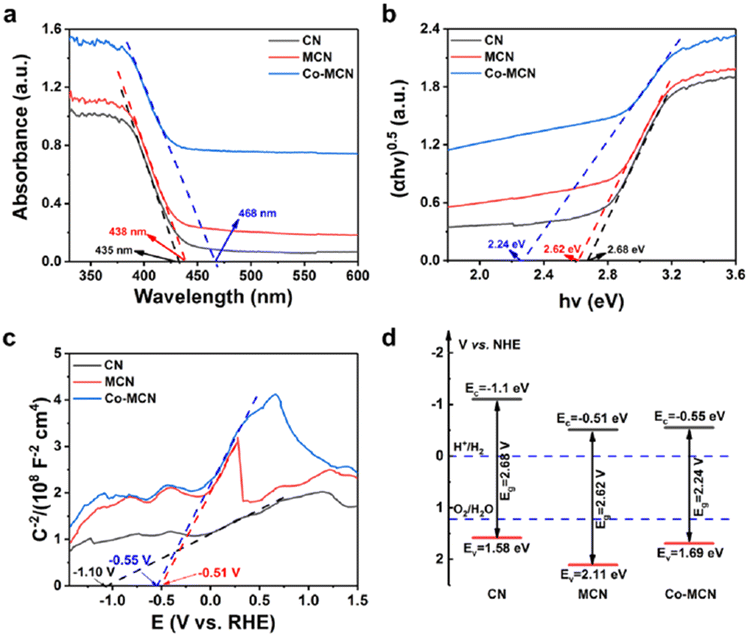
In addition, the light absorption ability and band positions are also pivotal factors in the photocatalytic hydrogen production of photocatalysts. An enhanced light absorption ability is often associated with more photogenerated electrons after excitation of the photocatalyst; thus, leading to better H2 production.35Fig. 8a shows the UV-Vis absorption spectra of the selected photocatalysts. It can be seen that pure CN, MCN, and Co–MCN have strong absorption capacities in the UV region, where the incorporation of MoSx and Co–MoSx red-shifted the absorption edge of CN from 435 nm to 438 nm and 468 nm, respectively. This indicates better photon absorption capability of the modified photocatalyst, as well as their much facile excitation for various photoreactions. The Tauc plot presented in Fig. 8b also illustrates the gap-narrowing tendency of the modifier, reducing that of pure CN from 2.68 eV to 2.62 eV and 2.24 eV upon incorporation of MoSx and Co–MoSx, respectively. Furthermore, the band positions for each photocatalyst were also estimated using a Mott–Schottky (MS) plot, as shown in Fig. 8c. An apparent linear zone with a positive gradient was identified in the profile of all tested photocatalysts, thereby confirming their N-type attribute, possessing a higher number of electron donors in the composition. Flat-band potential for each photocatalyst can be obtained by determining respective X-intercept values, which are −1.10 eV, −0.51 eV, and −0.55 eV for pure CN, MCN, and Co–MCN, respectively. For N-type semiconductors, their flat band potentials are known to be positioned at the vicinity of their conduction band minimum, therefore, providing a rather accurate estimation of the energy of the photo-electron. In this regard, MCN and Co–MCN secure better charge separation (Fig. 7) at the cost of sacrificing a portion of the reductive energy of their electrons. However, the ultimate electron energy of both the mentioned photocatalysts is still sufficient to prompt H2 from water, which required only 0 eV vs. RHE. Summarizing the results from Fig. 8a–c, the corresponding band positions of CN, MCN, and Co–MCN are illustrated in Fig. 8d, along with the identification of respective band gaps. The digital picture of the photocatalyst is shown in Fig. S9.† Pure CN is initially in pale yellow color, and progressively darkened after loading with MoSx and Co–MoSx. At varied MoSx content (2nd row in Fig. S9†), similar darkening effects could also be observed, therefore, explaining the gap-narrowing phenomena confirmed in Fig. 8b. As for the 3rd row of Fig. S9,† the color tone of resultant powder is visibly lightened at high Co:MoSx ratio, probably due to lower content of MoSx.
 | ||
| Fig. 8 (a) UV-vis diffuse reflection spectra; (b) Tauc plot; (c) Mott–Schottky (MS) and (d) energy band structure diagrams of CN, MCN, and Co–MCN. | ||
Interestingly, the PL spectra shown in Fig. 9 indicate a distinctive peak ranging from 400–600 nm for all photocatalysts, where the intensity of the peak indicates the rate of charge recombination in each photocatalyst. It can be seen that pure CN exhibits the strongest fluorescence response, therefore, generating much lower H2 during photoreaction. Meanwhile, the suppressed PL signals by MCN and Co–MCN also confirmed positive effects from MoSx and Co–MoSx modifiers, with the latter being more robust judging from its lowest PL intensity.
 | ||
| Fig. 9 Photoluminescence spectra (PL) of CN, MCN, and Co–MCN. | ||
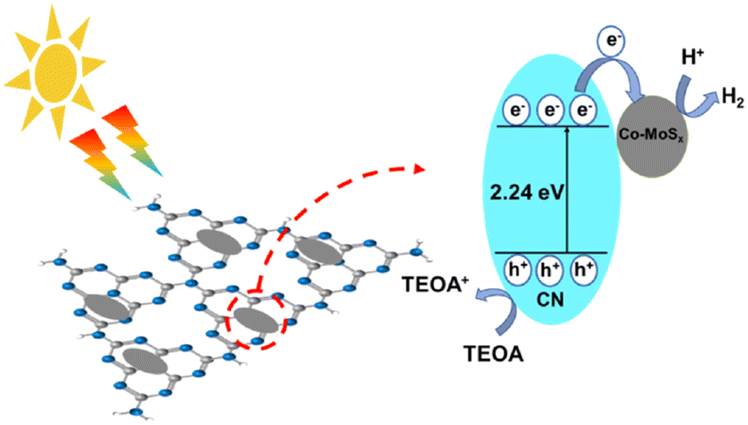
Concluding from the results from photo-electrochemical and electrochemical evaluations, it was found that MoSx can accelerate the migration of electrons after their generation in CN, ascribed to the tight interface between the constituents. This establishes facilitated channels for interfacial shuttling of electrons (as confirmed by XPS), thereby giving rise to a much lower impedance environment within the photocatalytic particles. While Co–MoSx permits similar effects, the presence of Co further regulated MoSx in terms of electronic properties, enabling even lower impedance for electron shuttlings. Also, Co could also activate MoSxvia the formation of the “Co–Mo–S” active structure; it attracts H+ while serving as an active site for hydrogen production in the process. Therefore, electrons generated by CN in the composite system upon absorbing light would first migrate to the surface of the CN, followed by the Co–MoSx modifier across the intimate interface. This prompts spatial separation of photo-charges, thereby explaining the suppressed PL signal shown in Fig. 9.
At the same time, holes that resided at the valence band of CN will be captured by sacrificial reagents (TEOA), further plummeting the occurrence of charge recombination of electron holes. The synergy of these mechanisms has consequently led to an enhanced H2 generation kinetics, resulting in an optimal hydrogen production capacity of 420.3 μmol g−1 h−1, which outperforms that reported in most studies, as listed in Table S2.†Fig. 10 illustrates the entire mechanism of H2 generation from water over our Co–MoSx-modified CN.
 | ||
| Fig. 10 Schematic diagram of visible-light-driven Co–MoSx/CN hydrogen production. | ||
In this study, it has been determined that Co–MoSx can be used as an effective cocatalyst to improve the photocatalytic hydrogen production performance of CN under visible light irradiation. MoSx can be loaded onto CN by photodeposition, and can act as an electron trap in the photocatalytic hydrogen production reaction, capturing photogenerated electrons from the CN. Further, Co can be used to activate MoSx and construct a “Co–Mo–S” active center, providing a large number of active sites for hydrogen production reactions, while accelerating the electron flow between the components of the composite catalyst, thereby improving photocatalytic activity. However, this may lead to a relatively weak binding force between the cocatalyst Co–MoSx and CN, and the particle size may change during the synthesis process, forming larger particles, resulting in lower material utilization, or partial detachment of the cocatalyst during the hydrogen production process, resulting in a reduction in hydrogen production performance. In future research, these issues should be addressed to further improve the efficiency of Co–MoSx/CN photocatalysts.
Conflicts of interest
There are no conflicts to declare.Acknowledgements
The authors thank the projects of International Cooperation and Exchanges NSFC (22361162607), National Natural Science Foundation of China (22075046 and 22375047), Doctoral Research Initiation Grant Program (KYQD2023015), National Key Research and Development Program of China (2022YFB3804905, 2022YFB3804900), Natural Science Foundation of Fujian Province (2022J01568).Notes and references
- L. Yang, Y. Peng, X. Luo, Y. Dan, J. Ye, Y. Zhou and Z. Zou, Chem. Soc. Rev., 2021, 50, 2147–2172 RSC.
- X. Ruan, C. Huang, H. Cheng, Z. Zhang, Y. Cui, Z. Li, T. Xie, K. Ba, H. Zhang, L. Zhang, X. Zhao, J. Leng, S. Jin, W. Zhang, W. Zheng, S. K. Ravi, Z. Jiang, X. Cui and J. Yu, Adv. Mater., 2023, 35, 2209141 CrossRef CAS PubMed.
- Y. Lei, K. Ng, Y. Zhu, Y. Zhang, Z. Li, S. Xu, J. Huang, J. Hu, Z. Chen, W. Cai and Y. Lai, Chem. Eng. J., 2023, 452, 139325 CrossRef CAS.
- Y. Lei, T. Zhao, K. Ng, Y. Zhang, X. Zhang, X. Li, W. Cai, J. Huang, J. Hu and Y. Lai, Acta Phys.-Chim. Sin., 2023, 39, 2206006 Search PubMed.
- T. Sun, C. Li, Y. Bao, J. Fan and E. Liu, Acta Phys.-Chim. Sin., 2023, 39, 2212009 Search PubMed.
- S. Xu, J. Huang, Z. Li, Y. Lei, Y. Zhang, K. Ng and Y. Lai, J. Cleaner Prod., 2023, 402, 136672 CrossRef CAS.
- R. Sharma, M. Almáši, S. P. Nehra, V. S. Rao, P. Panchal, D. R. Paul, I. P. Jain and A. Sharma, Renewable Sustainable Energy Rev., 2022, 168, 112776 CrossRef CAS.
- J. Cai, J. Huang, S. Wang, J. Iocozzia, Z. Sun, J. Sun, Y. Yang, Y. Lai and Z. Lin, Adv. Mater., 2019, 31, 1806314 CrossRef PubMed.
- Z. Lei, X. Ma, X. Hu, J. Fan and E. Liu, Acta Phys.-Chim. Sin., 2022, 38, 2110049 Search PubMed.
- R. Shen, L. Hao, Q. Chen, Q. Zheng, P. Zhang and X. Li, Acta Phys.-Chim. Sin., 2022, 38, 2110014 Search PubMed.
- M. Liu, P. Xia, L. Zhang, B. Cheng and J. Yu, ACS Sustain. Chem. Eng., 2018, 6, 10472–10480 CrossRef CAS.
- Y. Lv, P. Chen, J. J. Foo, J. Zhang, W. Qian, C. Chen and W.-J. Ong, Mater. Today Nano, 2022, 18, 100191 CrossRef CAS.
- U. Sharma, S. Karazhanov, R. Jose and S. Das, J. Mater. Chem. A, 2022, 10, 8626–8655 RSC.
- Y. Xu, R. Ge, J. Yang, J. Li, S. Li, Y. Li, J. Zhang, J. Feng, B. Liu and W. Li, J. Energy Chem., 2022, 74, 45–71 CrossRef CAS.
- P.-W. Chen, K. Li, Y.-X. Yu and W.-D. Zhang, Appl. Surf. Sci., 2017, 392, 608–615 CrossRef CAS.
- B. Wang, X. Li, H. Wu, G. Xu, X. Zhang, X. Shu, J. Lv and Y. Wu, ChemCatChem, 2020, 12, 911–916 CrossRef CAS.
- W. Liu, X. Wang, H. Yu and J. Yu, ACS Sustain. Chem. Eng., 2018, 6, 12436–12445 CrossRef CAS.
- N. H. Attanayake, L. Dheer, A. C. Thenuwara, S. C. Abeyweera, C. Collins, U. V Waghmare and D. R. Strongin, ChemElectroChem, 2020, 7, 3606–3615 CrossRef CAS.
- H. Zhao, Q. Mao, L. Jian, Y. Dong and Y. Zhu, Chin. J. Catal., 2022, 43, 1774–1804 CrossRef CAS.
- M. Wang, J. Cheng, X. Wang, X. Hong, J. Fan and H. Yu, Chin. J. Catal., 2021, 42, 37–45 CrossRef CAS.
- J. Wang, Y. Fan, R. Pan, Q. Hao, Y. Wu, T. van Ree and R. Holze, ACS Appl. Energy Mater., 2021, 4, 13288–13296 CrossRef CAS.
- X. Liu, Y. Xue, Y. Lei, F. Wang and S. Min, ACS Appl. Nano Mater., 2018, 1, 6493–6501 CrossRef CAS.
- Y. Zhu, Q. M. Ramasse, M. Brorson, P. G. Moses, L. P. Hansen, H. Topsøe, C. F. Kisielowski and S. Helveg, Catal. Today, 2016, 261, 75–81 CrossRef CAS.
- Y. Lei, Y. Zhang, Z. Li, S. Xu, J. Huang, K. Hoong Ng and Y. Lai, Chem. Eng. J., 2021, 425, 131478 CrossRef CAS.
- L. Dong, H. Chu, S. Xu, Y. Li, S. Zhao and D. Li, Nanophotonics, 2022, 11, 139–151 CrossRef CAS.
- Y. Guo, Q. Zhou, X. Chen, Y. Fu, S. Lan, M. Zhu and Y. Du, J. Mater. Sci. Technol., 2022, 119, 53–60 CrossRef CAS.
- S. Sivakumar, T. Daniel Thangadurai, N. Manjubaashini and D. Nataraj, Colloids Surf., A, 2022, 654, 130090 CrossRef CAS.
- Y. Hou, A. B. Laursen, J. Zhang, G. Zhang, Y. Zhu, X. Wang, S. Dahl and I. Chorkendorff, Angew. Chem., Int. Ed., 2013, 52, 3621–3625 CrossRef CAS PubMed.
- B. Xu, M. B. Ahmed, J. L. Zhou, A. Altaee, G. Xu and M. Wu, Sci. Total Environ., 2018, 633, 546–559 CrossRef CAS PubMed.
- S. Reddy, R. Du, L. Kang, N. Mao and J. Zhang, Appl. Catal., B, 2016, 194, 16–21 CrossRef CAS.
- L. Li, D. Gao, F. Chen, X. Wang and H. Yu, Appl. Surf. Sci., 2023, 608, 155173 CrossRef CAS.
- J. Staszak-Jirkovský, C. D. Malliakas, P. P. Lopes, N. Danilovic, S. S. Kota, K.-C. Chang, B. Genorio, D. Strmcnik, V. R. Stamenkovic, M. G. Kanatzidis and N. M. Markovic, Nat. Mater., 2016, 15, 197–203 CrossRef PubMed.
- Q. Jiang, L. Sun, J. Bi, S. Liang, L. Li, Y. Yu and L. Wu, ChemSusChem, 2018, 11, 1108–1113 CrossRef CAS PubMed.
- Y. Lei, Y. Xue, Y. Li, X. Liu, W. Deng, L. Tian, F. Wang and S. Min, J. Photochem. Photobiol., A, 2018, 367, 226–235 CrossRef CAS.
- Y. Wu, P. Xiong, J. Wu, Z. Huang, J. Sun, Q. Liu, X. Cheng, J. Yang, J. Zhu and Y. Zhou, Nano-Micro Lett., 2021, 13, 48 CrossRef PubMed.
Footnote |
| † Electronic supplementary information (ESI) available: Experimental section, additional Fig. S1–S8, Tables S1 and S2. See DOI: https://doi.org/10.1039/d3se01515g |
| This journal is © The Royal Society of Chemistry 2024 |