
Carbon quantum dot-mediated binary metal–organic framework nanosheets for efficient oxygen evolution at ampere-level current densities in proton exchange membrane electrolyzers†
Qianjia
Ni‡
a,
Shiyuan
Zhang‡
b,
Kang
Wang
a,
Huazhang
Guo
a,
Jiye
Zhang
 c,
Minghong
Wu
bd and
Liang
Wang
*ae
c,
Minghong
Wu
bd and
Liang
Wang
*ae
aInstitute of Nanochemistry and Nanobiology, School of Environmental and Chemical Engineering, Shanghai University, Shanghai, 200444, P. R. China. E-mail: wangl@shu.edu.cn
bShanghai Institute of Applied Radiation, Key Laboratory of Organic Compound Pollution Control Engineering (MOE), School of Environmental and Chemical Engineering, Shanghai University, Shanghai, 200444, P. R. China
cSchool of Materials Science and Engineering, Shanghai University, Shanghai, 200444, P. R. China
dCollege of Environment & Safety Engineering, Fuzhou University, Fuzhou, 350108, P. R. China
eShanghai Engineering Research Center of Organ Repair, Joint International Research Laboratory of Biomaterials and Biotechnology in Organ Repair (Ministry of Education), Shanghai University, Shanghai, 200444, P. R. China
First published on 1st November 2024
Abstract
The widespread utilization of noble metal-based catalysts for the oxygen evolution reaction (OER) is hindered by their rarity and substantial expense, posing significant challenges for large-scale applications. Therefore, developing an efficient OER electrocatalyst for proton exchange membrane (PEM) water electrolyzers remains a significant challenge. Here, we present a bottom-up synthesis strategy utilizing ultrasound-assisted exfoliation to design nickel–iron bimetallic organic framework (NiFe-MOF) nanosheets with high electrooxidation activity, in situ induced by carbon quantum dots (CQDs). This approach eliminates the reliance on intricate and inefficient exfoliation techniques, producing NiFe-MOF nanosheets with a regulated thickness of just 10 nm. This enhanced electron transport induced by CQDs plays a pivotal role in improving the OER performance of NiFe-MOF, achieving a current density of 10 mA cm−2 with an overpotential of only 280 mV, with a Tafel slope of 71.98 mV dec−1, lower Rct, and larger ECSA. In situ FTIR spectroscopy suggests that the OER mechanism in NiFe-MOF-CQD mainly follows the adsorbate evolution mechanism. The NiFe-MOF-CQD catalyst demonstrates remarkable durability and resilience during PEM water electrolysis, reaching industrially relevant current densities of 2 A cm−2 at 2 V. This research's results not only promote green and low-carbon development but also inject new vitality into the development of hydrogen energy technologies.
1. Introduction
Hydrogen energy holds immense potential as a leading contender among clean energy alternatives, and is instrumental in facilitating the global shift towards sustainable energy systems and ultimately contributing to the achievement of carbon neutrality goals. Water electrolysis for hydrogen production only consumes water, offering key advantages such as high flexibility and rapid response times, making it an environmentally benign alternative. Proton exchange membrane (PEM) water electrolysis stands out as a promising hydrogen production technology when compared to traditional alkaline electrolysis (AE), which operates at higher current densities, with improved overall efficiency and hydrogen purity. Its rapid dynamic response further makes it suitable for coupling with intermittent renewable energy sources, solidifying its position as a key technology in the hydrogen economy.1–3Despite the advantages of PEM water electrolysis, anode polarization presents a critical challenge, especially at high current densities, where it directly impacts the overall efficiency. Most researches have focused on anode electrocatalysts that are active at relatively low current densities (e.g., 10 mA cm−2) under laboratory conditions, but industrial-scale applications demand catalysts that can perform reliably at much high current densities (>1000 mA cm−2). In addition, the harsh oxidative conditions at the anode limit the selection of catalysts to a few precious metals such as Ir and Ru or their oxides, significantly increasing the cost of water electrolysis.4–9 Therefore, the development of cost-effective, highly efficient, and stable non-precious metal-based electrocatalysts is crucial for advancing PEM electrolysis technology on an industrial scale.10,11
Metal–organic frameworks (MOFs) hold significant potential in electrocatalysis owing to their exceptional characteristics, including a high specific surface area, porosity, and the ability to tailor their structure according to specific requirements.12–15 In parallel, carbon quantum dots (CQDs) have emerged as versatile materials with excellent photogenerated electron transfer properties, stability, and ease of synthesis.16,17 Recent studies have demonstrated that the integration of CQDs with other materials can modulate the internal electronic structure of a composite, leading to enhanced physicochemical properties, making them attractive candidates for the in situ synthesis of advanced two-dimensional (2D) materials.18–22 In this work, we focus on the design of a highly efficient and stable anode electrocatalyst for PEM water electrolysis. Using an ultrasound-assisted synthesis, we successfully prepare nickel-iron bimetallic organic framework (NiFe-MOF) nanosheets with high electrooxidation activity induced by CQDs without the need for complex exfoliation processes. Our results show long-term stability at ampere-level current densities, achieving a performance of 2 A cm−2 at 2 V in PEM water electrolysis. These findings not only advance the development of sustainable hydrogen energy but also establish a robust platform for future industrialization of clean energy technologies.
2. Experimental section
2.1 Chemicals and materials
p-Phenylenediamine (≥99%), ammonium hydroxide (25–28%), sodium trifluoromethanesulfonate (≥98%), ferric nitrate nonahydrate (Fe(NO3)3·9H2O, 99.9%), nickel acetate tetrahydrate (Ni(Ac)2·4H2O, ≥98%), 1,4-naphthalenedicarboxylic acid (H2NDC, 98%), trolamine (TEA, 99.5%), and N,N-dimethylacetamide (DMAC, 99.8%) were purchased from Adamas. Potassium hydroxide (KOH, ≥90%), ethanol (≥99.7%) and isopropanol (≥99.7%) were sourced from Greagent. Deionized (DI) water was used throughout this study. The Nafion solution (5 wt%) was obtained from Sigma Alpha, the proton exchange membrane (PEM) was from Nafion 115 (DuPont™), and Hispec 4100 Pt/C (40 wt% Pt) was from Johnson Matthey.2.2 Synthesis of CQDs
For the synthesis of CQDs, 500 mg p-phenylenediamine and 500 mg sodium trifluoromethanesulfonate were dispersed in 50 mL of isopropanol to create a uniform solution by means of ultrasonic dispersion. After preparation, the solution was carefully transferred into a 100 mL Teflon-lined stainless-steel autoclave, where it underwent a controlled heating process. This involved gradually raising the temperature to 200 °C at a rate of 8 °C per minute and maintaining this temperature for a duration of 12 hours. After cooling down to approximately 20 °C, the solution was filtered using a 0.22 μm organic phase microporous membrane to remove impurities. Following rotary evaporation, CQDs were thoroughly dialyzed with deionized water for approximately 7 days. Finally, a portion of the obtained CQD stock solution was stored for further synthesis, while the remainder was dried at 80 °C for measurements.2.3 Synthesis of NiFe-MOF
To synthesize NiFe-MOF following the standard procedure, two separate solutions were prepared. Solution A consisted of 0.7 mmol of Ni(Ac)2·4H2O (174.3 mg) and 0.1 mmol of Fe(NO3)3·9H2O (40.4 mg), which were dissolved in 35 mL deionized water and magnetically stirred for 30 minutes. Meanwhile, solution B was formulated by dissolving 0.3 mmol of H2NDC (64.8 mg) in 35 mL DMAC and stirring for an equal duration of 30 minutes. Subsequently, solution A was added to solution B while stirring for 15 minutes, followed by the rapid injection of 1.6 mL of TEA. After that, the beaker was continuously subjected to ultrasonic treatment for 6 hours at normal temperature. Subsequently, the product was isolated through centrifugation, thoroughly rinsed with ethanol at least three times to remove impurities, and finally dried at 60 °C for 12 hours to ensure complete removal of moisture. In accordance with the above synthesis procedures, the sole difference lies in that in the metal solution for Ni-MOF synthesis, there is only 0.7 mmol of Ni(Ac)2·4H2O present, whereas in the metal solution for Fe-MOF synthesis, only 0.1 mmol of Fe(NO3)3·9H2O is present.2.4 Synthesis of NiFe-MOF-CQD
The synthesis method of NiFe-MOF-CQD is similar to that of NiFe-MOF. The sole difference lies in injecting 1 mL of the original carbon quantum dot solution into solution B. The subsequent processing steps are identical to those for the synthesis of NiFe-MOF.2.5 Synthesis of PEM-WE
The Nafion 115 membrane (127 μm) is adopted as the solid polymer electrolyte membrane for preparing membrane electrodes. The synthesized NiFe-MOF-CQD is used as the oxygen evolution electrocatalyst, and Pt/C is employed as the hydrogen evolution electrocatalyst. For preparing the catalyst ink, a certain amount of catalyst powder is dispersed in a mixture of isopropanol, deionized water, and Nafion solution (isopropanol![[thin space (1/6-em)]](https://www.rsc.org/images/entities/char_2009.gif) :water:5% Nafion = 25:25:4), and then ultrasonic treatment is carried out for 1 hour. A uniform layer of anode and cathode catalyst inks is applied to opposite sides of the Nafion 115 membrane via an ultrasonic spraying technique, resulting in the creation of a catalyst-coated membrane (CCM) electrode. The loading amount of the anode catalyst is 2.5 mg cm−2 of catalyst, and the loading amount of the cathode catalyst is 0.25 mgPt cm−2. The effective area of the prepared membrane electrode is 58 cm2.
:water:5% Nafion = 25:25:4), and then ultrasonic treatment is carried out for 1 hour. A uniform layer of anode and cathode catalyst inks is applied to opposite sides of the Nafion 115 membrane via an ultrasonic spraying technique, resulting in the creation of a catalyst-coated membrane (CCM) electrode. The loading amount of the anode catalyst is 2.5 mg cm−2 of catalyst, and the loading amount of the cathode catalyst is 0.25 mgPt cm−2. The effective area of the prepared membrane electrode is 58 cm2.
3. Results and discussion
NiFe-MOF nanosheets, regulated by CQDs, were successfully synthesized via a straightforward bottom-up process, avoiding the need for conventional high-temperature pyrolysis. This approach effectively retains the inherent structural diversity of the material, and circumvents the need for additional complex and low-yield exfoliation steps (Fig. 1). Firstly, CQDs were synthesized using a straightforward and scalable one-step hydrothermal process, leveraging sodium trifluoromethanesulfinate and p-phenylenediamine as efficient precursors due to their high synthesis yield. For the synthesis of NiFe-MOF, we innovatively employed 1,4-naphthalenedicarboxylic acid as a cost-effective and novel ligand. In the synthesis process, the CQD stock solution was introduced into a ligand-containing solution, followed by the addition of a metal salt solution composed of Ni(Ac)2·4H2O and Fe(NO3)3·9H2O. After a 6 hour ultrasonic reaction, uniformly dispersed ultrathin NiFe-MOF nanosheets were obtained.13,23 | ||
| Fig. 1 Schematic illustration of the synthesis process of the NiFe-CQD-MOF sample. | ||
The original NiFe-MOF exhibits a yellow color, while introducing CQDs transforms the sample into a brownish-black color, as shown in Fig. S1.† The obvious color change serves as a clear indication of the successful combination of CQDs into the NiFe-MOF. In addition, the fluorescence properties of the synthesized CQDs were evaluated, revealing a bright blue fluorescence in isopropanol solvent. To further characterize the optical properties of the CQDs, both ultraviolet-visible (UV-Vis) absorption and photoluminescence (PL) spectra were analyzed. The UV-Vis absorption spectrum reveals a prominent absorption band, while the PL spectrum indicates that the maximum excitation wavelength for the CQDs is 385 nm, with a corresponding emission peak at 545 nm (Fig. S2†).24
To delve into the morphological and structural features of the synthesized materials, advanced imaging techniques such as transmission electron microscopy (TEM) and atomic force microscopy (AFM) were employed. As shown in Fig. 2a, the CQDs exhibit good monodispersity, with a lateral size of about 3.07 nm.20 The AFM image (Fig. 2b) further confirms this, revealing that the CQDs have a height varying from 2 to 6 nm. These measurements highlight the uniformity and nanoscale dimensions of the CQDs. Fig. 2c demonstrates that pure NiFe-MOF presents a relatively thick sheet-like stacked structure, with a measured thickness of about 150 nm (Fig. 2d). However, with the introduction of CQDs, the morphology of the MOF undergoes a significant transformation. As shown in Fig. 2e, the NiFe-MOF changes from bulky/stacked sheets to ultrathin nanosheets, indicating the crucial role of CQDs in regulating the framework's structural evolution. TEM images of the NiFe-MOF-CQD composite reveal uniformly dispersed CQD particles on the surface of the MOF nanosheets. Further examination using high-resolution TEM (HRTEM) reveals distinct lattice fringes with a precise spacing of 0.21 nm (Fig. 2e).25 In addition, similar lattice fringes with a spacing of 0.24 nm, observed in pure NiFe-MOF (Fig. S3†), are also observed in NiFe-MOF-CQD, further confirming the successful incorporation of CQDs into the MOF structure. AFM analysis of the NiFe-MOF-CQD shows a marked reduction in thickness to approximately 10 nm (Fig. 2f), in stark contrast to bulk NiFe-MOF. This significant reduction underscores the effectiveness of CQDs in modulating the structural properties of the MOF, facilitating the formation of thinner nanosheets and improving the overall material dispersion.18,19 The elemental mapping conducted using energy dispersive spectroscopy (EDS) in Fig. 2g provides additional evidence for the uniform distribution of C, O, Ni, and Fe throughout the NiFe-MOF-CQD structure. In addition, trace amounts of N, F, S and Na, originating from the CQDs, were detected, corroborating the successful integration of CQDs into the MOF matrix.26
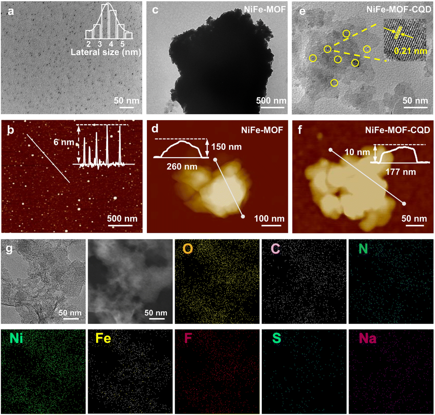 | ||
| Fig. 2 TEM images of (a) CQD, (c) NiFe-MOF and (e) NiFe-MOF-CQD. AFM images of (b) CQD, (d) NiFe-MOF and (f) NiFe-MOF-CQD. (g) EDS mapping images of the NiFe-MOF-CQD sample. | ||
To investigate the crystalline nature of NiFe-MOF and NiFe-MOF-CQD, X-ray diffraction (XRD) analysis was carried out. As shown in Fig. 3a, the XRD patterns of both materials show three strong diffraction peaks centered at 33°, 38°, and 59°, indicating a well-crystallized structure. Moreover, the introduction of CQDs does not affect the crystalline framework of the NiFe-MOF. In addition, the broad peak observed at around 20° in the NiFe-MOF-CQD sample, which corresponds to the (002) plane of CQDs (Fig. S4a†), is more intense than that of pure NiFe-MOF, confirming the successful integration of CQDs into the MOF matrix.24,27 The successful formation of the NiFe-MOF-CQD composite is additionally corroborated by Raman spectroscopy analysis. As shown in Fig. 3b, the Raman spectra of both NiFe-MOF and NiFe-MOF-CQD display consistent peak positions. The peak at approximately 1092 cm−1 corresponds to the Ni–O bond in the MOF, whereas peaks at around 1386 and 1612 cm−1 are attributed to COO− groups from the 1,4-naphthalenedicarboxylic acid ligand.28 The enhanced intensity of these peaks in the NiFe-MOF-CQD, particularly in comparison to the pure NiFe-MOF, can be attributed to the presence of CQDs (Fig. S4b†).29
 | ||
| Fig. 3 (a) XRD patterns of NiFe-MOF and NiFe-MOF-CQD. (b) Raman spectra of NiFe-MOF and NiFe-MOF-CQD. (c) FT-IR spectra of NiFe-MOF and NiFe-MOF-CQD. High-resolution XPS spectra of (d) O 1s, (e) Ni 2p and (f) Fe 2p of NiFe-MOF and NiFe-MOF-CQD. | ||
Fourier transform infrared (FT-IR) spectroscopy was utilized to identify functional groups within the samples. The FT-IR spectrum of CQDs displays absorption peaks at 1124 cm−1 and 824 cm−1, which are attributed to the stretching vibrations of C–F and C–F2 bonds, respectively. This observation confirms the successful incorporation of fluorine from the precursor material (Fig. S4c†).30 In addition, the wide absorption band identified in the range of 3000 to 3500 cm−1 is associated with the stretching vibrations of –OH groups, while the distinct peak at 3190 cm−1 is attributed to the presence of –NH2 groups, further reflecting the influence of the precursor on the functionalization of the CQDs.18 As is shown in Fig. 3c, the characteristic symmetric and asymmetric stretching vibrations of carboxyl groups coordinated with metal centers can be seen at 1620 and 1396 cm−1, respectively. Additionally, the C–H vibrational modes associated with the benzene ring appear at 740 cm−1, while the detection of the Ni–O stretching vibration at 594 cm−1 suggests the establishment of metal–oxygen bonds between the Ni/Fe atoms and the carboxyl groups of 1,4-naphthalenedicarboxylic acid.28 The FT-IR results further suggest the presence of exposed carboxylate groups on the surface of the NiFe-MOF-CQD composite due to their hydrophilic nature, which are likely to enhance water availability during the oxygen evolution reaction (OER). The hydrophilicity of both NiFe-MOF and NiFe-MOF-CQD was further demonstrated by static contact angle measurements (Fig. S5†).
To explore how the composite structure affects its electrocatalytic activity, X-ray photoelectron spectroscopy (XPS) was utilized to examine the valence states of the elements. The XPS survey spectrum (Fig. S6a†) detected signals corresponding to C, N, O, Na, F and S elements, originating from the CQDs, which were derived from the two precursors, o-phenylenediamine and sodium trifluoromethanesulfinate. High-resolution XPS spectra of each element further reveal the detailed elemental composition (Fig. S6b–g†), providing deeper insight into the chemical environment and bonding states within the composite. The XPS survey spectrum (Fig. S7a†) detected signals for C, O, Ni and Fe in both NiFe-MOF and NiFe-MOF-CQD, with additional signals for N, F, S and Na in NiFe-MOF-CQD, originating from the CQDs.31,32 These findings are consistent with the elemental mapping results and confirm the successful incorporation of CQDs. The high-resolution C 1s spectra of NiFe-MOF and NiFe-MOF-CQD (Fig. S7b†) were analyzed and deconvoluted into three distinct peaks observed at 284.8, 286.1, and 288.4 eV. These peaks are attributed to the presence of C![[double bond, length as m-dash]](https://www.rsc.org/images/entities/char_e001.gif) C, C–O, and O–CO functional groups, respectively.23,33 The O 1s spectrum (Fig. 3d) exhibits three main peaks, derived from C–O (532.68 eV), M–O–R (531.06 eV), and M–O (529.13 eV).34,35 Notably, the peaks corresponding to M–O–R and M–O bonds in NiFe-MOF-CQD shift slightly towards higher binding energy than those in NiFe-MOF, suggesting that the introduction of CQDs leads to a reduction in the local electron density around carbon atoms, increasing the binding energy.
C, C–O, and O–CO functional groups, respectively.23,33 The O 1s spectrum (Fig. 3d) exhibits three main peaks, derived from C–O (532.68 eV), M–O–R (531.06 eV), and M–O (529.13 eV).34,35 Notably, the peaks corresponding to M–O–R and M–O bonds in NiFe-MOF-CQD shift slightly towards higher binding energy than those in NiFe-MOF, suggesting that the introduction of CQDs leads to a reduction in the local electron density around carbon atoms, increasing the binding energy.
The high-resolution Ni 2p spectra reveal binding energy peaks at 855.6 eV and 873.2 eV, which are attributed to Ni 2p3/2 and Ni 2p1/2, respectively (Fig. 3e).36 After coupling with CQDs, the Ni 2p3/2 peak of NiFe-MOF shifts toward a higher binding energy, reflecting a partial oxidation of Ni2+ to Ni3+, suggesting enhanced catalytic properties.19,37 Similarly, in the Fe 2p image of NiFe-MOF-CQD (Fig. 3f), the Fe 2p3/2 and Fe 2p1/2 peaks belonging to Fe3+ in FeOOH shift by 0.66 eV and 0.31 eV toward lower binding energies, respectively.38–41 The above XPS results collectively show the electron -donating and -withdrawing effects induced by the CQDs, which promote electron transfer between metal and oxygen atoms in the composite, thereby optimizing the electronic structure. This enhanced electron transport significantly contributes to the improved OER performance, underscoring the role of CQDs in modulating the catalytic efficiency of NiFe-MOF.
To further investigate the catalytic effects of NiFe-interacting bimetals, we synthesized pure Ni-MOF and Fe-MOF under the same conditions as NiFe-MOF and tested their electrooxidation performance. The results, as shown in Fig. S8,† demonstrate that the water oxidation performance of pure Ni-MOF and Fe-MOF is far inferior to that of NiFe-MOF. These findings underscore the synergistic effect between Ni and Fe in the bimetallic structure. While Ni primarily drives the water oxidation process, Fe plays an auxiliary role, at a relatively low concentration (Fe:Ni = 1:7) in the composite. This confirms that the combination of Ni and Fe is critical to achieving the enhanced OER activity observed in NiFe-MOF. To assess the electrocatalytic performance of the NiFe-MOF-CQD sample, which consists of ultrathin nanosheets combined with CQDs, linear sweep voltammetry (LSV) was performed. This measurement took place in a 1.0 M KOH solution saturated with O2, conducted at a rotation speed of 1600 rpm using a standard three-electrode system. As illustrated in Fig. 4a, the OER activity of NiFe-MOF-CQD significantly surpasses that of pure NiFe-MOF. This improvement is attributed to the presence of electron-withdrawing groups in the CQDs. NiFe-MOF-CQD achieves a current density of 10 mA cm−2 with an overpotential of only 280 mV, considerably lower than that of NiFe-MOF (330 mV).42–44 To gain further insights into the OER kinetics, Tafel slope analysis was carried out (Fig. 4b). The results demonstrate that NiFe-MOF-CQD exhibits a smaller Tafel slope of 71.98 mV dec−1, indicating faster reaction kinetics compared to NiFe-MOF (90.11 mV dec−1).45–47 Additionally, cyclic voltammetry (CV) curves (Fig. S9†) reveal reversible redox peaks at around 1.31 and 1.50 V vs. RHE for NiFe-MOF-CQD, and at 1.35 and 1.51 V vs. RHE for NiFe-MOF. These peaks correspond to the redox process of Ni species between Ni2+ and Ni3+.48,49 Notably, the redox peaks of NiFe-MOF-CQD are more obvious, highlighting the positive effect of CQDs on Ni redox reactions.
 | ||
| Fig. 4 (a) LSV curves of NiFe-MOF and NiFe-MOF-CQD in 1.0 M KOH and (b) corresponding Tafel plots. (c) Nyquist plots at 1.50 V vs. RHE of NiFe-MOF and NiFe-MOF-CQD. (d) The CV curves for NiFe-MOF-CQD at different scan rates: 20, 40, 60, 80 and 100 mV s−1 from inside to outside. (e) Cdl curves of NiFe-MOF and NiFe-MOF-CQD. (f) The In situ Raman spectra of NiFe-MOF-CQD at 1.50 V vs. RHE in 1.0 M KOH. In situ FT-IR spectra of (g) NiFe-MOF and (h) NiFe-MOF-CQD at 1.50 V vs. RHE in 1.0 M KOH. | ||
Electrochemical impedance spectroscopy (EIS) was utilized to examine the charge transfer characteristics. As illustrated in Fig. 4c, the charge transfer resistance (Rct) of NiFe-MOF-CQD is notably lower than that of pure NiFe-MOF. This finding suggests that the incorporation of CQDs enhances charge transfer at the electrode/electrolyte interface.50 Additionally, the electrochemical active surface area (ECSA) was estimated by assessing the electrochemical double-layer capacitance (Cdl) within the non-faradaic region, specifically from 0.92 to 1.02 V vs. RHE (Fig. 4d and S10†). As demonstrated in Fig. 4e, NiFe-MOF-CQD exhibits a higher Cdl value, which indicates an increased ECSA and a greater number of active catalytic sites that promote the OER process. From the LSV curve normalized by the electrochemical surface area (Fig. S11†), it is evident that the potential of NiFe-MOF-CQD at a given current density is lower than that of NiFe-MOF. This is consistent with the LSV results from the three-electrode system, further proving the excellent electrochemical performance of NiFe-MOF-CQD.35,51,52
To further explore potential dynamic surface reconstruction during the OER process, in situ Raman spectroscopy was performed on NiFe-MOF-CQD. At an applied voltage of 1.5 V vs. RHE, distinct Raman signals emerged near 496 and 588 cm−1, attributable to Ni–O bending and NiOOH stretching vibrations, respectively, as depicted in Fig. 4f.53,54 Moreover, in situ FTIR spectroscopy was carried out to explore the reaction pathways (Fig. 4g and h). At 1.5 V vs. RHE, a significant absorption peak at 1224 cm−1, attributed to the superoxide (O2−) intermediate, was detected. The gradual increase in the intensity of the absorption peak over time indicates the evolution of the *OOH intermediate, suggesting that the OER mechanism in NiFe-MOF-CQD mainly follows the adsorbate evolution mechanism rather than the lattice oxygen evolution mechanism. This observation suggests enhanced structural stability during the OER process. Furthermore, the band around 1406 cm−1 corresponds to the O–O stretching mode of weakly adsorbed molecular oxygen. For comparison, the *OOH intermediate was also detected in NiFe-MOF at 1220 cm−1, further confirming the involvement of the adsorbate evolution mechanism in both systems.50,55
To further evaluate the practical applicability of the NiFe-MOF-CQD catalyst for water electrolysis, we built a PEM electrolyzer utilizing Nafion 115 as the membrane (Fig. 5a and b). In this setup, the NiFe-MOF-CQD acted as the anode catalyst for the OER, while the cathode catalyst for hydrogen evolution was commercial Pt/C. The prepared membrane electrode was prepared with a catalyst loading of 2.5 mg cm−2 for the anode and 0.25 mgPt cm−2 for the cathode, with an effective area of 58 cm2 (Fig. 5a).11 The current–voltage (I–V) curve of the NiFe-MOF-CQD/PEM/Pt/C electrolyzer (without iR compensation) demonstrates impressive water electrolysis activity (Fig. 5c).56,57 At an operating temperature of 60 °C, the electrolyzer based on NiFe-MOF-CQD achieves current densities of 0.5, 1.0, 1.5, and 2 A cm−2 at corresponding voltages of 1.68, 1.79, 1.89, and 2.00 V, respectively. These results highlight the high catalytic efficiency of NiFe-MOF-CQD in water-splitting applications.3,58 Furthermore, we have added a comparison of the electrochemical performance of our catalyst with other reported OER catalysts, further proving the superiority of NiFe-MOF-CQD (Fig. S12†). These comparisons and the additional information will provide readers with a clearer understanding of the catalyst's outstanding performance and robustness.
 | ||
| Fig. 5 (a) Photographs of the assembled reactor and membrane electrode assembly. (b) Schematic of the PEM–WE device. (c) Polarization curves of the PEM water electrolyzers. (d) Chronopotentiometry testing of NiFe-MOF-CQD catalysts at 2 A cm−2 in the PEM water electrolyzers. (e) Chronocurrent testing of NiFe-MOF-CQD catalysts at 2 V in the PEM water electrolyzers. | ||
Stability is a critical factor in practical applications, even more important than its catalytic activity. To evaluate the durability of the NiFe-MOF-CQD-based electrolyzer, we conducted long-term electrolysis under industrially relevant high current conditions. As shown in Fig. 5d, the electrolyzer maintains a stable voltage of approximately 2 V for over 7 hours at a constant current density of 2 A cm−2, confirming the excellent OER stability of the NiFe-MOF-CQD catalyst under real-world operating conditions.59 Furthermore, chronoamperometric measurements conducted at 60 °C under an applied voltage of 2 V further corroborate this stability. As illustrated in Fig. 5e, the current density remains constantly at 2 A cm−2 over the 7 hour testing period, proving the long-term stability and robustness of NiFe-MOF-CQD for industrial water electrolysis applications.60
4. Conclusions
In summary, we employed a bottom-up synthesis strategy involving ultrasound-assisted exfoliation to fabricate ultrathin NiFe-MOF nanosheets, with high electrooxidation activity, in situ induced by CQDs. This innovative catalyst was further applied in industrial PEM water electrolysis, showcasing remarkable performance. The original NiFe-MOF nanosheets with a thickness of 150 nm were reduced to just 10 nm upon CQD incorporation, fully exposing electrochemically active sites. XPS analysis revealed that the electron-donating and -withdrawing effects of CQDs enhanced electron transfer between metal and oxygen atoms in the composite, optimizing the electronic structure. This improved electron transport significantly boosted the OER performance, demonstrating CQDs' vital role in modulating the catalytic activity of NiFe-MOF. The NiFe-MOF-CQD composite demonstrates exceptional performance, achieving a current density of 10 mA cm−2 with a significantly reduced overpotential of just 280 mV, which is notably lower than the 330 mV required by NiFe-MOF alone. Furthermore, its Tafel slope of 71.98 mV dec−1 signifies faster electrochemical reaction kinetics, outperforming NiFe-MOF's slope of 90.11 mV dec−1. Furthermore, NiFe-MOF-CQD has a larger Cdl value, indicating a larger ECSA and more active catalytic sites, which are favorable for the OER process. In situ FTIR spectroscopy revealed a gradual increase in the intensity of the absorption peak over time, indicating the evolution of the *OOH intermediate. This suggests that the OER mechanism in NiFe-MOF-CQD mainly follows the adsorbate evolution mechanism rather than the lattice oxygen evolution mechanism. The electrolytic cell using NiFe-MOF-CQD as the anode catalyst demonstrated remarkable performance, requiring only 1.79, 1.89, and 2.00 V to reach industrial-level current densities of 1.0, 1.5, and 2.0 A cm−2 respectively. Moreover, under an applied voltage of 2 V, the current density was maintained at 2 A cm−2 for over 7 hours, demonstrating the excellent electrochemical activity and long-term stability of NiFe-MOF-CQD. This research achievement not only advances the dual-carbon goal but also lays a solid foundation for the development of hydrogen energy and chemical industries, setting a milestone for the application of 2D materials in industrial-scale PEM water electrolysis.Data availability
The data supporting this article have been included as part of the ESI.†Author contributions
Liang Wang: conceptualization, supervision, project administration, writing – review & editing, funding acquisition. Qianjia Ni: methodology, investigation, validation, formal analysis, data curation, writing – original draft, writing – review & editing. Shiyuan Zhang: methodology, investigation, data curation, formal analysis. Kang Wang: writing – review & editing. Huazhang Guo: writing – review & editing. Jiye Zhang: writing – review & editing. Minghong Wu: writing – review & editing.Conflicts of interest
There are no conflicts to declare.Acknowledgements
The project was funded by the Shanghai Pujiang Program (21PJD022) and Changsha Natural Science Foundation (kq2208392). This work is Supported by the Shanghai Technical Service Center of Science and Engineering Computing, Shanghai University.References
- L. Zhu, H. Zhang, A. Zhang, T. Tian, Y. Shen, M. Wu, N. Li and H. Tang, Adv. Powder Mater., 2024, 3, 100020 Search PubMed
.
- H. Gao, Z. Xiao, S. Du, T. Liu, Y. C. Huang, J. Shi, Y. Zhu, G. Huang, B. Zhou, Y. He, C. L. Dong, Y. Li, R. Chen and S. Wang, Angew. Chem., Int. Ed., 2023, 62, e202313954 Search PubMed
- Z.-Y. Wu, F.-Y. Chen, B. Li, S.-W. Yu, Y. Z. Finfrock, D. M. Meira, Q.-Q. Yan, P. Zhu, M.-X. Chen, T.-W. Song, Z. Yin, H.-W. Liang, S. Zhang, G. Wang and H. Wang, Nat. Mater., 2022, 22, 100–108 CrossRef PubMed
- P. Guo, L. Shi, D. Liu, X. Wang, F. Gao, Y. Ha, J. Yin, M. Liu, H. Pan and R. Wu, Mater. Today Catal., 2023, 1, 100002 Search PubMed
- K. Yu, H. Yang, H. Zhang, H. Huang, Z. Wang, Z. Kang, Y. Liu, P. W. Menezes and Z. Chen, Nano-Micro Lett., 2023, 15, 186 Search PubMed
- G. Shen, R. Zhang, L. Pan, F. Hou, Y. Zhao, Z. Shen, W. Mi, C. Shi, Q. Wang, X. Zhang and J. J. Zou, Angew. Chem., Int. Ed., 2019, 59, 2313–2317 CrossRef PubMed
- K. H. Yue, J. L. Liu, Y. T. Zhu, C. F. Xia, P. Wang, J. W. Zhang, Y. Kong, X. Y. Wang, Y. Yan and B. Y. Xia, Energy Environ. Sci, 2021, 14, 6546–6553 RSC
- M. Li, H. Li, X. Jiang, M. Jiang, X. Zhan, G. Fu, J.-M. Lee and Y. Tang, J. Mater. Chem. A, 2021, 9, 2999–3006 RSC
- Y. Zhu, X. Wang, X. Zhu, Z. Wu, D. Zhao, F. Wang, D. Sun, Y. Tang, H. Li and G. Fu, Small, 2022, 19, 2206531 CrossRef PubMed
- X. Gao, S. Dai, Y. Teng, Q. Wang, Z. Zhang, Z. Yang, M. Park, H. Wang, Z. Jia, Y. Wang and Y. Yang, Nano-Micro Lett., 2024, 16, 108 CrossRef CAS PubMed
- R.-T. Liu, Z.-L. Xu, F.-M. Li, F.-Y. Chen, J.-Y. Yu, Y. Yan, Y. Chen and B. Y. Xia, Chem. Soc. Rev., 2023, 52, 5652–5683 RSC
- K. Rui, G. Q. Zhao, Y. P. Chen, Y. Lin, Q. Zhou, J. Y. Chen, J. X. Zhu, W. P. Sun, W. Huang and S. X. Dou, Adv. Funct. Mater., 2018, 28, 1801554 CrossRef
- L. Q. Ji, Y. Kong, C. Wang, H. Tan, H. L. Duan, W. Hu, G. N. Li, Y. Lu, N. Li, Y. Wang, J. Tian, Z. M. Qi, Z. H. Sun, F. C. Hu and W. S. Yan, ACS Catal., 2020, 10, 5691–5697 Search PubMed
- M. J. Wang, X. Dong, Z. D. Meng, Z. W. Hu, Y. G. Lin, C. K. Peng, H. S. Wang, C. W. Pao, S. Y. Ding, Y. Y. Li, Q. Shao and X. Q. Huang, Angew. Chem., Int. Ed., 2021, 60, 11190–11195 CrossRef CAS PubMed
- F. Wang, L. Hu and Y. Jing, J. Mater. Chem. A, 2024, 12, 28764–28770 Search PubMed
- H. Guo, Y. Lu, Z. Lei, H. Bao, M. Zhang, Z. Wang, C. Guan, B. Tang, Z. Liu and L. Wang, Nat. Commun., 2024, 15, 4843 CrossRef CAS PubMed
- H. Zhang, H. Guo, D. Li, Y. Zhang, S. Zhang, W. Kang, C. Liu, W. Le, L. Wang, D. Li and B. Dai, Nat. Commun., 2024, 15, 2980 CrossRef CAS PubMed
- B. Hu, K. Huang, B. Tang, Z. Lei, Z. Wang, H. Guo, C. Lian, Z. Liu and L. Wang, Nano-Micro Lett., 2023, 15, 217 CrossRef CAS PubMed
- D. Song, H. Guo, K. Huang, H. Zhang, J. Chen, L. Wang, C. Lian and Y. Wang, Mater. Today, 2022, 54, 42–51 CrossRef CAS
- H. Liao, K. Huang, W. Hou, H. Guo, C. Lian, J. Zhang, Z. Liu and L. Wang, Adv. Powder Mater., 2024, 3, 100243 CrossRef
- M. Cai, Y. Zhang, Y. Zhao, Q. Liu, Y. Li and G. Li, J. Mater. Chem. A, 2020, 8, 20386–20392 RSC
- S. Chen, Z. Zheng, Q. Li, H. Wan, G. Chen, N. Zhang, X. Liu and R. Ma, J. Mater. Chem. A, 2023, 11, 1944–1953 RSC
- K. Rui, G. Q. Zhao, M. M. Lao, P. X. Cui, X. S. Zheng, X. B. Zheng, J. X. Zhu, W. Huang, S. X. Doug and W. P. Sun, Nano Lett., 2019, 19, 8447–8453 CrossRef CAS PubMed
- L. Xie, C. Liang, Y. Wu, K. Wang, W. Hou, H. Guo, Z. Wang, Y. M. Lam, Z. Liu and L. Wang, Small, 2024, 20(37), 2401253 CrossRef CAS PubMed
- W. Liu, B. Tang, K. Huang, Z. Zhang, Z. Wang, G. An, M. Zhang, K. Wang, S. Fu, H. Guo, T. Han, C. Lian, B. Zhang, T. Wu, Z. Lei and L. Wang, Small, 2024, 2408688 Search PubMed
- L. Zhao, B. L. Dong, S. Z. Li, L. J. Zhou, L. F. Lai, Z. W. Wang, S. L. Zhao, M. Han, K. Gao, M. Lu, X. J. Xie, B. Chen, Z. D. Liu, X. J. Wang, H. Zhang, H. Li, J. Q. Liu, H. Zhang, X. Huang and W. Huang, ACS Nano, 2017, 11, 5800–5807 CrossRef CAS PubMed
- Y. Xu, W. Hou, K. Huang, H. Guo, Z. Wang, C. Lian, J. Zhang, D. Wu, Z. Lei, Z. Liu and L. Wang, Adv. Sci., 2024, 11(28), 2403607 CrossRef CAS PubMed
- F. Z. Sun, G. Wang, Y. Q. Ding, C. Wang, B. B. Yuan and Y. Q. Lin, Adv. Energy Mater., 2018, 8, 1800584 CrossRef
- G. An, K. Wang, Z. Wang, M. Zhang, H. Guo and L. Wang, ACS Appl. Mater. Interfaces, 2024, 16, 29060–29068 CrossRef CAS PubMed
- J. Liang, L. Liang, B. Zeng, B. Feng, L. Du, X. Qiu, Y. Wang, H. Song, S. Liao, M. Shao and Z. Cui, Angew. Chem., Int. Ed., 2024, e202412825 Search PubMed
- P. M. L. Le, T. D. Vo, H. Pan, Y. Jin, Y. He, X. Cao, H. V. Nguyen, M. H. Engelhard, C. Wang, J. Xiao and J. G. Zhang, Adv. Funct. Mater., 2020, 30, 001151 Search PubMed
- K. Lakshmanan, W. H. Huang, S. A. Chala, B. W. Taklu, E. A. Moges, J. F. Lee, P. Y. Huang, Y. C. Lee, M. C. Tsai, W. N. Su and B. J. Hwang, Adv. Funct. Mater., 2022, 32, 2109310 CrossRef CAS
- F. Z. Sun, G. Wang, Y. Q. Ding, C. Wang, B. B. Yuan and Y. Q. Lin, Adv. Energy Mater., 2018, 8, 1800584 CrossRef
- Z. Liu, Z. Zhao, Y. Wang, S. Dou, D. Yan, D. Liu, Z. Xia and S. Wang, Adv. Mater., 2017, 29, 1606207 CrossRef PubMed
- Z. H. Zou, T. T. Wang, X. H. Zhao, W. J. Jiang, H. R. Pan, D. Q. Gao and C. L. Xu, ACS Catal., 2019, 9, 7356–7364 CrossRef CAS
- A. Bovas and T. P. Radhakrishnan, J. Mater. Chem. A, 2024, 12, 24872–24877 RSC
- F. J. Li, M. H. Jiang, C. G. Lai, H. F. Xu, K. Y. Zhang and Z. Jin, Nano Lett., 2022, 22, 7238–7245 CrossRef CAS PubMed
- Z. Li, X. Wu, X. Jiang, B. Shen, Z. Teng, D. Sun, G. Fu and Y. Tang, Adv. Powder Mater., 2022, 1, 100020 CrossRef
- D. Zhang, Y. Wang, Y. Peng, Y. Luo, T. Liu, W. He, F. Chen and M. Ding, Adv. Powder Mater., 2023, 2, 100129 CrossRef
- J. Zhou, L. Chen, J. Wu, Z. Lu, F. Liu, X. Chen, P. Xue, C. Li, L. Wei, G. Wu, Q. Li and Q. Zhang, Nano Lett., 2023, 23, 11297–11306 CrossRef CAS PubMed
- H. Liu, X. Lu, Y. Hu, R. Chen, P. Zhao, L. Wang, G. Zhu, L. Ma and Z. Jin, J. Mater. Chem. A, 2019, 7, 12489–12497 RSC
- Y. Zhang, M. Du, Y. Ma, J. Shang and B. Qiu, Mater. Today Catal., 2023, 3, 100027 CrossRef
- X. Zou, Q. Lu, M. Tang, J. Wu, K. Zhang, W. Li, Y. Hu, X. Xu, X. Zhang, Z. Shao and L. An, Nano-Micro Lett., 2024, 17, 6 CrossRef PubMed
- Z. Li, X. Zhang, Z. Zhang, P. Chen, Y. Zhang and X. Dong, Appl. Catal., B, 2023, 325, 122311 CrossRef CAS
- Y. Hao, X. Cao, C. Lei, Z. Chen, X. Yang and M. Gong, Mater. Today Catal., 2023, 2, 100012 CrossRef
- L. Wang, J. Huang, Z. Huang, H. Li, T. Taylor Isimjan and X. Yang, Chem. Eng. J., 2023, 472, 144924 CrossRef CAS
- Z. Ye, C. Qin, G. Ma, X. Peng, T. Li, D. Li and Z. Jin, ACS Appl. Mater. Interfaces, 2018, 10, 39809–39818 CrossRef CAS PubMed
- J. Liang, X. T. Gao, B. Guo, Y. Ding, J. W. Yan, Z. X. Guo, E. C. M. Tse and J. X. Liu, Angew. Chem., Int. Ed., 2021, 60, 12770–12774 CrossRef CAS PubMed
- F. L. Li, P. T. Wang, X. Q. Huang, D. J. Young, H. F. Wang, P. Braunstein and J. P. Lang, Angew. Chem., Int. Ed., 2019, 58, 7051–7056 CrossRef CAS PubMed
- N. Zhang, Y. Hu, L. An, Q. Li, J. Yin, J. Li, R. Yang, M. Lu, S. Zhang, P. Xi and C. H. Yan, Angew. Chem., Int. Ed., 2022, 61, e202207217 CrossRef CAS PubMed
- Y. Hu, L. Li, J. Zhao, Y.-C. Huang, C.-y. Kuo, J. Zhou, Y. Fan, H.-J. Lin, C.-L. Dong, C.-W. Pao, J.-F. Lee, C.-T. Chen, C. Jin, Z. Hu, J.-Q. Wang and L. Zhang, Appl. Catal., B, 2023, 333, 122785 CrossRef CAS
- Z.-Y. Liu, Q.-Y. Wang, T. Xu and J.-M. Hu, J. Mater. Chem. A, 2024, 12, 27497–27505 RSC
- J. Zhang, Y. Ye, Z. Wang, Y. Xu, L. Gui, B. He and L. Zhao, Adv. Sci., 2022, 9, 2201916 CrossRef CAS PubMed
- J. Zhang, Y. Ye, B. Wei, F. Hu, L. Sui, H. Xiao, L. Gui, J. Sun, B. He and L. Zhao, Appl. Catal., B, 2023, 330, 122661 CrossRef CAS
- X. Lin, X. Li, L. Shi, F. Ye, F. Liu and D. Liu, Small, 2023, 20, 2308517 CrossRef PubMed
- J. K. Lee, G. Anderson, A. W. Tricker, F. Babbe, A. Madan, D. A. Cullen, J. D. Arregui-Mena, N. Danilovic, R. Mukundan, A. Z. Weber and X. Peng, Nat. Commun., 2023, 14, 4592 CrossRef CAS PubMed
- L. Yao, F. Zhang, S. Yang, H. Zhang, Y. Li, C. Yang, H. Yang and Q. Cheng, Adv. Mater., 2024, 36, 2314049 CrossRef CAS PubMed
- Y. F. Cui, S. D. Jiang, Q. Fu, R. Wang, P. Xu, Y. Sui, X. J. Wang, Z. L. Ning, J. F. Sun, X. Sun, A. Nikiforov and B. Song, Adv. Funct. Mater., 2023, 33, 2306889 CrossRef CAS
- C. Zhao, J. Wang, Y. Gao, J. Zhang, C. Huang, Q. Shi, S. Mu, Q. Xiao, S. Huo, Z. Xia, J. Zhang, X. Lu and Y. Zhao, Adv. Funct. Mater., 2023, 34, 2307917 Search PubMed
- X. Guo, H. Zhang, W. Xia, M. Ma, D. Cao and D. Cheng, Adv. Funct. Mater., 2024, 2316539 CrossRef CAS
Footnotes |
| † Electronic supplementary information (ESI) available. See DOI: https://doi.org/10.1039/d4ta06855f |
| ‡ These authors contributed equally. |
| This journal is © The Royal Society of Chemistry 2024 |