 Open Access Article
Creative Commons Attribution 3.0 Unported Licence
Correction: A novel nanomissile targeting two biomarkers and accurately bombing CTCs with doxorubicin
Yu
Gao
,
Xiaodong
Xie
,
Fengqiao
Li
,
Yusheng
Lu
,
Tao
Li
,
Shu
Lian
,
Yingying
Zhang
,
Huijuan
Zhang
,
Hao
Mei
and
Lee
Jia
*
Cancer Metastasis Alert and Prevention Center, and Pharmaceutical Photocatalysis of State Key Laboratory of Photocatalysis on Energy and Environment, and Fujian Provincial Key Laboratory of Cancer Metastasis Chemoprevention and Chemotherapy, Fuzhou University, Fuzhou 350108, China. E-mail: pharmlink@gmail.com
First published on 30th April 2018
Abstract
Correction for ‘A novel nanomissile targeting two biomarkers and accurately bombing CTCs with doxorubicin’ by Yu Gao et al., Nanoscale, 2017, 9, 5624–5640.
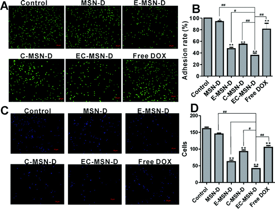
The authors have discovered two errors in Fig. 6 and 7. In the previously published Fig. 6A, the fluorescent microscopic image of free DOX was improperly presented as the same image as MSN-D. Furthermore, in the previously published Fig. 7C, an incorrect image was provided for panel f, and a caption was also not provided. In the revised version of Fig. 7C, Fig. 7C-f has been replaced with the correct image and the caption has been modified. In addition, Fig. 7C-a has been replaced with the H&E staining image, which more accurately represents the average change caused by free DOX treatment. Although these errors do not affect the conclusions and findings of this research paper, the authors sincerely apologize for these errors and any confusion caused.
Please find below the corrected versions of Fig. 6 and 7 and their corrected captions below.
The Royal Society of Chemistry apologises for these errors and any consequent inconvenience to authors and readers.
| This journal is © The Royal Society of Chemistry 2018 |


