
Artificial spidroin bioelectronic dressings for intelligent wound management
Shuhuan
Li
ab,
Baoyang
Lin
b,
Yongji
Xiong
b,
Qian
Zhou
a,
Bingbing
Gao
 *a and
Bingfang
He
ab
*a and
Bingfang
He
ab
aSchool of Pharmaceutical Sciences, Nanjing Tech University, Nanjing 211816, China. E-mail: gaobb@njtech.edu.cn
bCollege of Biotechnology and Pharmaceutical Engineering, Nanjing Tech University, Nanjing 211816, China
First published on 30th July 2024
Abstract
Wound infection has always been a huge threat to human health. Most of the existing wound medical materials are made of composite materials with poor biocompatibility and may contain harmful chemicals, which may cause allergic reactions and secondary wound infection, becoming a hidden danger to human health. Therefore, there is an urgent need to develop innovative medical materials for wound treatment. At present, the intelligent application of high-performance materials prepared from artificial spider silk protein (spidroin) in wound healing has been widely discussed. Its advantages are good biocompatibility, biodegradability, cell adhesion and excellent bioelectronic performance. In this paper, the biological application of various high-performance medical materials prepared from artificial spidroins in wound healing was introduced, and the sequence design and synthesis methods of artificial spidroins were briefly discussed. Finally, the application prospects of high-performance medical materials prepared from artificial spidroins in wound treatment are discussed.
1. Introduction
Skin is the largest organ of the human body, and the rupture or defect of the skin tissue caused by trauma is called a wound,1 and a wound that cannot be repaired normally and completely is defined as a chronic wound.2 Chronic wounds are affected by pathological factors such as hyperglycemia,3 related growth factor deficiency,4 and peripheral vascular disease,5 and are more difficult to heal than ordinary wounds. Non-healing wounds can cause pain, inflammation, bacterial infection and even death, posing a major threat to human health. Therefore, with the aim to treat chronic wounds in a timely and effective manner, efforts have been made to develop new multifunctional wound dressings. At present, most traditional wound dressings are mainly made of high polymer or composite bio-based materials as raw materials,6–9 which can cover and bandage the wound, absorb wound exudate,10 and provide protection against microbial and mechanical damage to the wound surface.11 These dressings are widely used because they are easy to make and easy to use. However, due to the lack of good biodegradability, such traditional dressings may release microplastics during use, which can accumulate in the human body and the environment and become a hidden danger to human health.12–14 In addition, allergic reactions,15 secondary skin lesions16 and secondary wound infections occur during wound healing,17 which may be related to harmful chemicals contained in the dressing. Due to the excellent biocompatibility and biodegradation properties of spider silk proteins, they have great potential in the medical field as targeted drug delivery systems and controlled release vectors. Especially in the field of cardiovascular disease treatment, nanofiber scaffolds prepared from spider silk proteins have made breakthroughs. By blending recombinant spider silk proteins with biodegradable polymers such as polylactide (PLA-co-GA) and using electrospinning technology,18 the nanofiber scaffold has significantly improved its tensile strength and elongation compared with traditional materials, providing a more ideal scaffold material for cardiovascular tissue engineering. In addition, the hydrogel system based on the recombinant spider silk protein not only shows excellent self-healing ability, but also has excellent cell affinity and strong antibacterial properties, which makes it a key material for the development of advanced wound dressings.19 The hydrogel can speed up the wound healing process, reduce the risk of infection, and improve treatment results. However, the current production efficiency of recombinant spider silk proteins is still challenging, and their production is not enough to meet the needs of small-scale clinical applications, including medical wound dressings. Therefore, optimizing the heterologous expression sequence design of spider silk proteins and improving the expression efficiency and yield have become the key research direction to promote their industrial application. This includes, but is not limited to, strategies such as genetic engineering modification, optimization of expression systems, and improvement of fermentation processes in order to achieve efficient and large-scale production of spider silk proteins to meet the growing needs of medical applications.In recent years, wound dressings prepared from artificial spidroins have attracted wide attention due to their unique advantages such as good biocompatibility, biodegradability, high mechanical properties20 and excellent bioelectronic performance. With the increasing demand for personalized medicine, the demand for smart wound dressings is also increasing, and spidroin smart wound dressings have great potential in the next generation of wound management. Spidroin bioelectronic dressings, with their excellent bioelectronic properties, show extraordinary potential in the field of wound management. The dressing can naturally generate appropriate current stimulation,21 and this unique bioelectrical activity greatly promotes the wound healing process, opening up a new research direction for the development of intelligent wound dressings without an external power supply. In addition, spider silk, as its main component, gives the dressing excellent biocompatibility, ensuring that it not only effectively promotes tissue repair in the treatment of various wounds, but also exhibits extremely low immunogenicity, significantly reducing the risk of adverse reactions, making it a promising biomedical material.22 In addition, Spidroin electronic dressings, by virtue of their high strength, high toughness and good elasticity, ensure the stability and durability of the dressings in the dynamic wound environment, effectively cope with the wound tension changes caused by joint activity, muscle contraction, etc., and provide solid physical support for the smooth healing of the wound.23 At the same time, thanks to the good biodegradability of spidroin bioelectronic dressings, they can effectively reduce the impact of environmental problems on human health.24 In conclusion, smart wound dressings prepared from spidroins provide a promising approach for smart wound management.
A variety of multi-functional intelligent wound dressings prepared from artificial spidroins have been applied to wound intelligent management, such as spidroin films with biochemical sensing function that have been used in wound pH monitoring.25 The high performance spider silk membrane based on the Kirigami structural design shows great potential for wound treatment in movable joints due to its significantly improved tensile properties. This innovative material combines the geometric cutting art of Kirigami with the excellent mechanical properties of the spider silk itself, through precision patterning cutting and folding, the material achieves high flexibility while significantly increasing its tensile strength and durability.26 The spidroin films with the function of nano-friction power generation are used to accelerate wound healing.21 At the same time, thanks to the excellent plasticity and biocompatibility of spider silk proteins, the surface can be precisely integrated with ordered microarray structures with controllable size and fine capillary characteristics. This design cleverly utilizes the principle of capillary force to achieve natural guidance and effective management of wound exudate, significantly improving the accumulation of secretions that are prone to occur in traditional dressings, thereby optimizing the wound microenvironment and promoting the healing process.27 Furthermore, by optimizing the ordered microarray structure into a microneedle array with high puncture force, the dressing can significantly improve the efficiency and precision of drug delivery. Microneedle (MN) technology enables drugs to directly penetrate the skin barrier and act quickly and accurately on wound tissue, not only improving the bioavailability of drugs, but also reducing side effects caused by drug leakage or uneven distribution. In addition, combined with the innovative application of microfluidic technology and immunoimaging substances, the dressing also has the capability of real-time biochemical detection of wound surfaces. This function enables medical staff to instantly obtain the biochemical index data of the wound, such as the level of inflammatory factors, bacterial load, etc.,28 which provides a scientific basis for the formulation and adjustment of personalized treatment plans, and further improves the intelligent and accurate level of wound management. However, a comprehensive review of the multifunctional preparation methods of wound dressings prepared from artificial spidroins and their application to intelligent monitoring in various aspects remains to be proposed. This paper reviews the multifunctional preparation methods of various artificial spidroin intelligent wound dressings and the application of intelligent wound monitoring in various aspects, as is shown in Fig. 1. Firstly, we introduced the preparation methods of artificial spidroin smart wound dressings, such as electrospinning, wet spinning and film forming. Secondly, we detailed the role of artificial spidroin smart wound dressings in accelerating wound healing. We then describe their function for intelligent monitoring of various aspects of the wound. Finally, the future development of artificial spidroin smart wound dressings in the further treatment of chronic wounds is briefly presented.
 | ||
| Fig. 1 Schematic diagram of spidroin bioelectronic dressings and their applications. | ||
2. Design and preparation of artificial spidroins
Spider silk, as a high molecular weight protein fiber, has unique mechanical properties and excellent biocompatibility indicate a wide range of potential applications in many fields. At present, the scientific community has a comprehensive and in-depth understanding of the genetic sequences of various spider silk proteins, which has laid a solid foundation for the precise regulation of the synthesis and performance optimization of spider silk proteins through genetic engineering technology.29 Although there has been a lot of interest in the sequence design, expression strategies, and application development of recombinant spider silk proteins, the only way to obtain significant quantities of spider silk is through heterologous expression because, unlike silkworms, spiders are cannibalistic arthropods and cannot be bred on a large scale. Extensive exploratory investigations have been carried out by researchers to analyze and alter the structural properties and sequence motifs of spider silk proteins.In order to create the chimera recombinant spidroin, Cheng et al.30 chose the short repetitive region (Rep) from E. australis, the N-terminal domain (NT) from Euprosthenops. australis MaSp1, the C-terminal domains (CT) from Araneus. ventricosus MiSp with high pH sensitivity and solubility, and the amino acid of spidroin, which is displayed in Fig. 2(a). Following the refinement of the inducing conditions, a noteworthy yield of highly soluble recombinant spidroin (6.4 mg mL−1) was obtained. It is often possible to increase the amount of microcrystalline areas in order to obtain improved mechanical performance. With the same NheI and SpeI tails, the vector was exposed to single- and double-enzyme digestion in Xiong et al.'s recursive directed genetic assembly approach.26 The cloning vector pse380 was used to produce the octoploid repeat sequence (Fig. 2(b)). High-purity NT8RepCT was achieved following elution. Fig. 2(c) shows the obtained target protein bands. The protein sizes on the SDS-PAGE gel were in excellent agreement with the 54 kDa predicted molecular weight. The prepared dressing made of spidroin MN exhibited a high tensile strength and flexibility of about 80%. Although this process improves the synthetic spider silk protein's mechanical qualities, a significant yield cannot be guaranteed. Lin et al.21 developed a novel and viable strategy for improving self-assembly performance while producing high-yield artificial spider silk protein. NT from MaSp1 of E. Australis, and CT from MiSp of A. Ventricosus, bracketing a brief repeated section from E. australis, was utilized as the template. NT-4RepCT (40.4 kDa) is a mosaic minicarachnid protein that is already accessible in our laboratory. The number of polyalanine motifs dropped to seven and the number of polyalanine blocks increased to six when the sequence length was kept constant. The result was an enhanced microcrystalline area density Fig. 2(f)-(i) and the bionic spidroin NT-Cs-6rep-CT (39.8 kDa). Amyloid was used to replace the microcrystalline region of NT-Cs-6rep-CT. This amyloid was able to produce a highly organized cross-β-spine structure, as seen in Fig. 2(d). The self-assembly performance of Amy-6rep greatly improved and its expression increased by approximately 200 percent as a result of sequence change (Fig. 2(e)). Following Amy-6rep electrospinning, the nanofibers demonstrate a strong ability to generate tribopower. This article describes a biomimetic spidroin sequence design that exhibits good spinning performance and a high yield. This design may open up new avenues for the commercial manufacture of artificial spider silk and provide further ideas for related uses.
 | ||
| Fig. 2 The molecular makeup and SDS-PAGE examination of synthetic spidroin. (a) Diagram showing the amino acid sequence composition of NT, Rep, and CT as well as the molecular makeup of artificial spidroin. (b) A schematic representation of the artificial spidroin's molecular makeup. (c) SDS-PAGE analysis of refined biomimetic artificial spirite. M stands for protein marker; rows 1 and 2 contain purified spidroin MW; rows 3 and 4 are empty pSE380 vectors bearing the recombinant spidroin gene. (c) Schematic representation of the artificial spidroin composition changed into the microcrystalline zone. (d) A schematic representation of the artificial spidroin's molecular makeup changed into a microcrystalline zone. The poly-Ala-β-sheet-crystal structure (accession code: ma-cs24y from ModelArchive, old pdb id: 2slk) and the cross-β-spine structure (pdb id:5e61). (e) Artificial spidroin solid powder. (f) SDS-PAGE examination of synthetic spidroin. Note: M: protein marker; line 1: 4rep (the primitive biomimetic spidroin, 40.4 kDa); line 2: crystal structure-6rep (the biomimetic spidroin with increased microcrystalline region density, 39.8 kDa); line 3: amyloid-6rep (the biomimetic spidroin with increased microcrystalline region density and amyloid polypeptide replacement (39.6 kDa)) (i). | ||
3. Preparation of artificial spidroins dressing
The mechanical and biocompatibility qualities of artificial spider silk are exceptional. Nevertheless, the main goal of current research on spider silk protein functional materials is to stack many modules from the same spider.31–33 In order to improve the mechanical characteristics of spider silk fibers. The application of composite microstructure design in increasingly complex and varied scenarios is hampered by the paucity of research in this area, which significantly limits the utilization of spider silk protein materials.To create the cathode polydimethylsiloxane (PDMS) template, Lin et al.23 replicated the silicon microcolumn template to obtain the cathode PDMS template, subsequently poured in the Spidroin-PU (polyurethane) (SP) solution, dried it at room temperature, and peeled it off to produce the SP film. The microscopic image of the SP film with a microcolumn array structure is shown in Fig. 3(a)-(i). The size and interval of the microarray can be adjusted by molding to obtain SP films with different microcolumns. Microscopic images of the SP film viewed from the side (Fig. 3(a)-(ii), (iii)) show that an SP film with a microstructure can be obtained simply and accurately by this method. Ordered microarrays with controllable size and capillary features can guide liquid flow through capillary forces to improve wound secretion deposition, thus enabling patterned SP films with the ability to control pump-free fluid flow for biochemical sensing. However, this kind of micro-column structure often has relatively low puncture force and has no obvious effect on drug delivery. Therefore, SPs have been used in MN manufacturing because they have sufficient mechanical strength to meet the hardness requirements and exhibit puncture ability.34–36
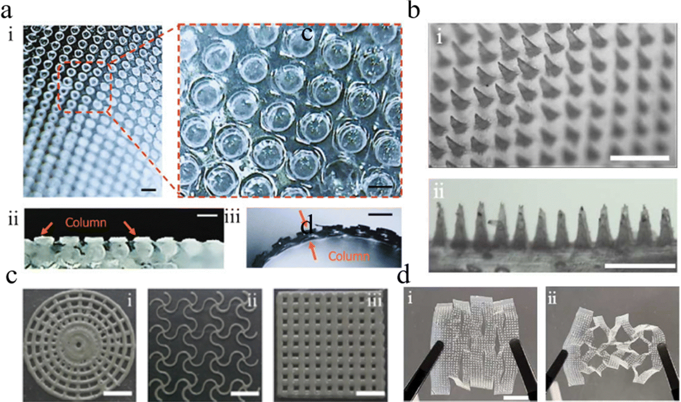 | ||
| Fig. 3 An image of the artificial spider silk material's microstructure based on proteins. microscopically images of a substance based on artificial spidroin. (a) Top view microscope pictures of the spider/PU composite (SP) showing the photonic crystal (PC) structure and micropillar (i) as well as a slightly enlarged view (ii). (c) Side view microscope images of SP: (i) just single-layer SP; (ii) pink line represents a PC put onto SP. (b) Vertical and side views of the microneedle scaffold (MNs) captured using a microscope. Vaseline anti-structure grooves I and II and the accompanying images captured with a Vaseline microscope (iii). After stretching, microscope pictures of the MNs film based on SP-MXene: i nonstretched; ii extended. | ||
As shown in Fig. 2(b), the needle sizes are 0.8 mm in height and 0.4 mm in diameter, which exhibits uniform morphology and neatness of arrangement. Further, in order to apply more scenes, shape plasticity becomes particularly important.37 Zhang et al.38 prepared a mixed emulsion composed of spirochete protein, PU, and aloe vera gel, and EGaIn, and then dispersed EGaIn into the emulsion by ultrasound. This emulsion was subsequently printed using a programmed 3D printer and stacked onto a PDMS microneedle mold, forming a microneedle dressing consisting of microneedles and hollow scaffold backing. It was observed that ESMN could be printed into various complex planar patterns using a programmed printer, as shown in Fig. 3(c), indicating that the hybrid emulsion has excellent rheology and formability, is suitable for continuous 3D printing, and is beneficial for different applications. However, despite the remarkable mechanical strength of spidroin materials, the low flexibility39,40 makes it difficult to adapt to long-term dynamic motion. In addition, motion sensing shows better performance on the flexible base,41 contributing to its excellent tensile recovery performance. To solve this problem, Li et al.42 created the flexible layer of KSM-TENG(triboelectric nanogenerator) patches by using a kirigami structure and PU to address this issue. With the use of a kirigami structure, a planar structure can be made into a three-dimensional structure that can stretch and remain consistent enough to adjust to intricate motion processes when subjected to external force.
In the field of artificial spider silk protein wound dressing preparation, most of the research focuses on the use of polymer (such as polyurethane) and artificial spidroins blending system as the base material,9,23 through a variety of molding technologies (including but not limited to film forming technology and electrospinning technology) to prepare high-performance spidroin dressing. On this basis, in order to further improve the intelligent level of dressing, researchers cleverly combined the inherent spontaneous electrical properties of spider silk protein with excellent biocompatibility, and realized the development of intelligent spider silk protein wound dressing by integrating multifunctional pathways on its surface. What is particularly noteworthy is that Lin et al.21 pioneered the use of pure spider silk protein as the only basic raw material, abandoning the polymer component in the traditional blending system. Using high pressure electrospinning technology, they successfully processed pure spider silk proteins into nanofiber films with self-electric properties. The experimental results in a mouse wound model showed that the pure spider silk protein self-electric nanofiber film showed a remarkable therapeutic effect, opening up a new path for the preparation of wound dressings based on spider silk protein. Compared with the spidroins-polymer mixed blending system, the use of pure spider silk protein raw material may simplify the preparation process, while avoiding the potential effect of polymer additives on the spontaneous electrical and mechanical properties of spider silk protein. Therefore, this method is expected to produce spider silk protein wound dressings with superior performance and better biocompatibility, providing a more efficient and safe solution for clinical wound treatment. A summary of preparation methods and functions to fabricate artificial spidroin bioelectronic dressings in wound management is shown in Table 1.
| Material | Form | Fabrication method | Properties | Application | Ref. |
|---|---|---|---|---|---|
| Artificial spidroins | Composite woven film, microcolumn array structure, patch | Mold curing, laser cutting, crimping | Spontaneous liquid flow properties | Chronic wound | 23 and 30 |
| Composite multilayer film, microneedle, kirigrami structure, patch | Mold curing, laser cutting, | Nanofriction generation | Friction nanogenerator, arthrophlogosis | 42 | |
| Nano spider silk fibers | Electrostatic spinning | Nanofriction generation | Friction nanogenerator | 21 | |
| Composite woven film, microcolumn array structure, patches | Mold curing, laser cutting, crimping | Immunofluorescence electric conduction | Wound healing | 43 | |
| Microcolumn array, electronic skin, composite multilayer film | Mold curing, 3D printing, laser cutting | Capillary fluidity | Wound healing, capacitor | 44 | |
| Microneedles, patches | 3D printing, mold curing | Self-healing property | Wound healing | ||
| Patch, kirigrami structure | Mold curing, laser cutting | Immunofluorescence, conduction | Wound healing | 26 | |
| Microneedles, patches, scaffolds, multi-layer supports | 3D printing | Photothermal conversion | Wound healing | 38 |
4. Functionalization of artificial spidroin dressings
Investigators have added more properties to artificial spider protein materials, such as conduction45–47 or spontaneous electric properties,48–51 controlling pump-less fluid flow,52–54 photothermal conversion effect,55 fluorescent properties,56,57 pH responsiveness,58 and self-healing properties,59–61etc.; these properties have allowed spider protein materials to have a wider range of applications.Numerous technical disciplines have made considerable use of pumpless fluid flow in microarrays. In order to confirm the SP's ability to produce spontaneous liquid flow, Cheng et al.30 put SP strips on a glass plate at various diameters and intervals (Fig. 4(a)). By measuring the water droplet flow distance and the accompanying time consumption, the flow rate was computed. As seen in Fig. 4(b), the liquid flows through the SP on its own due to capillary force. It has been demonstrated that SP film regulates pumpless liquid flow properties. Consequently, by using microarray SPs with various diameters and intervals, the flow rate may be changed (Fig. 4(c)). Ordered microarray with the controllable size and capillary is often used for microfluidic chips.62–65 The design of the array can improve the deposition of the wound secretion.66–68 However, the characteristics of pump free fluid alone can only reduce deposits, which is not enough for the analysis of wound healing. The deposits secreted at different stages of the wound contain important signals for wound healing.6 This signal is often reflected in pH changes. The pH value in the microenvironment of the wound area represents the pH of part of the liquid.69,70 The specific driving mechanism behind the acute or chronic wound healing process is unclear, but it is generally believed that the acidic microenvironment can promote wound healing.71–75 Therefore, it is necessary to determine the extent of early bacterial infection in patients by monitoring the pH value of wound secretions. Cheng et al.30 demonstrated that a significant amount of carboxyl and amino groups could collect on the surface of synthetic PC, and the PC film demonstrated pH-dependent structural color and reacted to hydrogen or hydroxyl ions. (Fig. 4(d)). The PC film looks blue when the ambient solution's pH value is between 4 and 9. When the pH value fell between 10 and 12, the color changed to green. These findings showed that SP including PC elastic membrane complied with the requirements for wound care pH monitoring.
 | ||
| Fig. 4 (a) Properties of artificial spidroin materials related to drug release. One method is to simulate the capillary flow of water on a micropillar array using finite elements. (b) The velocity of water capillary flow on the array of micropillars with varying sizes and intervals. (c) A digital schematic showing how pumpless liquid flow occurs on woven SP. (d) Digital photos (i), the matching love pattern reflectance spectra in response to different pH solution values (ii). At 450 nm, the reflectance of the love pattern changes in response to the solution's pH value (iii). (e) Fluorescence pictures of microneedles loaded with FITC-BSA that release during a sequence of temperature changes. (f) Electronic skin with a photonic crystal pattern. (g) Grid scaffold-like hydrogel self-healing (i, ii); dye infiltration following the healing of the dyed hydrogel film (i, ii); (iii, iv, v). (h) The process of the patch being heated by infrared light. | ||
Bending, twisting, and external forces can easily rip or damage microneedle dressings.76–78 Moreover, the long-term usage of microneedle excipients in the wound healing process necessitates the consumption of high-value biomaterials throughout the microneedle fabrication process.64 Because microneedle dressings shatter and get damaged easily, it is not cost-effective for patients to change them during the wound-healing process. In order to address this issue, the self-healing feature can shield the microneedle dressing from unintentional harm, resulting in steady long-term performance and lower use costs. Zhang et al.38 reported a self-healing material—esmn, which is made up of spider elements, aloe vera gel, and Pu, which can form a gel network with a large number of non-covalent hydrogen bonds by amino, carboxy, and hydroxyl groups, allowing microneedles to be reconnected by dynamic hydrogen bonds after cleavage (Fig. 4(e)).
In addition, external pharmacological intervention is required to eradicate bacterial infection and expedite the wound healing process because it may arise throughout the healing process.3 A good way to control the release of drugs is through near-infrared response.79 Mxene's near-infrared photothermal effect allows near-infrared light to produce local surface plasmon resonance, which can subsequently be converted into thermal energy (Fig. 4(h)). This allows for a volume phase transition to occur in response to changes in ambient temperature, among other things. Shao et al.44 constructed a near-infrared responsive intelligent drug release system using NIPAM (N-isopropyl acrylamide) hydrogel and Mxene, and integrated it into a microneedle scaffold to achieve controllable drug release. In addition, the quantitative fluorescence statistics of esmns irradiated by near-infrared light cycle showed that the fluorescence intensity of microneedles slowly weakened after each cycle, indicating that the drug could be slowly released (Fig. 4(g)). These findings point to the possibility of precise and regulated medication release applications.
5. Functionalization of artificial spidroin bioelectronic dressings
The tensile properties of patches and dressings prepared with spidroins are guaranteed, but these materials may be too simple, which greatly limits their application. However, by plating a layer of metal on the surface or using conductive materials to form electrodes and add circuits or other microelectronic devices, such as micro-generators80 and sensors.23,81 Microelectronic technology can be introduced into materials to give them relevant electrical characteristics.82 Therefore, the integrated system can produce amazing effects and can be used for treatment and sensing detection.TENGs have garnered a lot of interest for self-powered motion sensing applications because of their ability to effectively convert mechanical excitation into electrical signals.83 Li et al.42 adopted a TENG device that was flexible enough to conform to the shape of a human body by fusing SP and PEFE (polytetrafluoroethene) films with kirigami structure. The flexible device was affixed to various body postures (Fig. 5(a)). In order to investigate Teng's motion detection capabilities. KSM-TENG patches can be found on the elbows, wrists, and fingers. Subsequently, biomechanical movement bending at various angles generates electrical energy; as a result, the associated voltage variations can be steadily recorded and assessed. Furthermore, the voltage variations in the twisting and nodding modes show that they have enough detecting sensitivity to keep an eye on breathing. The patch was applied to the knee to record the signal of bending release behavior in addition to tracking the minuscule dynamic movement, allowing for the real-time response at the joint.26,34,84 For SP thin films, Cheng et al.30 combined microchannels and mxene-based microcircuits. We created and wove a set of patterned microcircuit i-SPTs. For motion detection, they were directly attached to the volunteers’ fingers, wrists, and elbow joints (Fig. 5(b)). Real-time resistance rises in tandem with the bending angle's steady increase. What's similar is that a trend and good sensing abilities were seen when SP was adhered to the wrist, and the resistance of the SP membrane changed quickly and steadily in response to the deformation brought on by the elbow and wrist. These findings show that, in dynamic activities, SP has a high sensitivity and stable motion detecting ability. Spider silk protein is categorized as a positive triboelectric material due to its electrical conductivity and strong electron-losing ability. It also frequently generates electricity through friction when combined with other positive triboelectric materials, as shown in Fig. 5(c).85 KSM-TENG was created by Cheng et al. Cu tape is bonded to SP and PTFE, which are positive and negative triboelectric pairs, respectively, as the lower positive and upper negative layers. By compressing and releasing external force, the electrostatic potential difference is created using the vertical separation mode, which causes electrons to flow from the top Cu electrode to the bottom Cu electrode (Fig. 5(d)). It has been demonstrated that compared to flat SP film, the potential distribution of ksm-teng with Mn structure is substantially larger. The “bending friction deformation” behavior of nanoparticles, which increases the contact area between triboelectric materials, is primarily responsible for this higher output performance. Lin et al.21 created a biomimetic spider silk protein called Amy-6rep, which charges a 50 NF capacitor (Fig. 5(e)). This electrospun fiber works better than the others.
 | ||
| Fig. 5 (a) Artificial spidroin materials’ conductance and triboelectric generation propertiesa. KSM-TENG patch's relative voltage response to bending, nodding, and twisting of the fingers, wrists, elbows, and knees. (b) The conductivity response of a composite, programmable, artificial spider silk protein (spidroin) woven textile (i-spt) on fingers at various bending angles. (c) The KSM-TENG's structure, is based on Cu, PTFE, and the spider/PU microneedle scaffold (SP-MNs) film. (d) A schematic image that shows how the KSM-TENG operates in the vertical contact-separation mode. (e) Applying slow and rapid hand rubbing to a nanospider fiber to charge a 50 nF capacitor (ix). | ||
6. Application of artificial spidroin bioelectronic dressings
Therapeutic medications that are stable and long-lasting are necessary for wound healing, particularly in some cases of chronic wounds. High permeability, biocompatibility, flexibility, and scalability are all exhibited by spider silk protein-based materials.86,87 Additionally, some intelligently controlled drug release systems with near-infrared light response are present. These systems are capable of withstanding a variety of movement-induced deformations, including deformation, stretching, and folding, as well as controlled drug release energy.10,88–90 It is therefore possible to use it as a medication carrier to enhance wound healing.Shao et al.44 used hEGF (human epidermal growth factor) and mupirocin-encapsulated MNs in mice in vivo tests to validate the function of 3D printed MNs in wound healing. Five groups of mice were randomly assigned: control (nontreated), MN blank (blank MN treatment), hEGF and mupirocin coated, no NIR laser irradiated MN group (treated with hEGF and MNs loaded with mupirocin), and NIR laser irradiated MN group (treated with hEGF and mupirocin-loaded MN treatment and NIR laser). Mice treated with medicated MNs exposed to near-infrared light showed considerably improved skin wound repair as compared to the other four groups (Fig. 6(a)). This suggests that the creation of intelligent dressings capable of responding to infrared light can have very positive therapeutic effects.91–94 Comparably, Haoxi Luo et al.43 used electronic skin coated with vascular endothelial growth factor (VEGF) to conduct in vivo tests using VEGF E-skin, a drug-loading electronic skin. On days 0, 3, 6, 9, and 12, pictures were taken of the wound closure (Fig. 6(b)). Furthermore, its therapeutic benefits are superior. Frictional electricity can potentially regulate drug release for more effective therapeutic results.95–97 In order to achieve effective medication dose release through electrostatic repulsion stimulation, Shuhuan Li et al.42 designed a novel KSM-TENG patch, in which SP-MNs have the potential to generate charges while in frictional contact with negative frictional materials. Periodic electrical stimulation reduced inflammation and expedited medication release behavior in the treatment of animal models. The KSM-TENG patches have been shown to have an effective therapeutic impact on wound healing based on the photos of various healing processes taken on days 0, 10, 14, 16, 18, and 20 (Fig. 6(c)). ESMNs were investigated for their potential use in wound management by Haifeng Zhang et al.38 VEGF can regulate medication release to increase therapeutic efficacy and aid wound healing. The impact of microneedles causes VEGF to better penetrate the mouse body, and mice treated with ESMNs loaded with VEGF have a little higher healing rate than those directly coated with VEGF (Fig. 6(d)). The most stable concentration and maximum medication release are obtained with ESMN in conjunction with near-infrared drug release management. Lin et al.21 successfully prepared nanofibers based on Amy-6rep artificial spidroin through innovative electrospinning technology. These nanofibers show excellent efficacy in the wound treatment of diabetic mice, providing strong biological support for overall wound healing by finely regulating the proliferation, differentiation and migration of wound cells, and significantly accelerating the process of tissue repair. The experimental observation showed that compared with other treatment groups, the experimental group using spider silk combined with tribopower generation technology had the most significant performance in reducing the wound area. Further quantitative analysis showed that the group using spider fiber alone had a significant increase in wound healing rate compared with the control group. The spider fiber group with the introduction of self-friction power generation mechanism has a better therapeutic effect than the simple spider fiber group, which strongly proves that the Amy-6rep bionic spider silk protein electrospinning fiber generated by self-friction has higher efficiency and advantages as a new wound repair strategy.
 | ||
| Fig. 6 An illustration of synthetic scar tissue used in wound care. (a) Pictures showing forelegs of mice with circular wounds treated differently; 500 mm scale bar. (b) An optical image of the skin lesions in each of the following groups: control, control 2, vascular endothelial growth factor (VGEF), and electronic skin (VGEF E-skin) loaded with VGEF after 12 days. (c) After 20 days, pictures showing the profile of the rear paw. 500 μm is the scale bar. (d) An optical picture taken within 12 days of the skin wounds in the following groups: control, microneedles (MNs) based on EGaIn and spidroin (ESMNs), VGEF, VGEF&ESMNs, VEGF&ESMNs, and near infrared (NIR). (e) Representative digital images of wounds from each group on days 0, 3, 5 and 12 (mean ± SD; n = 6) (scale bar: 6 mm). (f) Contours of each group at days 0, 3, 5 and 12 (mean ± SD; n = 6) (scale bar: 6 mm). (g) Quantification of the wound area (means ± SDs; n = 6). Bar of scale: 200 mm. | ||
7. Conclusion and perspectives
At present, most medical textiles are mainly made of composite biological materials, which not only promote the wide application of biological materials in the medical field, but also bring some worrying problems. On the one hand, most of the existing textiles contain harmful chemicals, which may cause allergic reactions, secondary skin damage and wound secondary infection, becoming a hidden danger to human health. On the other hand, most medical textiles have limited biodegradability and release plastic microfibers that accumulate in our environment throughout the life of the product, causing damage to the environment. There is therefore an urgent need for new sustainable, high-performance medical materials that can replace these textiles.Due to the good biocompatibility, biodegradability, cell adhesion and excellent bioelectronic performance of artificial spidroins, wound dressings based on spidroins have unique advantages in the field of medical applications and wound management. In this paper, the application of medical materials prepared by artificial spidroins in wound healing was reviewed. The main types of medical materials prepared by artificial spidroins include film, array microneedles, and nanofibers prepared by electrospinning, wet spinning and bionic spinning. Thanks to the characteristics of artificial spidroins, these materials have significant advantages compared with traditional materials in wound management and treatment, such as excellent mechanical properties, wound permeability and hemostasis. This means more diverse applications of wound management materials. In addition, by mixing photonic crystals or Mxene in the artificial spidroin solution, medical materials with biochemical sensing, microfluidic, motion monitoring, and triboelectric can be prepared to achieve more accurate wound treatment. Although the novel medical materials prepared by artificial spidroins have considerable research prospects in the field of wound treatment, there are still some challenges in the further application of the novel medical materials prepared from artificial spidroins in the field of wound treatment.
Firstly, as a new type of high-performance medical material, artificial spidroins have significant advantages such as good formability, excellent mechanical properties and good biocompatibility. However, due to its direct contact with human skin as a wound medical material, heterologous pathogenic factors such as endotoxin carried by its preparation through the alien host may cause rejection by the human body. Therefore, it is necessary to optimize the purification method in the preparation process of artificial spidroins in order to obtain artificial spidroins with high purification efficiency and excellent purity. Secondly, since the multi-functionalization of multi-functional artificial spidroins wound medical materials depends on the loaded functional materials, the shedding phenomenon of these materials in the substrate prepared by the artificial spidroins may lead to the reduction of the monitoring sensitivity of various functions. This can be done through sequence design or protein chemical engineering, in the preparation stage of artificial spidroins, the required functional group is introduced into the protein side chain or immobilized with the protein, so as to meet the needs of multi-functional spidroin medical materials for long-term stable functional monitoring. In addition, the artificial spidroin material with certain tensile strength and high ductility can be obtained by rationally designing the proportion of spidroin microcrystalline region and flexible region, which can be used for diabetic wound dressings which is prone to secondary wound injury. Or by improving the composition of the microcrystalline region sequence in spidroins, materials with high tensile strength can be obtained for cervical spine fixation nails requiring high strength. In addition, similar structural sequences can be replaced for the flexible regions with high glycine content of spidroins, and more suitable sequences for heterologous expression vectors can be designed, so that the production of spidroins can reach an industrial scale for a wider range of fields in the future. In conclusion, the wound application materials prepared by artificial spidroins have great prospects for wound healing. The sequence modification strategy of spidroins will undoubtedly better tap into the functional potential of artificial spidroins and further expand the application range of artificial spidroins.
Data availability
Data will be available upon request to corresponding authors.Conflicts of interest
The authors declare that they have no conflicts of interest.Acknowledgements
The authors gratefully acknowledge the financial support from the National Key R&D Program of China (2019YFA0905200), the National Natural Science Foundation of China (32371435 and 32101118), the Jiangsu Government Scholarship for Overseas Studies (Bingbing Gao), the Postgraduate Research & Practice Innovation Program of Jiangsu Province (KYCX24_1553 and KYCX24_1537), the Nanjing Tech University Teaching Reform Project (20230248) and the Discipline Fund of Nanjing Tech University School of Pharmaceutical Sciences (2023), supported by the Cultivation Program for The Excellent Doctoral Dissertation of Nanjing Tech University (2024-18).References
- H. Pratsinis, E. Mavrogonatou and D. Kletsas, Scarless wound healing: From development to senescence, Adv. Drug Delivery Rev., 2019, 146, 325–343 CrossRef CAS PubMed.
- G. Orive and M. F. Desimone, Biopolymers Take Center Stage in Wound Healing Advancements, Pharmaceutics, 2024, 16, 755 CrossRef PubMed.
- Y. Liang, Y. Liang, H. Zhang and B. Guo, Antibacterial biomaterials for skin wound dressing, Asian J. Pharm. Sci., 2022, 17, 353–384 CrossRef PubMed.
- M. Konop, Biomaterials in Skin Wound Healing and Tissue Regenerations—An Overview, Pharmaceutics, 2022, 14, 1291 CrossRef PubMed.
- W. Hu, et al., High Flexible and Broad Antibacterial Nanodressing Induces Complete Skin Repair with Angiogenic and Follicle Regeneration, Adv. Healthcare Mater., 2020, 9, 2000035 CrossRef CAS PubMed.
- Z.-H. Xu, et al., Construction of self-healing gallium(III)-cross-linked konjac glucomannan/polyacrylamide hydrogels for efficiently killing bacteria and accelerating wound healing, J. Appl. Polym. Sci., 2024, 141, e55748 CrossRef CAS.
- F. Xing, et al., Multifunctional metal–organic frameworks for wound healing and skin regeneration, Mater. Des., 2023, 233, 112252 CrossRef CAS.
- T. T.-P. Ho, et al., Fabrication of chitosan oligomer-coated electrospun polycaprolactone membrane for wound dressing application, Mater. Sci. Eng., C, 2021, 120, 111724 CrossRef CAS PubMed.
- A. Foroozandeh, et al., Electrospun nylon 6/hyaluronic acid/chitosan bioactive nanofibrous composite as a potential antibacterial wound dressing, J. Biomed. Mater. Res., Part B, 2024, 112, e35370 CrossRef CAS PubMed.
- J. Tian, et al., Water-dispersible and soluble porous organic polymers for biomedical applications, Aggregate, 2022, 3, e187 CrossRef CAS.
- J. Jayram, S. S. Kondaveeti, A. Balu, Y. Madhavan and M. Kalachaveedu, Polymer blended Acalypha indica bioactive for potential wound dressing applications through solvent casting – an experimental interrogation, Biomass Convers. Biorefin., 2023 DOI:10.1007/s13399-023-04667-y.
- J. Jiang, Z. Shu and L. Qiu, Adverse effects and potential mechanisms of polystyrene microplastics (PS-MPs) on the blood-testis barrier, Environ. Geochem. Health, 2024, 46, 238 CrossRef CAS PubMed.
- M. Gong, et al., Aerogel-hydrogel biphase gels based on physically crosslinked β-lactoglobulin fibrils/polyvinyl alcohol for skin wound dressings: In vitro and in vivo characterization, Chem. Eng. J., 2023, 473, 145394 CrossRef CAS.
- S. A. Bhat, Z. M. Han, S. K. Dewi, Y. Wei and F. Li, Effect of conventional and biodegradable microplastics on earthworms during vermicomposting process, Environ. Geochem. Health, 2024, 46, 189 CrossRef CAS PubMed.
- R. C. Op’t Veld, et al., Thermosensitive biomimetic polyisocyanopeptide hydrogels may facilitate wound repair, Biomaterials, 2018, 181, 392–401 CrossRef PubMed.
- Z. Abdali, S. Logsetty and S. Liu, Bacteria-Responsive Single and Core–Shell Nanofibrous Membranes Based on Polycaprolactone/Poly(ethylene succinate) for On-Demand Release of Biocides, ACS Omega, 2019, 4, 4063–4070 CrossRef CAS PubMed.
- W. Xu, Q. Song, J.-F. Xu, M. J. Serpe and X. Zhang, Supramolecular Hydrogels Fabricated from Supramonomers: A Novel Wound Dressing Material, ACS Appl. Mater. Interfaces, 2017, 9, 11368–11372 CrossRef CAS PubMed.
- Y. Zhou, Q. Shen, Y. Lin, S. Xu and Q. Meng, Evaluation of the potential of chimeric spidroins/poly(L-lactic-co-ε-caprolactone) (PLCL) nanofibrous scaffolds for tissue engineering, Mater. Sci. Eng., C, 2020, 111, 110752 CrossRef CAS PubMed.
- Z. Liu, et al., Spider silk inspired strong yet tough composite hydrogels, Compos. Sci. Technol., 2024, 252, 110613 CrossRef CAS.
- B. Schmuck, et al., Strategies for Making High-Performance Artificial Spider Silk Fibers, Adv. Funct. Mater., 2023, 2305040 CrossRef.
- B. Lin, J. Xie, B. Gao and B. He, Efficient Biosynthetic Fabrication of Spidroins with High Spinning Performance, Adv. Sci., 2024, 11, 2400128 CrossRef CAS PubMed.
- S. Withanage, et al., Native Spider Silk-Based Antimicrobial Hydrogels for Biomedical Applications, Polymers, 2021, 13, 1796 CrossRef CAS PubMed.
- B. Lin, L. Yuan, B. Gao and B. He, Patterned Duplex Fabric Based on Genetically Modified Spidroin for Smart Wound Management, Adv. Healthcare Mater., 2023, 12, 2202213 CrossRef CAS PubMed.
- J. Johansson and A. Rising, Doing What Spiders Cannot—A Road Map to Supreme Artificial Silk Fibers, ACS Nano, 2021, 15, 1952–1959 CrossRef CAS PubMed.
- K. Dong, M. Wei, Q. Zhou, B. He and B. Gao, Bionic Diffractive Meta-Silk Patch for Visually Flexible Wearables, Laser Photonics Rev., 2024, 18, 2300972 CrossRef.
- Y. Xiong, Y. Xu, B. Lin, B. He and B. Gao, Kirigami-inspired artificial spidroin microneedles for wound patches, Int. J. Biol. Macromol., 2024, 268, 131838 CrossRef CAS PubMed.
- Microfluidics, Nat. Biotechnol., 2023, 41, 469 Search PubMed.
- Y. Hu, et al., Smart colloidal photonic crystal sensors, Adv. Colloid Interface Sci., 2024, 324, 103089 CrossRef CAS PubMed.
- M. Humenik, A. M. Smith and T. Scheibel, Recombinant Spider Silks—Biopolymers with Potential for Future Applications, Polymers, 2011, 3, 640–661 CrossRef CAS.
- C. Cheng, et al., Artificial Spider Silk Based Programmable Woven Textile for Efficient Wound Management, Adv. Funct. Mater., 2022, 32, 2107707 CrossRef CAS.
- C. H. Bowen, et al., Recombinant Spidroins Fully Replicate Primary Mechanical Properties of Natural Spider Silk, Biomacromolecules, 2018, 19, 3853–3860 CrossRef CAS PubMed.
- X.-X. Xia, et al., Native-sized recombinant spider silk protein produced in metabolically engineered Escherichia coli results in a strong fiber, Proc. Natl. Acad. Sci. U. S. A., 2010, 107, 14059–14063 CrossRef CAS PubMed.
- N. Weichert, V. Hauptmann, C. Helmold and U. Conrad, Seed-Specific Expression of Spider Silk Protein Multimers Causes Long-Term Stability, Front. Plant Sci., 2016, 7, 6 Search PubMed.
- Q. Zhou, K. Dong, M. Wei, B. He and B. Gao, Rolling Stone Gathers Moss: Rolling Microneedles Generate Meta Microfluidic Microneedles (MMM), Adv. Funct. Mater., 2024, 2316565 CrossRef CAS.
- M. Guo, Y. Wang, B. Gao and B. He, Shark Tooth-Inspired Microneedle Dressing for Intelligent Wound Management, ACS Nano, 2021, 15, 15316–15327 CrossRef CAS PubMed.
- J. Yang, X. Liu, Y. Fu and Y. Song, Recent advances of microneedles for biomedical applications: drug delivery and beyond, Acta Pharm. Sin. B, 2019, 9, 469–483 CrossRef PubMed.
- J. M. Loh, et al., Design and fabrication of customizable microneedles enabled by 3D printing for biomedical applications, Bioact. Mater., 2024, 32, 222–241 CAS.
- H. Zhang, Y. Shao, B. Gao and J. Li, Spidroin-based multifunctional microneedles with controlled drug release for efficient wound management, Eur. Polym. J., 2023, 198, 112429 CrossRef CAS.
- W. He, et al., Establishing superfine nanofibrils for robust polyelectrolyte artificial spider silk and powerful artificial muscles, Nat. Commun., 2024, 15, 3485 CrossRef CAS PubMed.
- G. Tan, T. Jia, Z. Qi and S. Lu, Regenerated Fiber’s Ideal Target: Comparable to Natural Fiber, Materials, 2024, 17, 1834 CrossRef CAS PubMed.
- X. Bo, et al., Surfactant Self-Assembly Enhances Tribopositivity of Stretchable Ionic Conductors for Wearable Energy Harvesting and Motion Sensing, Adv. Mater., 2024, 2403905 CrossRef CAS PubMed.
- S. Li, S. Cao, H. Lu, B. He and B. Gao, Kirigami triboelectric spider fibroin microneedle patches for comprehensive joint management, Mater. Today Bio, 2024, 26, 101044 CrossRef CAS PubMed.
- H. Luo, et al., Spidroin Composite Biomimetic Multifunctional Skin with Meta-Structure, Adv. Mater. Technol., 2022, 7, 2101097 CrossRef CAS.
- Y. Shao, K. Dong, X. Lu, B. Gao and B. He, Bioinspired 3D-Printed MXene and Spidroin-Based Near-Infrared Light-Responsive Microneedle Scaffolds for Efficient Wound Management, ACS Appl. Mater. Interfaces, 2022, 14, 56525–56534 CrossRef CAS PubMed.
- X. Lin, et al., A Multifunctional Biosensor via MXene Assisted by Conductive Metal–Organic Framework for Healthcare Monitoring, Adv. Funct. Mater., 2024, 34, 2311637 CrossRef CAS.
- Y.-C. Yang, et al., MXene Nanosheet-Based Microneedles for Monitoring Muscle Contraction and Electrostimulation Treatment, ACS Appl. Nano Mater., 2021, 4, 7917–7924 CrossRef CAS.
- R. Qin, et al., Recent Advances in Flexible Pressure Sensors Based on MXene Materials, Adv. Mater., 2024, 2312761 CrossRef CAS PubMed.
- G. Wang, et al., Energy Harvesting and Sensing Integrated Woven Structure Kneepad Based on Triboelectric Nanogenerators, Adv. Mater. Technol., 2023, 8, 2200973 CrossRef CAS.
- D. P. Pabba, et al., MXene-Based Nanocomposites for Piezoelectric and Triboelectric Energy Harvesting Applications, Micromachines, 2023, 14, 1273 CrossRef PubMed.
- M. V. Paranjape, et al., Phosphor-Loaded Triboelectric Film-Based Multipurpose Triboelectric Nanogenerators for Highly-Efficient Energy Harvesting, Sensing, and Self-Illumination Applications, Adv. Funct. Mater., 2024, 2405838 CrossRef.
- Z. Li, A. Yu, Q. Zhang and J. Zhai, Recent advances in fabricating high-performance triboelectric nanogenerators via modulating surface charge density, Int. J. Extreme Manuf., 2024, 6, 052003 CrossRef.
- L. Zhou, et al., A Novel Self-Pumping Janus Dressing for Promoting Wound Immunomodulation and Diabetic Wound Healing, Adv. Healthcare Mater., 2024, 13, 2303460 CrossRef CAS PubMed.
- J. Lan, L. Shi, W. Xiao, X. Zhang and S. Wang, A Rapid Self-Pumping Organohydrogel Dressing with Hydrophilic Fractal Microchannels to Promote Burn Wound Healing, Adv. Mater., 2023, 35, 2301765 CrossRef CAS PubMed.
- W. Xiao, et al., A Viscous-Biofluid Self-Pumping Organohydrogel Dressing to Accelerate Diabetic Wound Healing, Adv. Mater., 2024, 2401539 CrossRef CAS PubMed.
- I. Ullah, et al., Silver incorporated SeTe nanoparticles with enhanced photothermal and photodynamic properties for synergistic effects on anti-bacterial activity and wound healing, RSC Adv., 2024, 14, 18871–18878 RSC.
- Z. Liu, Z. Chen, S. Yang, H. Jia and J. Wei, Dual-Mode Multicolor Display Based on Structural and Fluorescent Color CdS Photonic Crystal Hydrogel, Langmuir, 2024 DOI:10.1021/acs.langmuir.4c01383.
- Y. Zhang, et al., Glucose-Responsive Gold Nanocluster-Loaded Microneedle Patch for Type 1 Diabetes Therapy, ACS Appl. Bio Mater., 2020, 3, 8640–8649 CrossRef CAS PubMed.
- J. Liao, et al., Multiresponsive Elastic Colloidal Crystals for Reversible Structural Color Patterns, Adv. Funct. Mater., 2019, 29, 1902954 CrossRef.
- Y. Peng, S. Gu, Q. Wu, Z. Xie and J. Wu, High-Performance Self-Healing Polymers, Acc. Mater. Res., 2023, 4, 323–333 CrossRef CAS.
- S.-D. Wu, W.-T. Chuang, J.-C. Ho, H.-C. Wu and S. Hsu, Self-Healing of Recombinant Spider Silk Gel and Coating, Polymers, 2023, 15, 1855 CrossRef CAS PubMed.
- S. Wang and M. W. Urban, Self-healing polymers, Nat. Rev. Mater., 2020, 5, 562–583 CrossRef CAS.
- X. Z. Niu, S. L. Peng, L. Y. Liu, W. J. Wen and P. Sheng, Characterizing and Patterning of PDMS-Based Conducting Composites, Adv. Mater., 2007, 19, 2682–2686 CrossRef CAS.
- S. K. Tiwari, S. Bhat and K. K. Mahato, Design and Fabrication of Low-cost Microfluidic Channel for Biomedical Application, Sci. Rep., 2020, 10, 9215 CrossRef CAS PubMed.
- C. Sun, et al., Design and fabrication of a microfluidic chip to detect tumor markers, RSC Adv., 2020, 10, 39779–39785 RSC.
- A.-G. Niculescu, C. Chircov, A. C. Bîrcă and A. M. Grumezescu, Fabrication and Applications of Microfluidic Devices: A Review, Int. J. Mol. Sci., 2021, 22, 2011 CrossRef CAS PubMed.
- L. Barnum, et al., 3D-Printed Hydrogel-Filled Microneedle Arrays, Adv. Healthcare Mater., 2021, 10, 2001922 CrossRef CAS PubMed.
- R. Li, et al., 3D-printed microneedle arrays for drug delivery, J. Controlled Release, 2022, 350, 933–948 CrossRef CAS PubMed.
- J. L. Paris, L. K. Vora, M. J. Torres, C. Mayorga and R. F. Donnelly, Microneedle array patches for allergen-specific immunotherapy, Drug Discovery Today, 2023, 28, 103556 CrossRef CAS PubMed.
- D. Huang, et al., Injectable Hydrogels with Integrated Ph Probes and Ultrasound-Responsive Microcapsules as Smart Wound Dressings for Visual Monitoring and On-Demand Treatment of Chronic Wounds, Adv. Healthcare Mater., 2024, 13, 2303379 CrossRef CAS PubMed.
- O. Eskilson, et al., Nanocellulose composite wound dressings for real-time pH wound monitoring, Mater. Today Bio, 2023, 19, 100574 CrossRef CAS PubMed.
- P. Sim, Y. Song, G. N. Yang, A. J. Cowin and S. Garg, In Vitro Wound Healing Properties of Novel Acidic Treatment Regimen in Enhancing Metabolic Activity and Migration of Skin Cells, Int. J. Mol. Sci., 2022, 23, 7188 CrossRef CAS PubMed.
- P. Sim, X. L. Strudwick, Y. Song, A. J. Cowin and S. Garg, Influence of Acidic pH on Wound Healing In Vivo: A Novel Perspective for Wound Treatment, Int. J. Mol. Sci., 2022, 23, 13655 CrossRef CAS PubMed.
- T. Cui, et al., Micro-Gel Ensembles for Accelerated Healing of Chronic Wound via pH Regulation, Adv. Sci., 2022, 9, 2201254 CrossRef CAS PubMed.
- J. Li, et al., Wound microenvironment self-adaptive all-in-one hydrogel for rapid healing of the diabetic wound, J. Mater. Chem. B, 2024, 12, 2070–2082 RSC.
- Z. Shao, et al., Wound microenvironment self-adaptive hydrogel with efficient angiogenesis for promoting diabetic wound healing, Bioact. Mater., 2023, 20, 561–573 CAS.
- K. Xu, J. Weng, J. Li and X. Chen, Advances in Intelligent Stimuli-Responsive Microneedle for Biomedical Applications, Macromol. Biosci., 2023, 23, 2300014 CrossRef CAS PubMed.
- S. Zhou, X. Li and B. Gao, Emerging Microelectronic Microneedles (eMN) for Biomedical Applications, J. Mater. Chem. C, 2024, 12, 9868–9887 RSC.
- Y. Wang, et al., Fiber-Reinforced Silk Microneedle Patches for Improved Tissue Adhesion in Treating Diabetic Wound Infections, Adv. Fiber Mater., 2024, 1–20 Search PubMed.
- K. Razdan, J. Garcia-Lara, V. R. Sinha and K. K. Singh, Pharmaceutical strategies for the treatment of bacterial biofilms in chronic wounds, Drug Discovery Today, 2022, 27, 2137–2150 CrossRef CAS PubMed.
- N. M. White, P. Glynne-Jones and S. P. Beeby, A novel thick-film piezoelectric micro-generator, Smart Mater. Struct., 2001, 10, 850 CrossRef.
- Y. Wang, B. Gao and B. He, Toward Efficient Wound Management: Bioinspired Microfluidic and Microneedle Patch (Small 3/2023), Small, 2023, 19, 2370016 CrossRef.
- J. Wu, Y. Sato and Y. Guo, Microelectronic fibers for multiplexed sweat sensing, Anal. Bioanal. Chem., 2023, 415, 4307–4318 CrossRef CAS PubMed.
- J. Shao, T. Jiang and Z. Wang, Theoretical foundations of triboelectric nanogenerators (TENGs), Sci. China: Technol. Sci., 2020, 63, 1087–1109 CrossRef.
- Y. Wang, et al., Personalized and Programmable Microneedle Dressing for Promoting Wound Healing, Adv. Healthcare Mater., 2022, 11, 2101659 CrossRef CAS PubMed.
- Y. Zhang, et al., “Genetically Engineered” Biofunctional Triboelectric Nanogenerators Using Recombinant Spider Silk, Adv. Mater., 2018, 30, 1805722 CrossRef PubMed.
- J. Hong, X. Han, H. Shi, L. Jin and J. Yao, Preparation of conductive silk fibroin yarns coated with polyaniline using an improved method based on in situ polymerization, Synth. Met., 2018, 235, 89–96 CrossRef CAS.
- M. Chu and Y. Sun, Self-assembly method for the preparation of near-infrared fluorescent spider silk coated with CdTe nanocrystals, Smart Mater. Struct., 2007, 16, 2453 CrossRef CAS.
- M. B. Schierling, E. Doblhofer and T. Scheibel, Cellular uptake of drug loaded spider silk particles, Biomater. Sci., 2016, 4, 1515–1523 RSC.
- H. M. Herold, A. Döbl, S. Wohlrab, M. Humenik and T. Scheibel, Designed Spider Silk-Based Drug Carrier for Redox- or pH-Triggered Drug Release, Biomacromolecules, 2020, 21, 4904–4912 CrossRef CAS PubMed.
- E. Agostini, G. Winter and J. Engert, Water-based preparation of spider silk films as drug delivery matrices, J. Controlled Release, 2015, 213, 134–141 CrossRef CAS PubMed.
- X. Xiao, et al., Triboelectric Nanogenerators for Self-Powered Wound Healing, Adv. Healthcare Mater., 2021, 10, 2100975 CrossRef CAS PubMed.
- T. Du, et al., Advances in Green Triboelectric Nanogenerators, Adv. Funct. Mater., 2024, 34, 2313794 CrossRef CAS.
- W. Wang, et al., Triboelectric Nanogenerators-Based Therapeutic Electrical Stimulation on Skin: from Fundamentals to Advanced Applications, ACS Nano, 2023, 17, 9793–9825 CrossRef CAS PubMed.
- S. Divya, et al., A review on the next generation of healing: Exploring the use of triboelectric nanogenerators in wound care, Chem. Phys. Lett., 2023, 826, 140648 CrossRef CAS.
- M. Ikram and M. A. P. Mahmud, Advanced triboelectric nanogenerator-driven drug delivery systems for targeted therapies, Drug Delivery Transl. Res., 2023, 13, 54–78 CrossRef PubMed.
- G. Liu, et al., Flexible Drug Release Device Powered by Triboelectric Nanogenerator, Adv. Funct. Mater., 2020, 30, 1909886 CrossRef CAS.
- P. Adhikary, M. A. P. Mahmud, T. Solaiman and Z. L. Wang, Recent advances on biomechanical motion-driven triboelectric nanogenerators for drug delivery, Nano Today, 2022, 45, 101513 CrossRef CAS.
| This journal is © The Royal Society of Chemistry 2024 |