SERS-active silver nanoparticle trimers for sub-attomolar detection of alpha fetoprotein†
Xiaoling Wu,
Pan Fu,
Wei Ma,
Liguang Xu,
Hua Kuang and
Chuanlai Xu*
State Key Lab of Food Science and Technology, School of Food Science and Technology, Jiangnan University, Wuxi, Jiangsu 214122, People's Republic of China. E-mail: xcl@jiangnan.edu.cn
First published on 24th August 2015
Abstract
SERS-active silver nanoparticle trimers were assembled in this study for the first time, through the aptamer of a cancer biomarker, alpha fetoprotein (AFP). It was successfully used for the sub-attomolar detection of AFP, with a limit of detection as low as 0.097 aM, which is the lowest reported so far.
Alpha-fetoprotein (AFP) is an oncogenic glycoprotein, a ‘molecular signature’ of the physiological state in adults and is frequently used as a suitable biomarker for hepatocellular carcinoma,1 yolk sac tumor,2 germ cell tumor,3 and certain gastric carcinomas.4 Currently, the most commonly used methods for AFP detection and quantification are based on electrochemiluminescence (ELC),5 enzyme-linked immunosorbent assay (ELISA),6 and time-resolved fluoroimmunoassay (TRFIA).7 However, these methods require expensive equipment as well as well-trained operators. Thus, rapid and simple diagnostic methods are needed, which are suitable for online detection. Besides, the detection of this biomarker at ultralow concentrations is crucial for the early diagnosis of cancerization of liver.8
Surface-enhanced Raman scattering (SERS) has unprecedented optical properties and has been shown to detect single molecules.9 Compared with the other techniques mentioned above, SERS has greater potential for biomarker analysis due to the narrower peak widths in the collected Raman spectra.10 SERS encoded plasmonic nanoparticle assemblies have a unique molecular fingerprint, high sensitivity, and have been investigated as SERS “tags” for the detection of environmental and clinical samples, and include AuNPs trimers,11 silver pyramids,12 silver nanowires,13 gold nanostars,14 chiral nanorods,15 and Au–Ag nanoshells,16 but have weak signals. Due to the strong surface plasma resonance of AgNPs,17 silver nanoparticle trimers (Ag-trimers) were assembled as the SERS substrate in this study, resulting in a large number of hot spots and an enhanced electromagnetic field. Herein, we demonstrate a new SERS biosensor based on Ag-trimers as a novel nano-assembly structure for the rapid, highly sensitive detection of AFP.
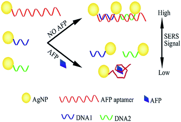
The assembly of Ag-trimers and their SERS sensor are shown in Scheme 1. First, AgNPs and ssDNA (Table S1†) were mixed in a ratio of 1![[thin space (1/6-em)]](https://www.rsc.org/images/entities/char_2009.gif) :
:![[thin space (1/6-em)]](https://www.rsc.org/images/entities/char_2009.gif) 2 to form the AgNP–aptamer,18 AgNP–DNA1, and AgNP–DNA2 conjugates, respectively. These three kinds of functional AgNPs were then mixed in a molar ratio of 1
2 to form the AgNP–aptamer,18 AgNP–DNA1, and AgNP–DNA2 conjugates, respectively. These three kinds of functional AgNPs were then mixed in a molar ratio of 1![[thin space (1/6-em)]](https://www.rsc.org/images/entities/char_2009.gif) :
:![[thin space (1/6-em)]](https://www.rsc.org/images/entities/char_2009.gif) 1.3
1.3![[thin space (1/6-em)]](https://www.rsc.org/images/entities/char_2009.gif) :
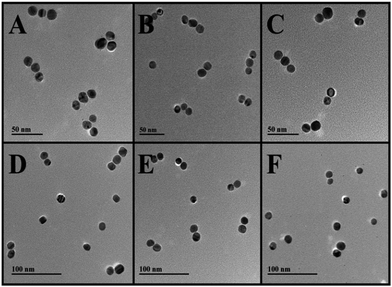
:![[thin space (1/6-em)]](https://www.rsc.org/images/entities/char_2009.gif) 1.3 (NP–aptamer, NP–DNA1, NP–DNA2), together for incubation to facilitate the formation of Ag-trimers. Due to high affinity of the AFP aptamers and their complementary sequences which conjugated to AgNPs, it could form the Ag-trimers. On the other hand, the Ag-trimers would disassemble when the free AFP target adding into the reaction solution. From the images of transmission electron microscope (TEM) in Fig. 1A, it clearly showed the assembly of Ag-trimer assemblies and the yield was about 92.6% by controlling the molar ratio of those conjugated AgNPs and the optimised hybridization conditions (see ESI†), without large aggregates obviously observed (Fig. S1 and S2†).
1.3 (NP–aptamer, NP–DNA1, NP–DNA2), together for incubation to facilitate the formation of Ag-trimers. Due to high affinity of the AFP aptamers and their complementary sequences which conjugated to AgNPs, it could form the Ag-trimers. On the other hand, the Ag-trimers would disassemble when the free AFP target adding into the reaction solution. From the images of transmission electron microscope (TEM) in Fig. 1A, it clearly showed the assembly of Ag-trimer assemblies and the yield was about 92.6% by controlling the molar ratio of those conjugated AgNPs and the optimised hybridization conditions (see ESI†), without large aggregates obviously observed (Fig. S1 and S2†).
 | ||
| Fig. 1 Typical TEM images of Ag-trimers with different concentration of AFP. (A) 0 aM, (B) 0.2 aM, (C) 1 aM, (D) 2 aM, (E) 10 aM, (F) 20 aM. | ||
Note to mention that the long sequence of AFP aptamer (72 bp) probably tended to a random coil-like ssDNA, which went against for the DNA hybridization. Therefore, it was necessary to heat the hybridization mixtures at high temperature (90 °C for 5 min) to open end of the long-chain DNA before the hybridization. However, for the case of the target protein addition, the long-chain DNA aptamer has already hybridized with the partial-complementary DNA sequences, and they would spontaneously bind to the target protein due to their higher affinity, without the adjustment of the temperature (ESI†).9a,19
Ag-trimers in the absence and presence of AFP were structurally characterized by UV-vis spectroscopy (Fig. S1†). A blue-shift of 3 nm in the plasmonic peak of Ag-trimers in the absence of AFP (from 398 to 395 nm) was observed, which were probably due to some extent disassembly of Ag-trimers, ruling out the factor of original change of dielectric constant by using the same buffer (5 mM PB solution, pH = 7.4).20 And the results of the dynamic light scattering (DLS, Fig. S2†), further confirmed the successful assembly.21 The DLS diameters (the length of the chain of NPs) of the Ag-trimers were 65 ± 3.1 and 35 ± 2.6 nm in the absence and presence of AFP, respectively (Fig. S2†) and without aggregations. Considering the AgNPs was functionalized with DNA, which was 39 ± 2.6 nm (AgNP– aptamer), 22 ± 2.1 nm (AgNP–DNA1), and 22 ± 2.3 nm (AgNP–DNA2), respectively. The gap between the adjacent NPs was 1 ± 0.2 nm (AgNP–aptamer and AgNP–DNA1), and 2 ± 0.5 nm (AgNP–DNA1 and AgNP–DNA2), respectively (Fig. 1A).
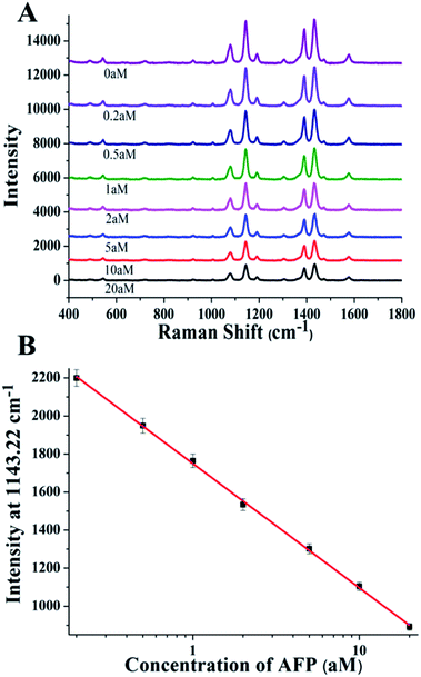
Taking into account of the high plasmonic intensity and the strong electromagnetic field enhancement of AgNP assemblies, which could show the SERS active Ag-trimers with high SERS intensity. Therefore, 4-aminothiophenol (4-ATP, Fig. S3†), as a model SERS reporter, was attached to the assembled Ag-trimers by a strong Ag–S bond. Since the SERS response of Ag-trimers gradually decreased as the 3D spatial geometries of the trimers altered accompanied by a longer gap length, which made it possible for the application of the AFP detection (Fig. 2A).
 | ||
| Fig. 2 AFP detection based on SERS technique with Ag-trimers. (A) SERS spectra for different concentration of AFP and (B) the corresponding SERS intensity calibration curve for AFP detection. | ||
Before the biosensing using the trimers, it was necessary to investigate the stability of the trimers in suspension. Therefore, the experiment for the stability test of the trimers was performed. From the results of DLS (Fig. S4†) and UV-Vis analysis (Fig. S5†), it was clearly showed that the trimers were stable in suspension for more than 8 hours (0.5 h for the target detection), without any sediment aggregate or spontaneously disassemble.
We then measured the SERS spectra of trimers produced in the presence of various concentrations of AFP. Following the addition of AFP to the SERS active assemblies, we found a marked change in the Raman spectra, which should be attributed to the stronger “hot spots” between adjacent AgNPs in trimers in the absence of AFP than of dispersed AgNPs in the presence of AFP target (Fig. S2†). Note to mention that, when the free AFP target competitively binded to the aptamer conjugated to AgNPs, the other two functionalized AgNPs (AgNP–DNA1 and AgNP–DNA2) would simultaneously disassemble from the trimers, leading to the stronger decrease of the electromagnetic field than that of AgNP dimers, and finally resulting in more significant change of the SERS signal. The Raman intensity at the frequency typical 4-ATP frequency of 1143.22 cm−1 was observed to be the highest, which was selected as the analytical band for the following experiments.11,22 When the concentration of AFP in the solution increased, the Raman signal at the band of 1143.22 cm−1 gradually decreased (Fig. 2A). The obtained calibration curve indicated an excellent linear range from 0.2 to 20 aM (Fig. 2B). The limit of detection (LOD) was calculated to be 0.097 aM, which is the lowest reported so far and was about 3 times lower than the best reported methods to date for AFP (Fig. 2B and Table S2†).23
The ultrasensitivity of the developed SERS sensor was probably due to multiple factors including below: (a) the strong plasmonic intensity of the Ag-trimers; (b) thousands of Raman labels for a single nanoparticle, which caused higher SERS change; (c) small interparticle distance resulted in high electromagnetic enhancement; and (d) competition advantages for the target against the designed complementary aptamer.24
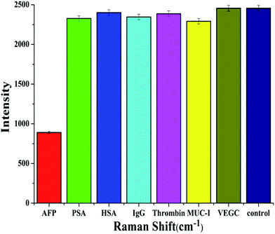
After the evaluation of sensitivity of the developed SERS sensor, specificity was assessed by using different types of biomarkers instead of AFP and a blank control without any biomarkers (Fig. 3). No significant changes among the six biomarkers and the blank group in the Raman spectra were observed, indicating the high selectivity of the developed sensor, due to its excellent specificity and affinity for AFP and its aptamer. We also added another negative control experiment where the trimers were prepared using a DNA sequence that was not able to recognize the target AFP protein. As showed in Fig. S6,† the Raman intensity remained nearly unchanged with and without the presence of AFP target, which further demonstrated the high specificity of the developed SERS assay. In addition, the SERS active Ag-trimers were selected for the detection of AFP in real human serum samples. As shown in Table 1, the results obtained by the established method was in good agreement with the standard clinical diagnostic assay method. These experimental results demonstrated that this developed SERS active Ag-trimers biosensor was robust in the complicated serum matrix, and could be applied to monitor AFP in fetal tissue and primary liver cancer.
 | ||
| Fig. 3 Specificity of the SERS-active platform for the detection of AFP. The concentration of AFP was 5 aM, and the concentration of other proteins were all 0.5 nM. | ||
| Serum samplesa | Original concentrationb (pM) | Diluted concentrationc (aM) | Detected concentrationd (aM) | CV (%) |
|---|---|---|---|---|
| a Serum sample No. 1 to No. 10 are human sera, which are sampling from ten donors at the Second People's Hospital of Wuxi, P.R.C.b Original concentrations of AFP in the serum were determined by the standard clinical diagnostic assay (ADVIA Centaur, Siemens).c Original serum samples No. 1 to No. 5 were serially diluted with bovine serum to a dilution factor of 8, while a dilution factor of 9 for samples No. 6 to No. 10. After the thorough mixing, the samples were then stood for at least 2 h before the determination.d SD was calculated based on five parallel experiments for each sample. | ||||
| 1 | 273.5 | 2.7 | 2.7 ± 0.1 | 3.7 |
| 2 | 234.6 | 2.3 | 2.5 ± 0.2 | 8.0 |
| 3 | 208.4 | 2.1 | 1.9 ± 0.2 | 10.5 |
| 4 | 455.9 | 4.6 | 4.4 ± 0.1 | 2.3 |
| 5 | 678.5 | 6.8 | 7.1 ± 0.4 | 5.6 |
| 6 | 1510.3 | 1.5 | 1.5 ± 0.1 | 6.7 |
| 7 | 3109.0 | 3.1 | 3.3 ± 0.2 | 6.1 |
| 8 | 2933.1 | 2.9 | 2.8 ± 0.2 | 7.1 |
| 9 | 5935.7 | 5.9 | 5.6 ± 0.3 | 5.4 |
| 10 | 6112.3 | 6.1 | 6.1 ± 0.4 | 6.6 |
In summary, we developed, for the first time, a sub-attomolar SERS biosensor for the detection of AFP through the assembly of Ag-trimers via the aptamer. Under the optimized conditions, the LOD of this established biosensor reached to attomolar, 0.097 aM. The SERS response of the assemblies was observed to be extremely sensitive, which was mainly due to the strong “hot spots” and the configuration changes of the NPs assemblies. The developed method has successfully achieved for the AFP determination in the complicated human serum. Therefore, this ultrasensitive SERS sensor will open up an avenue in the biological analysis field, especially for bio-diagnosis.
Acknowledgements
This work is financially supported by the National Natural Science Foundation of China (21371081, 21301073), and grants from Natural Science Foundation of Jiangsu Province (BK20140003).Notes and references
- N. G. Ladep, O. Noorullah, E. Boland, W. Y. Ding, T. Cross, C. Sieberhagen, R. Sturgess and N. Stern, Gastroenterology, 2015, 148, 639 CrossRef.
- D. F. Billmire, J. W. Cullen, F. J. Rescorla, M. Davis, M. G. Schlatter, T. A. Olson, M. H. Malogolowkin, F. Pashankar, D. Villaluna and M. Krailo, J. Clin. Oncol., 2014, 32, 465 CrossRef PubMed.
- A. L. Frazier, J. P. Hale, C. Rodriguez-Galindo, H. Dang, T. Olson, M. J. Murray, J. F. Amatruda, C. Thornton, G. S. Arul and D. Billmire, J. Clin. Oncol., 2014, 58, 3369 Search PubMed.
- W. Sun, Y. Liu, D. Shou, Q. Sun, J. Shi, L. Chen, T. Liang and W. Gong, Cancer Lett., 2015, 357, 43 CrossRef CAS PubMed.
- (a) G. Nie, C. Li, L. Zhang and L. Wang, J. Mater. Chem. B, 2014, 2, 8321 RSC; (b) F. Han, H. Jiang, D. Fang and D. Jiang, Anal. Chem., 2014, 86, 6896 CrossRef CAS PubMed; (c) S. Liu, J. Zhang, W. Tu, J. Bao and Z. Dai, Nanoscale, 2014, 6, 2419 RSC.
- X. Bi and Z. Liu, Anal. Chem., 2013, 86, 959 CrossRef PubMed.
- W. Zheng, D. Tu, P. Huang, S. Zhou, Z. Chen and X. Chen, Chem. Commun., 2015, 51, 4129 RSC.
- Z. Guo, T. Hao, J. Duan, S. Wang and D. Wei, Talanta, 2012, 89, 27 CrossRef CAS PubMed.
- (a) W. Yan, L. Xu, C. Xu, W. Ma, H. Kuang, L. Wang and N. A. Kotov, J. Am. Chem. Soc., 2012, 134, 15114 CrossRef CAS PubMed; (b) X. Xia, J. Zeng, B. McDearmon, Y. Zheng, Q. Li and Y. Xia, Angew. Chem., 2011, 123, 12750 CrossRef PubMed.
- Z. Zhang, Y. Wang, F. Zheng, R. Ren and S. Zhang, Chem. Commun., 2014, 51, 907 RSC.
- S. Li, L. Xu, W. Ma, H. Kuang, L. Wang and C. Xu, Small, 2015, 11, 3435 CrossRef CAS PubMed.
- L. Xu, W. Yan, W. Ma, H. Kuang, X. Wu, L. Liu, Y. Zhao, L. Wang and C. Xu, Adv. Mater., 2015, 27, 1706 CrossRef CAS PubMed.
- R. Liu, J.-F. Sun, D. Cao, L.-Q. Zhang, J.-F. Liu and G.-B. Jiang, Chem. Commun., 2015, 51, 1309 RSC.
- (a) L. Rodríguez-Lorenzo, R. de La Rica, R. A. Álvarez-Puebla, L. M. Liz-Marzán and M. M. Stevens, Nat. Mater., 2012, 11, 604 CrossRef PubMed; (b) W. Ma, M. Sun, L. Xu, L. Wang, H. Kuang and C. Xu, Chem. Commun., 2013, 49, 4989 RSC.
- W. Ma, H. Kuang, L. Xu, L. Ding, C. Xu, L. Wang and N. A. Kotov, Nat. Commun., 2013, 4, 2689 Search PubMed.
- Y. Wang, M. Salehi, M. Schütz and S. Schlücker, Chem. Commun., 2014, 50, 2711 RSC.
- (a) Y. Kitahama, T. Ikemachi, T. Suzuki, T. Miura and Y. Ozaki, Chem. Commun., 2014, 50, 9693 RSC; (b) X. Wu, L. Xu, L. Liu, W. Ma, H. Yin, H. Kuang, L. Wang, C. Xu and N. A. Kotov, J. Am. Chem. Soc., 2013, 135, 18629 CrossRef CAS PubMed.
- (a) Y.-H. Chen, H.-I. Lin, C.-J. Huang, S.-C. Shiesh and G.-B. Lee, Microfluid. Nanofluid., 2012, 13, 929 CrossRef CAS; (b) C.-J. Huang, H.-I. Lin, S.-C. Shiesh and G.-B. Lee, Biosens. Bioelectron., 2012, 35, 50 CrossRef CAS PubMed.
- (a) L. C. Bock, L. C. Griffin, J. A. Latham, E. H. Vermaas and J. J. Toole, Nature, 1992, 355, 564 CrossRef CAS PubMed; (b) Q. Wang, H. Wang, C. Lin, J. Sharma, S. Zou and Y. Liu, Chem. Commun., 2010, 46, 240 RSC.
- P. Jayaraman, S. Doss, H. Sridevi, K. Mathivanan and P. Arumugam, J. Bionanosci., 2013, 7, 432 CrossRef CAS PubMed.
- L. Xu, H. Kuang, C. Xu, W. Ma, L. Wang and N. A. Kotov, J. Am. Chem. Soc., 2012, 134, 1699 CrossRef CAS PubMed.
- (a) Y. Zhao, L. Xu, L. M. Liz-Marzán, H. Kuang, W. Ma, A. Asenjo-García, F. J. García de Abajo, N. A. Kotov, L. Wang and C. Xu, J. Phys. Chem. Lett., 2013, 4, 641 CrossRef CAS; (b) S. Pattanayak, A. Swarnkar, A. Priyam and G. M. Bhalerao, Dalton Trans., 2014, 11826 RSC.
- Z. Guo, T. Hao, S. Wang, N. Gan, X. Li and D. Wei, Electrochem. Commun., 2012, 14, 13 CrossRef CAS PubMed.
- (a) D. C. Harris, X. Chu and J. Jayawickramarajah, J. Am. Chem. Soc., 2008, 130, 14950 CrossRef PubMed; (b) E. Pérez-Ruiz, M. Kemper, D. Spasic, A. Gils, L. J. van IJzendoorn, J. Lammertyn and M. W. Prins, Anal. Chem., 2014, 86, 3084 CrossRef PubMed.
Footnote |
| † Electronic supplementary information (ESI) available: Details experimental section and additional experimental results. See DOI: 10.1039/c5ra12629k |
| This journal is © The Royal Society of Chemistry 2015 |

