Prediction of metastable energy level distribution of D3+ (D = Cr and Fe) doped phosphors based on machine learning
Jun
Li†
a,
Junkang
Sun†
a,
Yixiao
Wang
a and
Xiangfu
Wang
 *ab
*ab
aCollege of Electronic and Optical Engineering & College of Flexible Electronics (Future Technology), Nanjing University of Posts and Telecommunications, Nanjing, 210023, China. E-mail: xfwang@njupt.edu.cn
bState Key Laboratory of Luminescent Materials and Devices, South China University of Technology, Guangzhou 510641, China
First published on 20th June 2024
Abstract
The energy level transitions in phosphor materials critically determine their emission characteristics, and accurately predicting the energy level distribution of ions in these materials is critical for determining their luminescence behavior. However, reliance on multiple experimental methods to determine energy level distributions is inefficient, consuming both time and resources. There is an urgent need for a rapid and accurate method to predict the energy level distribution of ions in crystals. This paper employs regression models based on machine learning to propose a method for predicting the energy level distribution rules of Cr3+ and Fe3+ in various doped crystals, and identifies the position and distribution patterns of these levels in different doped crystals, as well as their impact on luminescence characteristics. Furthermore, a dataset detailing the energy level distributions of Cr3+ and Fe3+ doped into different phosphor materials was established. Eight machine learning regression algorithms were selected for model construction, and a comprehensive evaluation and comparison of these algorithms were conducted. The results demonstrate that robust regression delivers the best overall performance. Using trained models, predictions were made for the 2E and 4T1 energy levels in new Cr3+ and Fe3+ doped phosphor materials. The prediction errors of the optimal algorithms for these materials were all in the range of about 1%, with the best prediction error at just 0.0056%. This study introduces an innovative approach for predicting and optimizing the energy level structures and luminescence properties of phosphor materials.
1. Introduction
Transition metal ions, particularly Cr3+ and Fe3+, are crucial for the formation of optical properties in doped phosphor materials.1 Cr3+-activated near-infrared phosphor materials can achieve both narrow-band and broad-band emissions, finding applications across various scenarios.2 The d–d transitions of Cr3+ in crystals can generate broad photoluminescence from far-red (spin-forbidden 2E–4A2) to near-infrared (spin-allowed 4T2–4A2) regions, making Cr3+ one of the ideal activators for near-infrared light emission.3 Currently, there are numerous phosphor materials activated by Cr3+ that exhibit excellent properties, such as Mg4Ta2O9:Cr3+ with its long peak wavelength and broad-band near-infrared emission,4 and GaTaO4:Cr3+ which produces broad-band near-infrared emission under 460 nm blue light excitation.5 Besides Cr3+, Fe3+ also holds potential as a dopant. Based on its half-filled electronic configuration, Fe3+ ions can emit near-infrared light through 4T1–6A1 transitions.6 The abundance and non-toxic nature of Fe3+ ions make them an environmentally friendly doping option.7 The energy states of Cr3+ and Fe3+ ions in doped crystals, which involve specific electronic configurations and transitions, determine the material's optical properties, such as absorption and emission spectra.8 Therefore, studying the closely related energy levels of ions and the luminescence behavior of phosphor materials, such as the 2E energy level of Cr3+ and the 4T1 energy level of Fe3+, is essential, which not only elucidates the luminescence behavior of these materials, but also plays a key role in optimizing their functional performance in various applications. However, determining these specific energy levels through traditional experimental methods is challenging, often requiring significant time, resources, and complex procedures, thus presenting challenges in the research and optimization of Cr3+ and Fe3+-doped phosphor materials.9Benefiting from advancements in computer science, machine learning, as a data-driven approach, possesses the advantage of balancing development efficiency and cost. It is emerging as a novel paradigm in materials science research.10 The motivation for integrating machine learning algorithms into the study of doped phosphor materials stems from the vast amount of data generated over the past few decades related to these materials. Despite this abundance of data, certain fundamental aspects of the underlying physics of phosphors remain unclear. The data-driven nature of machine learning is particularly advantageous for addressing problems where the fundamental physics is not fully understood yet rich data are available. Consequently, this approach can accelerate the research and design of phosphor materials.11 Li et al.12 utilized a data-driven approach combining high-throughput DFT calculations and data mining of experimentally validated data to discover a monophase white-light phosphor (Sr2AlSi2O6N:Eu2+), which exhibited an exceptional emission bandwidth. Jiang et al.13 constructed wavelength and thermal stability machine learning models by extracting relevant features, and through an active learning-based multi-objective optimization approach, identified garnet-type phosphors Lu1.5Sr1.5Al3.5Si1.5O12:Ce that demonstrated excellent thermal stability at targeted wavelengths. Jang et al.14 created a high-quality dataset of inorganic phosphors and their optical properties using machine learning methods to predict optical characteristics such as the maximum photoluminescence (PL) emission wavelength and PL decay time, achieving significant predictive success. Kim et al.15 integrated machine learning models with an experimental dataset of synthesized Ba0.9−xSrxMgAl10O17:Eu2+, establishing a relationship between photoluminescence (PL) and crystal properties. However, there are currently few targeted studies on the metastable energy levels 2E and 4T1, which are closely related to the luminescence properties of Cr3+ and Fe3+ ions in doped crystals, and there is a lack of a method that can effectively predict the energy levels of these ions in doped crystals without the need for experimental measurements, and at very low time and resource costs.
Therefore, this paper aims to use machine learning methods to predict the 2E and 4T1 energy levels of Cr3+ and Fe3+ doped in various phosphor materials, which are closely related to the luminescence properties of the materials. Based on a detailed study of the luminescence behavior of Cr3+ and Fe3+ doped phosphor materials and factors influencing the transitions of these ions, this work has established a dataset necessary for the model through an in-depth literature review and rigorous data extraction methods. The dataset includes crystal field parameters Dq, Racah parameters B and C, and the 2E and 4T1 energy levels of Cr3+ and Fe3+. Based on this, eight machine learning regression algorithms (linear regression, robust regression, lasso regression, ridge regression, elastic net, decision tree, random forest, and gradient boosting) were selected to build models to predict the metastable energy levels 2E and 4T1 of Cr3+ and Fe3+ in doped crystals. A comprehensive evaluation of these algorithms’ predictions on known data was conducted. Subsequently, some new phosphor materials doped with Cr3+ and Fe3+ were selected, and the trained models were used to predict the 2E and 4T1 energy levels of Cr3+ and Fe3+ in these materials. Comparisons with actual values were made to further verify the effectiveness of the proposed prediction method.
2. Method
2.1. Machine learning in materials science
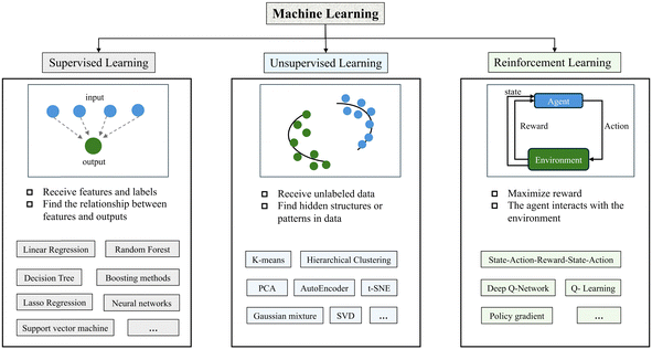
Machine learning aims to identify patterns in given data, subsequently making predictions or decisions. If we define a model function F(x,ω), where x is the input variable and ω represents the model parameters, the goal is to approximate an unknown true function g(x) that directly links input to output. During training, the model's performance is measured using a loss function L(y;F(x,ω)), with a mean squared error being a common choice, i.e., L(y;F(x,ω)) = ||y − F(x,ω)||2. The core of machine learning is to optimize the parameters ω to minimize the loss, thereby enhancing the model's predictive ability on unknown data. Based on the learning method used during training, machine learning is primarily divided into supervised learning, unsupervised learning, and reinforcement learning.16 Under this classification, typical machine learning models are shown in Fig. 1. The training data for supervised learning contain input samples and their corresponding output labels, and the goal is to use the training data and data feedback to learn the relationship between a given input and a given output. Unsupervised learning deals with unlabeled data and the goal is to discover hidden structures or patterns in the data. Reinforcement learning is a learning model of the decision-making process in which an agent learns how to act to maximize some cumulative reward by interacting with the environment. This research predominantly utilizes supervised learning, where the training data include input samples and their corresponding output labels, specifically the phosphor material parameters Dq, B, C, and the specific energy levels of ions in a doped environment; for Cr3+, this is the 2E level, and for Fe3+, the 4T1 level. The objective is to use the training data and feedback to learn the relationships between given inputs and outputs, thereby enabling the prediction of the 2E and 4T1 energy levels of doped ions.This study evaluates various machine learning tools that phosphor material researchers can utilize. The tools are categorized into several types: machine learning/deep learning (ML/DL) frameworks, comprehensive ML libraries, cloud computing platforms, data science and ML platforms, and scientific computing software. Additionally, for each category, several popular options commonly used by researchers are listed, and the characteristics or main functions of each software or tool are succinctly and clearly described, as shown in Table 1. This section enables researchers to more conveniently select the tools that best suit their specific research needs, thereby effectively advancing scientific research in the field of phosphor materials.
| Categories | Software/tools | Characteristics |
|---|---|---|
| ML/DL frameworks | PyTorch17 | Deeply integrated with Python |
| Dynamic computation graph and powerful GPU support | ||
| TensorFlow18 | Distributed computing and GPU acceleration | |
| Rich API multi-platform | ||
| Keras19 | The architecture is modular and scalable | |
| Can run on multiple underlying frameworks | ||
| Comprehensive ML libraries | Scikit-learn20 | Open source ML library, simple, and effective |
| A wide range of classic ML algorithms | ||
| Weka21 | Open source ML and data mining library | |
| Provide user-friendly GUI | ||
| Cloud computing platforms | Microsoft Azure ML22 | Full-process services from data preparation to model deployment |
| Google Cloud AI Platform23 | One-stop model training and tuning | |
| Integrate Google AI technology | ||
| Data science and ML platforms | RapidMiner24 | Integrated data science workflow designer |
| Supports a wide range of ML/DL algorithms | ||
| H2O25 | Open source ML platform that supports a wide range of statistical and ML algorithms | |
| Kaggle26 | Data set access and sharing, and open community | |
| Scientific computing software | MATLAB27 | Provide ML/DL toolbox |
| Strong mathematics and visualization skills |
2.2. Analysis of the luminescence behaviour of Cr3+ and Fe3+ doped phosphor materials and the main factors affecting ion transitions
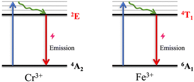
In compounds doped with Cr3+ ions, these ions typically manifest a 3d3 electronic configuration, giving rise to doublet states such as 2E and quartet states such as 4A2—the ground state of energy.28 Under the influence of a strong crystal field, the 2E level ascends to the position of the lowest excited state, undergoing transitions to 4A2via spin-forbidden processes, thereby emitting far-red light. The emission spectrum is characterized by a narrow-band peak, observable in materials such as La2ZnTiO6:Cr3+ and GdAlO3:Cr3+.29 Conversely, in a weak crystal field, the 4T2 state emerges as the lowest excited state, facilitating spin-allowed transitions to the ground state and resulting in a broadband near-infrared emission spectrum, as seen in LiInSi2O6:Cr3+ and ScF3:Cr3+. In environments of medium crystal field strength, both 2E–4A2 and 4T2–4A2 transitions occur concurrently in the Cr3+ electronic structure, with the emission spectrum displaying both peak and broadband emissions, exemplified by Sc4Zr3O12:Cr3+ and CaSc2O4:Cr3+.30 This investigation predominantly explores the 2E–4A2 transition of Cr3+ ions, known for producing distinct line emissions (R line). In contrast, Fe3+ ions exhibit a 3d5 electronic configuration with a solitary sextet state 6A1, serving as the ground energy state. The excited states predominantly comprise quartet states (4T1, 4T2, etc.), where transitions are also spin-forbidden. Upon doping into various matrices, Fe3+ may exhibit near-infrared photoluminescence due to 4T1–6A1 transition, underscoring its significant role in the absorption and emission processes of light in Fe3+.31Fig. 2 illustrates the schematic diagram of the 2E and 4T1 energy level transitions in Cr3+ and Fe3+ doped crystals, highlighting the 2E–4A2 and 4T1–6A1 transitions that are instrumental to the luminescence properties of these doped phosphors. Since the transitions of Cr ions are d–d transitions occurring in the d orbitals, the effect of electron cloud rearrangement on energy level transitions is minimal, and the influence of the crystal field dominates the transitions of Cr3+ ions.32 The Tanabe–Sugano diagram33 takes into account both the crystal field splitting Dq and the interactions between electrons (Racah parameters) affecting the transitions of Cr3+ energy levels. The crystal parameter Dq describes the splitting effect of the crystal field on electron states, while Racah parameters B and C represent the Coulomb interactions between electrons, playing a critical role in determining energy levels and transitions. The transitions of Fe3+ ions are also d–d transitions, with Dq, B, and C as the main factors influencing their energy level transitions.34 Based on this analysis, this study aims to explore, through machine learning methods, the complex relationships between parameters Dq, B, and C and the 2E energy levels of Cr3+ and the 4T1 energy levels of Fe3+, thereby predicting the energy levels of 2E and 4T1. | ||
| Fig. 2 Schematic diagram of 2E and 4T1 energy level transitions doped with Cr3+ and Fe3+ in crystals. | ||
2.3 Analysis of algorithms
This paper aims to use machine learning algorithms to predict the 2E and 4T1 energy levels of Cr3+ and Fe3+ doped in various phosphor materials. Specifically, this paper attempts to explore the complex relationship between the crystal parameter Dq, Racah parameters B and C, and the distribution of ion energy levels, so as to realize the prediction of ion energy levels in doped crystals. Regression models are supervised learning methods used to predict continuous value outputs in machine learning, mainly by analyzing the relationship between input features and the target variable. The goal of this model is to establish a mathematical equation that can describe the mapping relationship between inputs and outputs as accurately as possible. Therefore, in this paper, the regression models are chosen for the prediction of energy levels. This study selected eight typical regression algorithms for exploration, including linear regression, robust regression, lasso regression, ridge regression, elastic net, decision tree, random forest, and gradient boosting. In the field of machine learning, choosing the right algorithm is critical to the performance and accuracy of the model. These eight algorithms are all more classical and representative of regression algorithms, covering a wide range of techniques from simple to complex, and have the ability to handle different types of data, thus providing a comprehensive and effective analysis tool for research. The article conducts in-depth research and discussion on the basic principles, mathematical models, and applicability of these algorithms, and further analyzes and summarizes these models in this research context based on the final experimental results, with specific analysis content as follows.The primary objective of the linear regression algorithm35 is to determine the optimal weights of the independent variables to minimize the error between the model's predicted values and the actual observed values. The linear regression model is based on the fundamental assumption of a linear relationship, meaning that the dependent variable can be accurately predicted by a linear combination of the independent variables. This can be initially assessed through a scatter plot to judge whether a linear relationship exists between the variables. It is suitable for basic regression that does not focus on outliers, as shown in the following form:
 | (1) |
Robust regression36 is a method specifically designed to handle outliers in regression analysis. It employs robust loss functions to minimize the impact of outliers, thereby enhancing the robustness of the model and making it more effective in the presence of irregularities in the dataset. The mathematical model includes a robust loss function L(u), such as the Huber loss, which is formulated as follows:
 | (2) |
Lasso regression37 incorporates an L1 regularization term into traditional regression models. While fitting a generalized linear model and minimizing the sum of squared residuals, it constrains the complexity of the model through a penalty term. This approach facilitates variable selection and regularization, enhancing the model's predictive accuracy and interpretability. The formula is as follows:
 | (3) |
Ridge regression38 includes an L2 regularization term, which helps to suppress the size of the model coefficients and thus prevent overfitting. It minimizes the sum of squared residuals and imposes a penalty on large coefficients to balance bias and variance. The formula is as follows:
 | (4) |
Elastic net39 combines the features of ridge regression (L2 regularization) and lasso regression (L1 regularization). It is particularly suited for scenarios with multiple correlated independent variables. Adjusting the balance between the two types of regularization effectively controls the complexity and sparsity of the model, thereby optimizing the model's predictive performance and interpretability. The formula is as follows:
 | (5) |
In decision tree regression,40 each internal node represents a decision rule on a feature, which is based on maximizing the homogeneity (or minimizing the heterogeneity) of the data after each node split. Specifically, each split is chosen based on the feature that most reduces the variance of the target variable. The criterion typically used to select the best split is the mean squared error (MSE), which is calculated as follows:
 | (6) |
Random forest41 is an ensemble of decision trees, each built on a bootstrap sample of the original dataset. At each split point, a subset of features is randomly selected to determine the best split. The final prediction of the model is obtained by averaging the predictions from all the trees, which is expressed as follows:
 | (7) |
![[f with combining circumflex]](https://www.rsc.org/images/entities/i_char_0066_0302.gif) b(x) is the prediction result from the b-th tree. Random forest reduces the model's variance by increasing the independence between decision trees, effectively preventing overfitting. It is suitable for handling regression tasks with high-dimensional features and complex data structures.
b(x) is the prediction result from the b-th tree. Random forest reduces the model's variance by increasing the independence between decision trees, effectively preventing overfitting. It is suitable for handling regression tasks with high-dimensional features and complex data structures.
Gradient boosting regression42 optimizes the overall prediction accuracy by iteratively adding base models (typically weak learners, such as simple linear regression models or decision trees) to minimize the loss function. Specifically, the model update formula at each step can be expressed as:
 | (8) |
2.4. Experimental datasets and details
This article conducts a series of precise search strategies, providing a comprehensive literature review of relevant research fields. Specifically, the study focuses on the crystal field parameter Dq and the Racah parameters B and C, which play crucial roles in understanding and predicting the electronic energy levels of ions in doped phosphors. During data collection, we systematically extracted data from renowned academic databases such as Scopus, Web of Science, ScienceDirect, and IEEE Xplore, which is fundamental for constructing machine learning models. The dataset for Cr-doped samples is shown in Table 2,43–71 and the dataset for Fe-doped samples is presented in Table 3.72–96 These data, after preprocessing, serve as input features for the regression models listed in Section 2.3, enabling the prediction of Cr3+ and Fe3+ behavior in different environments.| Crystal | D q | B | C | 2E | Crystal | D q | B | C | 2E |
|---|---|---|---|---|---|---|---|---|---|
| KAl(MoO4)2 | 1494.8 | 585.5 | 3049 | 13![[thin space (1/6-em)]](https://www.rsc.org/images/entities/char_2009.gif) 517 517 |
CdO·P2O5 | 1545 | 700 | 3230 | 14![[thin space (1/6-em)]](https://www.rsc.org/images/entities/char_2009.gif) 815 815 |
| Y3Ga5O12 | 1626 | 645 | 2950 | 14![[thin space (1/6-em)]](https://www.rsc.org/images/entities/char_2009.gif) 472 472 |
PbO–Ga2O3–P2O5 | 1557 | 660 | 3061 | 14![[thin space (1/6-em)]](https://www.rsc.org/images/entities/char_2009.gif) 045 045 |
| LiAl5O8 | 1779 | 741 | 2875 | 14![[thin space (1/6-em)]](https://www.rsc.org/images/entities/char_2009.gif) 085 085 |
Pb3O4–ZnO–P2O5 | 1523 | 709 | 3232 | 14![[thin space (1/6-em)]](https://www.rsc.org/images/entities/char_2009.gif) 864 864 |
| Ba2Mg(BO3)2 | 1967 | 718 | 2756 | 14![[thin space (1/6-em)]](https://www.rsc.org/images/entities/char_2009.gif) 327 327 |
MgO | 1615 | 586 | 3249 | 14![[thin space (1/6-em)]](https://www.rsc.org/images/entities/char_2009.gif) 164 164 |
| NaAl(WO4)2 | 1548 | 615.6 | 3083 | 13![[thin space (1/6-em)]](https://www.rsc.org/images/entities/char_2009.gif) 822 822 |
KZnF3 | 1323 | 731 | 3437 | 15![[thin space (1/6-em)]](https://www.rsc.org/images/entities/char_2009.gif) 523 523 |
| YAl3(BO3)4 | 1680 | 672 | 3218 | 14![[thin space (1/6-em)]](https://www.rsc.org/images/entities/char_2009.gif) 663 663 |
LiNbO3 | 1355 | 644 | 3026 | 13![[thin space (1/6-em)]](https://www.rsc.org/images/entities/char_2009.gif) 772 772 |
| NaMg3Al(MoO4)5 | 1440 | 676 | 2945 | 13![[thin space (1/6-em)]](https://www.rsc.org/images/entities/char_2009.gif) 755 755 |
MgSrAl10O17 | 1549 | 677 | 3179 | 14![[thin space (1/6-em)]](https://www.rsc.org/images/entities/char_2009.gif) 506 506 |
| LiNbO3: ZnO | 1342 | 646 | 3022 | 13![[thin space (1/6-em)]](https://www.rsc.org/images/entities/char_2009.gif) 755 755 |
MgAl2O4 | 1814.9 | 544.7 | 3224.9 | 14![[thin space (1/6-em)]](https://www.rsc.org/images/entities/char_2009.gif) 577 577 |
| LiBP | 1577 | 707 | 3146 | 14![[thin space (1/6-em)]](https://www.rsc.org/images/entities/char_2009.gif) 600 600 |
SiO2:B2O3:Na2O:Al2O3:CaO:ZrO2 | 1574 | 792.2 | 3005 | 14![[thin space (1/6-em)]](https://www.rsc.org/images/entities/char_2009.gif) 684 684 |
| BaAl2O4 | 1811 | 533 | 2862 | 14![[thin space (1/6-em)]](https://www.rsc.org/images/entities/char_2009.gif) 180 180 |
GdScO3 | 1553 | 574 | 3211 | 13![[thin space (1/6-em)]](https://www.rsc.org/images/entities/char_2009.gif) 947 947 |
| Sc2(MoO4)3 | 1408 | 608 | 3054 | 13![[thin space (1/6-em)]](https://www.rsc.org/images/entities/char_2009.gif) 643 643 |
LiBaF3 | 1546 | 702 | 3300 | 15![[thin space (1/6-em)]](https://www.rsc.org/images/entities/char_2009.gif) 039 039 |
| LaSc3(BO3)4 | 1529 | 675 | 3448 | 14![[thin space (1/6-em)]](https://www.rsc.org/images/entities/char_2009.gif) 620 620 |
LiBaF3 | 1509 | 701 | 3292 | 15![[thin space (1/6-em)]](https://www.rsc.org/images/entities/char_2009.gif) 006 006 |
| YAl(BO3)4 | 1680 | 672 | 3225 | 14![[thin space (1/6-em)]](https://www.rsc.org/images/entities/char_2009.gif) 599 599 |
NaNH4SO4·2H2O | 1750 | 735 | 3220 | 15![[thin space (1/6-em)]](https://www.rsc.org/images/entities/char_2009.gif) 015 015 |
| GdAl(BO3)4 | 1695 | 673 | 3380 | 14![[thin space (1/6-em)]](https://www.rsc.org/images/entities/char_2009.gif) 599 599 |
(NH4)2Mg(SO4)2·6H2O | 2043 | 676 | 3371 | 13![[thin space (1/6-em)]](https://www.rsc.org/images/entities/char_2009.gif) 923 923 |
| Silicate glass | 1562 | 853 | 2898 | 14![[thin space (1/6-em)]](https://www.rsc.org/images/entities/char_2009.gif) 810 810 |
(CH3)4NCdCl3 | 2043 | 722 | 2845 | 13![[thin space (1/6-em)]](https://www.rsc.org/images/entities/char_2009.gif) 602 602 |
| CdO | 1540 | 619 | 3327 | 14![[thin space (1/6-em)]](https://www.rsc.org/images/entities/char_2009.gif) 595 595 |
YCrO3 | 1656.6 | 542.9 | 2962.3 | 13![[thin space (1/6-em)]](https://www.rsc.org/images/entities/char_2009.gif) 643 643 |
| SbPO | 1560 | 629 | 3171 | 14![[thin space (1/6-em)]](https://www.rsc.org/images/entities/char_2009.gif) 185 185 |
YAl3(BO3)4 | 1662 | 684 | 3194 | 14![[thin space (1/6-em)]](https://www.rsc.org/images/entities/char_2009.gif) 636 636 |
| Crystal | D q | B | C | 4T1 | Crystal | D q | B | C | 4T1 |
|---|---|---|---|---|---|---|---|---|---|
| KTaO3 | −640 | 640 | 2500 | 14![[thin space (1/6-em)]](https://www.rsc.org/images/entities/char_2009.gif) 569 569 |
ZnCdO | 920 | 840 | 2500 | 14![[thin space (1/6-em)]](https://www.rsc.org/images/entities/char_2009.gif) 595 595 |
| Mg2Al4Si5O18–Al2O3–MgAl2O4–SiO2 | 816 | 538 | 2944 | 14![[thin space (1/6-em)]](https://www.rsc.org/images/entities/char_2009.gif) 104 104 |
Al2O3 | 1527 | 650 | 3160 | 9450 |
| SrB4O7 | 790 | 700 | 3000 | 17![[thin space (1/6-em)]](https://www.rsc.org/images/entities/char_2009.gif) 852 852 |
ZnGa2O4 | 1560 | 600 | 3100 | 8400 |
| PVA capped ZnSe nanoparticles | 720 | 720 | 2500 | 16![[thin space (1/6-em)]](https://www.rsc.org/images/entities/char_2009.gif) 389 389 |
Li0.5Ga2.5O4 | 1550 | 560 | 3280 | 9200 |
| CdO nanopowders | 920 | 690 | 2750 | 14![[thin space (1/6-em)]](https://www.rsc.org/images/entities/char_2009.gif) 594 594 |
ZnS | 900 | 720 | 2700 | 14![[thin space (1/6-em)]](https://www.rsc.org/images/entities/char_2009.gif) 788 788 |
| K2SnCl4·H2O | 820 | 831 | 2198 | 13![[thin space (1/6-em)]](https://www.rsc.org/images/entities/char_2009.gif) 986 986 |
LiGa5O8 | 906 | 594 | 3737 | 18![[thin space (1/6-em)]](https://www.rsc.org/images/entities/char_2009.gif) 349 349 |
| Ca3Ga2Sn3O12 | 941 | 670 | 3047 | 14![[thin space (1/6-em)]](https://www.rsc.org/images/entities/char_2009.gif) 749 749 |
LiAl5O8 | 952 | 638 | 3868 | 19![[thin space (1/6-em)]](https://www.rsc.org/images/entities/char_2009.gif) 084 084 |
| Ca8Mg(SiO4)4Cl2 | 667 | 517 | 3267 | 17![[thin space (1/6-em)]](https://www.rsc.org/images/entities/char_2009.gif) 331 331 |
[NH4]2[Mg(H2O)6](SO4)2 | 740 | 757 | 2381 | 15![[thin space (1/6-em)]](https://www.rsc.org/images/entities/char_2009.gif) 267 267 |
| LiGa5O8 | 700 | 565 | 3000 | 15![[thin space (1/6-em)]](https://www.rsc.org/images/entities/char_2009.gif) 790 790 |
Humite | 760 | 690 | 2769 | 15![[thin space (1/6-em)]](https://www.rsc.org/images/entities/char_2009.gif) 240 240 |
| LiAl5O8 (ordered) | 800 | 644 | 2960 | 15![[thin space (1/6-em)]](https://www.rsc.org/images/entities/char_2009.gif) 255 255 |
NH4Cl | 675 | 645 | 2838 | 16![[thin space (1/6-em)]](https://www.rsc.org/images/entities/char_2009.gif) 291 291 |
| β-LiAlO2 | 883 | 630 | 3000 | 14![[thin space (1/6-em)]](https://www.rsc.org/images/entities/char_2009.gif) 750 750 |
Sr(NO3)2 | 1450 | 934 | 2059 | 12![[thin space (1/6-em)]](https://www.rsc.org/images/entities/char_2009.gif) 024 024 |
| β-LiGaO2 | 961 | 713 | 2995 | 14![[thin space (1/6-em)]](https://www.rsc.org/images/entities/char_2009.gif) 970 970 |
K2SO4-ZnSO4 | 1040 | 700 | 2800 | 14![[thin space (1/6-em)]](https://www.rsc.org/images/entities/char_2009.gif) 385 385 |
| KAlO2 | 771 | 593 | 3074 | 16![[thin space (1/6-em)]](https://www.rsc.org/images/entities/char_2009.gif) 030 030 |
Cadmium borosulphate glass | 1010 | 707 | 2778 | 14![[thin space (1/6-em)]](https://www.rsc.org/images/entities/char_2009.gif) 081 081 |
| CsAlO2 | 680 | 523 | 3152 | 16![[thin space (1/6-em)]](https://www.rsc.org/images/entities/char_2009.gif) 360 360 |
Y3Fe5O12 | 1336 | 783 | 2928 | 11![[thin space (1/6-em)]](https://www.rsc.org/images/entities/char_2009.gif) 357 357 |
| β-BaB2O4 | 915 | 680 | 2800 | 14![[thin space (1/6-em)]](https://www.rsc.org/images/entities/char_2009.gif) 702 702 |
Jadeites | 1290 | 566 | 3470 | 12![[thin space (1/6-em)]](https://www.rsc.org/images/entities/char_2009.gif) 563 563 |
After collecting and integrating data from relevant databases, a dataset suitable for machine learning models was constructed through a series of data preprocessing steps. In the data preprocessing phase, this study employed feature standardization techniques, specifically Z-score normalization. This method standardizes data features by adjusting their mean to 0 and standard deviation to 1. The core formula is as follows:
| Z = (X − μ)/σ | (9) |
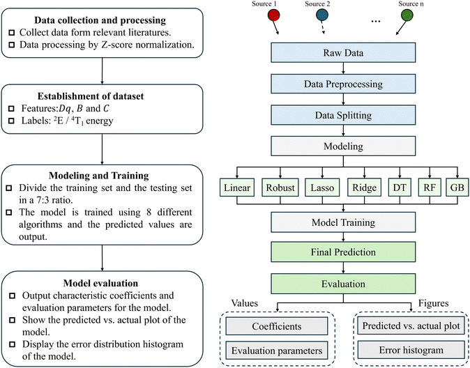
This article predicts the 2E and 4T1 energy levels of Cr3+ and Fe3+ doped phosphor materials using machine learning methods, with the experimental steps detailed in Fig. 3. It is particularly emphasized that appropriate division of the dataset is crucial for assessing the model's performance on unknown data, which directly relates to the model's generalization ability. This study employed a 7![[thin space (1/6-em)]](https://www.rsc.org/images/entities/char_2009.gif) :
:![[thin space (1/6-em)]](https://www.rsc.org/images/entities/char_2009.gif) 3 ratio to split the dataset into training and testing sets. Specifically, the training set comprises the majority of the data (70%), where the model learns to recognize or predict patterns, thus adjusting the model parameters. For the Cr3+-doped phosphor dataset, 23 samples were allocated to the training set, and for the Fe3+-doped dataset, 21 samples were designated for training. The remaining 30% of the data makes up the testing set, and is used to evaluate the model's performance on unknown data. The testing set contains 11 samples for the Cr3+-doped dataset and 9 samples for the Fe3+-doped dataset.
3 ratio to split the dataset into training and testing sets. Specifically, the training set comprises the majority of the data (70%), where the model learns to recognize or predict patterns, thus adjusting the model parameters. For the Cr3+-doped phosphor dataset, 23 samples were allocated to the training set, and for the Fe3+-doped dataset, 21 samples were designated for training. The remaining 30% of the data makes up the testing set, and is used to evaluate the model's performance on unknown data. The testing set contains 11 samples for the Cr3+-doped dataset and 9 samples for the Fe3+-doped dataset.
 | ||
| Fig. 3 Flow chart and step analysis of machine learning to predict the metastable energy level model of phosphor materials. | ||
3. Results and discussion
Building on the methodologies described, this paper employs machine learning regression algorithms to predict the 2E and 4T1 energy levels of Cr3+ and Fe3+ doped in phosphor materials. For this purpose, the study developed an ensemble model featuring eight different machine learning regression algorithms, allowing for the selection of the most suitable algorithm based on specific requirements. After prediction, the model outputs a predicted vs. actual plot based on the training and testing sets, and integrates an error distribution histogram into this plot. These visualizations facilitate a comprehensive assessment of model performance and aid in diagnosing potential issues. Additionally, the model provides the calculated parameter coefficients and performance evaluation metrics for the selected regression algorithms. Overall, the model developed in this paper effectively evaluates the performance of each algorithm in the scope of this study.3.1 Comparative analysis
In this article, the established model is used to predict the doping of Cr3+ and Fe3+, specifically focusing on the prediction of the 2E energy level for Cr3+ doping. The predicted vs. actual plot for the 2E energy level output is shown in Fig. 4. | ||
| Fig. 4 Different regression models based on training sets and test sets predicted vs. actual values of Cr3+ doped 2E energy (cm−1) scatter plots. | ||
Fig. 4 presents a comparative analysis of the actual and predicted values of the 2E energy level (cm−1) for Cr3+ doping, based on the training and testing datasets. The analysis utilizes eight different regression models: linear regression, robust regression, lasso regression, ridge regression, elastic net, decision tree, random forest, and gradient boosting. Additionally, a small error distribution histogram is embedded in Fig. 4. In the main chart, the x-axis represents the actual values of the 2E energy, while the y-axis displays the predicted values generated by the models, with different colors distinguishing the data points from the training set (purple squares) and the testing set (red squares). The black diagonal dashed line in the chart indicates the ideal prediction outcome, where the model's predicted values equal the actual values. The inset on the right side of the figure shows the distribution of relative errors for the testing set, with the horizontal axis representing the percentage of relative error and the vertical axis showing the probability density of errors.
Table 4 provides the coefficients of the parameters Dq, B, and C for Cr3+ doping, calculated using the training dataset across eight regression models. The larger the magnitude of the coefficients, the more significant the variable's impact on the predictions. Notably, the decision tree, random forest, and gradient boosting models do not provide traditional coefficients but offer feature importance scores instead. These scores reflect the contribution of each feature to the model's predictive outcomes.
| Parameter | Linear regression | Robust regression | Lasso regression | Ridge regression | Elastic net | Decision tree | Random forest | Gradient boosting |
|---|---|---|---|---|---|---|---|---|
| D q | −52.48 | 89.52 | −55.89 | −25.21 | −2.72 | 0.40 | 0.32 | 0.26 |
| B | 152.13 | 299.86 | 242.71 | 232.86 | 143.18 | 0.15 | 0.35 | 0.35 |
| C | 269.00 | 322.10 | 203.99 | 192.39 | 156.62 | 0.45 | 0.33 | 0.39 |
Additionally, the model provides common metrics for evaluating regression model performance, including mean absolute error (MAE), mean squared error (MSE), root mean squared error (RMSE), and the coefficient of determination (R2). The specific formulas for these coefficients are shown in Table 5. These metrics are essential for assessing the accuracy and reliability of the regression models used in the study.
where yi represents the actual values, and ŷi are the predicted values. For the first three metrics—MAE, MSE, and RMSE—the lower the values, the better the performance of the model. Conversely, for R2, values closer to 1 indicate a stronger explanatory power of the model. The specific values of these performance metrics for the Cr3+ doped model are presented in Table 6. These metrics quantify the model's performance in terms of accuracy and reliability.
| Model | MAE | MSE | RMSE | R 2 |
|---|---|---|---|---|
| Linear regressing | 306.93 | 109![[thin space (1/6-em)]](https://www.rsc.org/images/entities/char_2009.gif) 164.08 164.08 |
330.40 | 0.57 |
| Robust regression | 149.80 | 33![[thin space (1/6-em)]](https://www.rsc.org/images/entities/char_2009.gif) 603.59 603.59 |
183.31 | 0.82 |
| Lasso regression | 298.28 | 110![[thin space (1/6-em)]](https://www.rsc.org/images/entities/char_2009.gif) 731.08 731.08 |
332.76 | 0.59 |
| Ridge regression | 286.90 | 114![[thin space (1/6-em)]](https://www.rsc.org/images/entities/char_2009.gif) 974.23 974.23 |
339.08 | 0.58 |
| Elastic net | 359.05 | 164![[thin space (1/6-em)]](https://www.rsc.org/images/entities/char_2009.gif) 626.27 626.27 |
405.74 | 0.41 |
| Decision tree | 207.25 | 93![[thin space (1/6-em)]](https://www.rsc.org/images/entities/char_2009.gif) 820.50 820.50 |
306.30 | 0.56 |
| Random forest | 295.30 | 115![[thin space (1/6-em)]](https://www.rsc.org/images/entities/char_2009.gif) 887.01 887.01 |
340.42 | 0.64 |
| Gradient boosting | 244.91 | 104![[thin space (1/6-em)]](https://www.rsc.org/images/entities/char_2009.gif) 747.35 747.35 |
323.65 | 0.57 |
Table 6 clearly shows that robust regression significantly outperforms other algorithms in predicting Cr3+ doping. It achieves the lowest mean absolute error (MAE) at 149.80, indicating small prediction errors and excellent predictive performance. Its R2 value of 0.82 demonstrates that the model captures the primary trends in the data, reflecting its robustness and good fit. This may be due to robust regression's use of methods that minimize the impact of outliers, allowing the model to perform better with noisy data. In this task, linear regression has an MAE of 306.91 and an R2 of 0.57, indicating a moderate fit to the data. Linear regression assumes a linear relationship between features and the target variable, which can be limiting if the data contain non-linear relationships or outliers. Lasso regression does not stand out, with an MAE of 298.28 and an R2 value of 0.59. Lasso employs L1 regularization for feature selection, but the results suggest that this has limited effectiveness in this task. Ridge regression performs similarly to linear and lasso regression, with an MAE of 286.90 and an R2 of 0.58. It uses L2 regularization to reduce multicollinearity issues, smoothing weights to prevent overfitting, yet the performance improvement in this task is limited. Elastic net exhibits the largest error with an MAE of 359.05 and the lowest R2 at 0.41, indicating that the model struggles to handle complex relationships in the data effectively. Elastic net combines L1 and L2 regularization but performs less effectively than lasso or ridge regression separately, possibly due to an inadequate balance between the L1 and L2 regularization terms. The decision tree has a relatively low MAE of 207.25 and an R2 value of 0.56. Decision trees capture nonlinear relationships in data through a tree-like structure and, while they can achieve low errors, they are prone to overfitting. Random forest, which integrates multiple decision trees to reduce overfitting and enhance model robustness, achieves an MAE of 295.30 and a relatively higher R2 of 0.64, reflecting a better explanatory capability of the model. Gradient boosting, which incrementally adds weak learners to minimize errors, has an MAE of 244.91 and an R2 value of 0.57. This suggests that while the model reduces errors, its explanatory power could be further improved.
Similarly, Fig. 5 displays a comparative analysis between the actual and predicted values of the 4T1 energy level (cm−1) for Fe3+ doping, based on the training and testing datasets. This visualization aids in assessing the accuracy and performance of the predictive models used in this context.
 | ||
| Fig. 5 Different regression models based on training sets and test sets predicted vs. actual values of Fe3+ doped 4T1 energy (cm−1) scatter plots. | ||
Table 7 provides the coefficients (Dq, B, and C) calculated based on the training dataset for Fe3+ doping using eight different regression models. Table 8 displays the specific values of the performance evaluation metrics for models applied to Fe3+ doping. These tables offer critical insights into the effectiveness and predictive capabilities of the regression models under different doping conditions.
| Parameter | Linear regression | Robust regression | Lasso regression | Ridge regression | Elastic net | Decision tree | Random forest | Gradient boosting |
|---|---|---|---|---|---|---|---|---|
| Dq | −1845.00 | −2710.05 | −2174.64 | −1685.22 | −904.81 | 0.79 | 0.83 | 0.66 |
| B | 1015.04 | 1171.94 | 1492.22 | 845.08 | 333.58 | 0.04 | 0.05 | 0.03 |
| C | 1573.03 | 1748.95 | 1871.93 | 1273.70 | 462.92 | 0.17 | 0.12 | 0.31 |
| Model | MAE | MSE | RMSE | R 2 |
|---|---|---|---|---|
| Linear regression | 1044.85 | 1839![[thin space (1/6-em)]](https://www.rsc.org/images/entities/char_2009.gif) 904.42 904.42 |
1356.43 | 0.67 |
| Robust regression | 771.99 | 1220![[thin space (1/6-em)]](https://www.rsc.org/images/entities/char_2009.gif) 312.12 312.12 |
1104.68 | 0.81 |
| Lasso regression | 906.70 | 1552![[thin space (1/6-em)]](https://www.rsc.org/images/entities/char_2009.gif) 565.42 565.42 |
1246.02 | 0.58 |
| Ridge regression | 994.45 | 1973![[thin space (1/6-em)]](https://www.rsc.org/images/entities/char_2009.gif) 838.90 838.90 |
1404.93 | 0.69 |
| Elastic net | 1760.25 | 4318![[thin space (1/6-em)]](https://www.rsc.org/images/entities/char_2009.gif) 256.00 256.00 |
2078.04 | 0.39 |
| Decision tree | 1083.89 | 1815![[thin space (1/6-em)]](https://www.rsc.org/images/entities/char_2009.gif) 872.78 872.78 |
1347.54 | 0.71 |
| Random forest | 737.16 | 1100![[thin space (1/6-em)]](https://www.rsc.org/images/entities/char_2009.gif) 334.25 334.25 |
1048.97 | 0.72 |
| Gradient boosting | 1098.88 | 1696![[thin space (1/6-em)]](https://www.rsc.org/images/entities/char_2009.gif) 438.16 438.16 |
1302.47 | 0.82 |
The data from Table 8 indicate that compared to the Cr3+ doping scenario, the MAE significantly increased across all algorithms for Fe3+ doping, but some models were better at capturing the overall trends and complex relationships in the data, thus improving the R2 values. This reflects that model selection and optimization strategies need to vary based on different data characteristics.
For linear regression in the Fe3+ doping context, the MAE was notably high at 1044.85, suggesting a large error, yet the R2 value improved to 0.67, indicating a better fit than in some other models. Robust regression continued to perform well with a lower MAE of 771.99 and an excellent R2 of 0.81, demonstrating its robustness. Lasso regression still showed poor performance with an MAE of 906.7 and an R2 value of 0.58. Similar to linear regression, ridge regression saw some improvement in R2 to 0.69, but still had a high MAE of 994.45. Elastic net remained the least effective, with the highest MAE of 1760.25 and the lowest R2 value of 0.39, indicating poor predictive performance and failure to capture data trends. Both decision tree and random forest showed increases in R2 to 0.71 and 0.72, respectively. However, the decision tree had a higher MAE of 1083.89 compared to the random forest, which had an MAE of 737.16, demonstrating good predictive capabilities. Gradient boosting achieved the highest R2 value at 0.82, suggesting that it was better at explaining overall data trends, though its MAE of 1098.88 indicates that accuracy still needs to be improved.
3.2. Prediction of several new Cr3+ and Fe3+ doped phosphor materials
To validate the efficacy of the proposed models, additional new phosphor materials doped with Cr3+ and Fe3+ were selected for further testing, using trained models to predict their 2E or 4T1 energy levels. The newly selected Cr3+-doped phosphor materials include [C(NH2)3]M(HCOO)3(M = Mg2+),97 YScO3,98 and Zn2TiO4.99 The new Fe3+-doped phosphor materials are Sr9Ga(PO4)7100 and CaCdPH.101 The predictions for the 2E energy levels of these Cr3+-doped materials and the 4T1 energy levels of the Fe3+-doped materials were performed using the trained models. The comparison of the model predictions with the actual values of the 2E or 4T1 energy levels is shown in Table 9. This step provides a crucial test of the model's predictive capabilities on new and diverse materials.| Material | Actual value | Linear regression | Robust regression | Lasso regression | Ridge regression | Elastic net | Decision tree | Random forest | Gradient boosting | |
|---|---|---|---|---|---|---|---|---|---|---|
| Cr3+ (2E) | [C(NH2)3] M(HCOO)3 | 14![[thin space (1/6-em)]](https://www.rsc.org/images/entities/char_2009.gif) 552 552 |
14![[thin space (1/6-em)]](https://www.rsc.org/images/entities/char_2009.gif) 492.51 492.51 |
14![[thin space (1/6-em)]](https://www.rsc.org/images/entities/char_2009.gif) 560.59 560.59 |
14![[thin space (1/6-em)]](https://www.rsc.org/images/entities/char_2009.gif) 404.86 404.86 |
14![[thin space (1/6-em)]](https://www.rsc.org/images/entities/char_2009.gif) 442.04 442.04 |
14![[thin space (1/6-em)]](https://www.rsc.org/images/entities/char_2009.gif) 472.21 472.21 |
14![[thin space (1/6-em)]](https://www.rsc.org/images/entities/char_2009.gif) 600 600 |
14![[thin space (1/6-em)]](https://www.rsc.org/images/entities/char_2009.gif) 524.13 524.13 |
14![[thin space (1/6-em)]](https://www.rsc.org/images/entities/char_2009.gif) 604.73 604.73 |
| YScO3 | 14![[thin space (1/6-em)]](https://www.rsc.org/images/entities/char_2009.gif) 124 124 |
14![[thin space (1/6-em)]](https://www.rsc.org/images/entities/char_2009.gif) 143.37 143.37 |
14![[thin space (1/6-em)]](https://www.rsc.org/images/entities/char_2009.gif) 081.43 081.43 |
14![[thin space (1/6-em)]](https://www.rsc.org/images/entities/char_2009.gif) 250.16 250.16 |
14![[thin space (1/6-em)]](https://www.rsc.org/images/entities/char_2009.gif) 224.95 224.95 |
14![[thin space (1/6-em)]](https://www.rsc.org/images/entities/char_2009.gif) 259.16 259.16 |
13![[thin space (1/6-em)]](https://www.rsc.org/images/entities/char_2009.gif) 947 947 |
14![[thin space (1/6-em)]](https://www.rsc.org/images/entities/char_2009.gif) 132.19 132.19 |
14![[thin space (1/6-em)]](https://www.rsc.org/images/entities/char_2009.gif) 367.70 367.70 |
|
| Zn2TiO4 | 13![[thin space (1/6-em)]](https://www.rsc.org/images/entities/char_2009.gif) 966 966 |
13![[thin space (1/6-em)]](https://www.rsc.org/images/entities/char_2009.gif) 911.97 911.97 |
13![[thin space (1/6-em)]](https://www.rsc.org/images/entities/char_2009.gif) 993.46 993.46 |
14![[thin space (1/6-em)]](https://www.rsc.org/images/entities/char_2009.gif) 210.27 210.27 |
14![[thin space (1/6-em)]](https://www.rsc.org/images/entities/char_2009.gif) 165.20 165.20 |
14![[thin space (1/6-em)]](https://www.rsc.org/images/entities/char_2009.gif) 185.79 185.79 |
13![[thin space (1/6-em)]](https://www.rsc.org/images/entities/char_2009.gif) 923 923 |
14![[thin space (1/6-em)]](https://www.rsc.org/images/entities/char_2009.gif) 201.56 201.56 |
14![[thin space (1/6-em)]](https://www.rsc.org/images/entities/char_2009.gif) 342.69 342.69 |
|
| Fe3+ (4T1) | Sr9Ga(PO4)7 | 13![[thin space (1/6-em)]](https://www.rsc.org/images/entities/char_2009.gif) 333 333 |
13![[thin space (1/6-em)]](https://www.rsc.org/images/entities/char_2009.gif) 675.77 675.77 |
13![[thin space (1/6-em)]](https://www.rsc.org/images/entities/char_2009.gif) 332.26 332.26 |
13![[thin space (1/6-em)]](https://www.rsc.org/images/entities/char_2009.gif) 769.65 769.65 |
13![[thin space (1/6-em)]](https://www.rsc.org/images/entities/char_2009.gif) 918.46 918.46 |
14![[thin space (1/6-em)]](https://www.rsc.org/images/entities/char_2009.gif) 305.07 305.07 |
14![[thin space (1/6-em)]](https://www.rsc.org/images/entities/char_2009.gif) 385 385 |
15![[thin space (1/6-em)]](https://www.rsc.org/images/entities/char_2009.gif) 497.43 497.43 |
14![[thin space (1/6-em)]](https://www.rsc.org/images/entities/char_2009.gif) 634.82 634.82 |
| CaCdPH | 17![[thin space (1/6-em)]](https://www.rsc.org/images/entities/char_2009.gif) 726 726 |
16![[thin space (1/6-em)]](https://www.rsc.org/images/entities/char_2009.gif) 263.50 263.50 |
17![[thin space (1/6-em)]](https://www.rsc.org/images/entities/char_2009.gif) 242.89 242.89 |
15![[thin space (1/6-em)]](https://www.rsc.org/images/entities/char_2009.gif) 797.87 797.87 |
15![[thin space (1/6-em)]](https://www.rsc.org/images/entities/char_2009.gif) 867.27 867.27 |
15![[thin space (1/6-em)]](https://www.rsc.org/images/entities/char_2009.gif) 185.38 185.38 |
16![[thin space (1/6-em)]](https://www.rsc.org/images/entities/char_2009.gif) 389 389 |
16![[thin space (1/6-em)]](https://www.rsc.org/images/entities/char_2009.gif) 368.63 368.63 |
17![[thin space (1/6-em)]](https://www.rsc.org/images/entities/char_2009.gif) 546.85 546.85 |
Table 9 indicates that the models proposed in the article are effective in predicting the 2E or 4T1 energy levels of phosphor materials doped with Cr3+ and Fe3+. The prediction results from various algorithms are closely aligned with the actual energy levels.
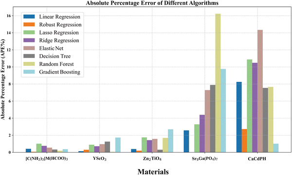
To provide a clearer comparison of the predictive performance of each algorithm, a bar chart depicting the percentage of absolute error in the predictions for these materials has been constructed, as shown in Fig. 6. This visualization helps to assess the precision of each model in practical applications, highlighting their strengths and potential areas for improvement.
 | ||
| Fig. 6 Histogram of the percentage error of different algorithms predicting the results of the materials. | ||
From Fig. 6, it is evident that, overall, the model's prediction errors for the energy levels under Fe3+ doping are higher than those for Cr3+ doping, which is consistent with the characteristics of the model predictions on known data discussed in Section 3.1. For [C(NH2)3]M(HCOO)3, robust regression achieved the best results, with an error of only 0.059%. For YScO3, random forest produced the best results, with an error of only 0.058%. For Zn2TiO4, robust regression was the most accurate, with an error of 0.1966%. For Sr9Ga(PO4)7, robust regression significantly outperformed other algorithms, with an error of only 0.0056%. For CaCdPH, gradient boosting performed the best, with an error of 1.01%. These results demonstrate that the machine learning models used in this study can predict the energy levels of ions in phosphor materials with a minimum error as low as 0.0056%, achieving highly precise predictions. Additionally, robust regression consistently showed high performance across all five materials. Linear regression, lasso regression, and ridge regression had similar levels of accuracy, while random forest and gradient boosting showed more variability and fluctuation in their prediction accuracy. Overall, the machine learning prediction models developed in this study achieved excellent results in predicting the 2E or 4T1 energy levels in phosphor materials doped with Cr3+ and Fe3+. Moreover, it can be found from the existing research results in this paper that robust regression performs outstandingly. Robust regression reduces the influence of outliers by adopting robust loss functions such as Huber's loss to improve the stability of the model, which makes the model less susceptible to the influence of a small portion of atypical sample points in the data, and thus has excellent prediction performance.
3.3. Challenge and limitations
Despite the considerable potential demonstrated by machine learning technologies in the research of doped phosphor materials, their application is still constrained by several challenges and limitations. These challenges primarily include issues concerning the availability and quality of data, the complexity and interpretability of models, difficulties in feature engineering, and the need for interdisciplinary knowledge.Firstly, regarding data availability and quality, the effectiveness of data-driven machine learning models heavily depends on having access to large, diverse, and accurate datasets. Thus, securing high-quality and diverse training datasets present a significant challenge. In the field of phosphor materials research, the complexity of material compositions, physical processes, and performance measurements makes it particularly difficult to obtain comprehensive and high-quality datasets. Secondly, the complexity and interpretability of models are major concerns. The doping processes in phosphor materials involve complex chemical and physical changes, often requiring sophisticated models for accurate simulation. Additionally, many efficient models, such as deep learning models, although precise in their predictions, lack sufficient interpretability. This makes understanding the underlying mechanisms behind model predictions challenging. Furthermore, feature engineering is crucial for enhancing model prediction accuracy. Effective feature extraction is vital for improving model accuracy, yet identifying which features best describe the intrinsic properties or external behaviours of materials remains a significant challenge in practical applications. Lastly, the demand for interdisciplinary knowledge cannot be overlooked. Research in this field not only requires deep knowledge of chemistry, physics, and materials science but also support from computer science and a thorough understanding of the chemical and physical characteristics of doped phosphor materials, which is itself a challenge. Facing these challenges, enhancing the quality of data collection, developing efficient yet more transparent and interpretable machine learning models, conducting in-depth feature engineering, and deeply integrating knowledge across these disciplines are key pathways to improving research outcomes.
5. Conclusions
This paper utilizes machine learning methods to predict the 2E and 4T1 energy levels of Cr3+ and Fe3+ ions doped in crystals, which are closely related to the luminescence properties of the phosphor materials doped with these ions. According to the task requirements, eight machine learning regression algorithms (linear regression, robust regression, ridge regression, elastic net, decision tree, random forest, and gradient boosting) were selected to build models for prediction. A comprehensive evaluation of each algorithm's performance on the dataset was conducted, and the results indicated that robust regression achieved excellent outcomes in predicting both Cr3+ and Fe3+. Subsequently, some new phosphor materials doped with Cr3+ and Fe3+, such as [C(NH2)3]M(HCOO)3:Cr3+, YScO3:Cr3+, Zn2TiO4:Cr3+, Sr9Ga(PO4)7:Fe3+, and CaCdPH:Fe3+ were also selected. The trained models were used to predict their 2E and 4T1 energy levels. The predictions show that for these materials, the best-performing algorithm in the ensemble model achieved a prediction error of only 0.0056%, and the optimal prediction errors for all materials were controlled to be approximately 1%, fully reflecting the effectiveness of the machine learning prediction models established in this study. This study provides a novel approach to predicting and optimizing the energy level structures and luminescence properties of phosphor materials. In the future, we will explore more possibilities of machine learning methods in phosphor material characterization, and we will try to use more complex and high-performance models or algorithms to accelerate the discovery of new materials and deeper study of complex properties in materials.Data availability
The data that support the findings of this study are available from the corresponding author upon reasonable request.Conflicts of interest
The authors declare that they have no conflicts of interest.Acknowledgements
This work was supported by the Natural Science Foundation of Jiangsu Higher Education Institutions of China (grant number 23KJA510005) and the Open Foundation of State Key Laboratory of Luminescent Materials and Devices, South China University of Technology (grant number 2023-skllmd-15).References
- Y. Wang, Q. Bu, D. Li, S. Yang, L. Li, G. Xiang, S. Jiang, Y. Chang, C. Jing, X. Zhou, L.-I. Bulyk and A. Suchocki, UV- and X-ray-activated broadband NIR garnet-type Ca3Ga2Sn3O12:Fe3+ phosphors with efficient persistent luminescence, Dyes Pigm., 2024, 225, 112091 Search PubMed.
- Q. Ma, T. Wang, W. Gao, B. Liu, H. Zhang, Z. Cui, H. Guo, L. Xiu, S. Wang, Z. Li, L. Guo, S. Yu, X. Yu, X. Xu and J. Qiu, Broadband, Enhanced, and Antithermally Quenched Near-Infrared Phosphors via a Cosubstitution Approach, Inorg. Chem., 2021, 60, 11616–11625 Search PubMed.
- S. Huang, Y. Yan, M. Shang, Y. Wang, Y. Sun, P. Dang and J. Lin, Super Broadband Near-Infrared Solid Solution Phosphors with Adjustable Peak Wavelengths from 1165 to 875 nm for NIR Spectroscopy Applications, Adv. Opt. Mater., 2023, 11, 2202291 Search PubMed.
- S. Wang, R. Pang, T. Tan, H. Wu, Q. Wang, C. Li, S. Zhang, T. Tan, H. You and H. Zhang, Achieving High Quantum Efficiency Broadband NIR Mg4Ta2O9:Cr3+ Phosphor Through Lithium-Ion Compensation, Adv. Mater., 2023, 35, 2300124 Search PubMed.
- J. Zhong, Y. Zhuo, F. Du, H. Zhang, W. Zhao, S. You and J. Brgoch, Efficient Broadband Near-Infrared Emission in the GaTaO4:Cr3+ Phosphor, Adv. Opt. Mater., 2022, 10, 2101800 Search PubMed.
- J. Su, R. Pang, T. Tan, S. Wang, X. Chen, S. Zhang and H. Zhang, A Novel Near-Infrared Emitting Sr2 LuSbO6:Fe3+ Phosphor with Persistent Luminescence Performance, Adv. Opt. Mater., 2024, 2303187 Search PubMed.
- E. L. Que, D. W. Domaille and C. J. Chang, Metals in Neurobiology: Probing Their Chemistry and Biology with Molecular Imaging, Chem. Rev., 2008, 108, 1517–1549 Search PubMed.
- F. Chi, W. Dai, S. Liu, L. Qiu, X. Wei, Y. Chen and M. Yin, Luminescence properties of Cr3+-doped Al6Ge2O13 broadband near-infrared phosphor, Opt. Mater., 2022, 126, 112218 Search PubMed.
- S. R. Kalidindi, D. B. Brough, S. Li, A. Cecen, A. L. Blekh, F. Y. P. Congo and C. Campbell, Role of materials data science and informatics in accelerated materials innovation, MRS Bull., 2016, 41, 596–602 Search PubMed.
- P. Friederich, F. Häse, J. Proppe and A. Aspuru-Guzik, Machine-learned potentials for next-generation matter simulations, Nat. Mater., 2021, 20, 750–761 Search PubMed.
- S. Behara, S. Rath and T. Thomas, Machine learning (ML) as a tool for phosphor design: A perspective, Mater. Lett., 2022, 308, 131061 Search PubMed.
- S. Li, Y. Xia, M. Amachraa, N. T. Hung, Z. Wang, S. P. Ong and R.-J. Xie, Data-Driven Discovery of Full-Visible-Spectrum Phosphor, Chem. Mater., 2019, 31, 6286–6294 Search PubMed.
- L. Jiang, X. Jiang, Y. Zhang, C. Wang, P. Liu, G. Lv and Y. Su, Multiobjective Machine Learning-Assisted Discovery of a Novel Cyan–Green Garnet: Ce Phosphors with Excellent Thermal Stability, ACS Appl. Mater. Interfaces, 2022, 14, 15426–15436 Search PubMed.
- S. Jang, G. S. Na, Y. Choi and H. Chang, Optical property dataset of inorganic phosphor, Sci. Rep., 2024, 14, 7639 Search PubMed.
- T.-G. Kim, D. Jurakuziev, M. S. Akhtar and O.-B. Yang, Machine learning investigation to predict the relationship between photoluminescence and crystalline properties of blue phosphor Ba0.9−xSrxMgAl10O17:Eu2+, J. Sci. Adv. Mater. Devices, 2023, 8, 100550 Search PubMed.
- E. Alpaydin, Introduction to Machine Learning, MIT Press, 4th edn, 2020 Search PubMed.
- A. Paszke, S. Gross, F. Massa, A. Lerer, J. Bradbury, G. Chanan, T. Killeen, Z. Lin, N. Gimelshein, L. Antiga, A. Desmaison, A. Köpf, E. Yang, Z. DeVito, M. Raison, A. Tejani, S. Chilamkurthy, B. Steiner, L. Fang, J. Bai and S. Chintala, PyTorch: an imperative style, high-performance deep learning library, Proceedings of the 33rd International Conference on Neural Information Processing Systems, No. 721, (Curran Associates Inc., 2019), 8026–8037.
- M. Abadi, A. Agarwal, P. Barham, E. Brevdo, Z. Chen, C. Citro, G. S. Corrado, A. Davis, J. Dean, M. Devin, S. Ghemawat, I. Goodfellow, A. Harp, G. Irving, M. Isard, Y. Jia, R. Jozefowicz, L. Kaiser, M. Kudlur, J. Levenberg, D. Mane, R. Monga, S. Moore, D. Murray, C. Olah, M. Schuster, J. Shlens, B. Steiner, I. Sutskever, K. Talwar, P. Tucker, V. Vanhoucke, V. Vasudevan, F. Viegas, O. Vinyals, P. Warden, M. Wattenberg, M. Wicke, Y. Yu and X. Zheng, TensorFlow: Large-Scale Machine Learning on Heterogeneous Distributed Systems, ( 2016).
- A. Gulli and S. Pal, Deep Learning with Keras, Packt Publishing Ltd, 2017 Search PubMed.
- F. Pedregosa, G. Varoquaux, A. Gramfort, V. Michel, B. Thirion, O. Grisel, M. Blondel, P. Prettenhofer, R. Weiss, V. Dubourg, J. Vanderplas, A. Passos and D. Cournapeau, Scikit-learn: Machine Learning in Python, Mach. Learn. PYTHON (n.d.).
- M. Hall, E. Frank, G. Holmes, B. Pfahringer, P. Reutemann and I. H. Witten, The WEKA data mining software: an update, ACM SIGKDD Explor. Newsl, 11, 10–18 ( 2009).
- J. Barnes, Microsoft Azure Essentials Azure Machine Learning, Microsoft Press, 2015 Search PubMed.
- E. Bisong, An Overview of Google Cloud Platform Services, Building Machine Learning and Deep Learning Models on Google Cloud Platform, Apress, Berkeley, CA, 2019, pp. 7–10 Search PubMed.
- M. Hofmann and R. Klinkenberg, RapidMiner: Data Mining Use Cases and Business Analytics Applications, CRC Press, 2016 Search PubMed.
- E. LeDell and S. Poirier, H2O AutoML: Scalable Automatic Machine Learning, (n.d.).
- C. S. Bojer and J. P. Meldgaard, Kaggle forecasting competitions: An overlooked learning opportunity, Int. J. Forecast., 2021, 37, 587–603 Search PubMed.
- R. V. Dukkipati, Matlab: An Introduction With Applications, New Age International, 2008 Search PubMed.
- S. Adachi, Luminescence Spectroscopy of 3d 3 (Mn4+, Cr3+) Ions in Multiple Octahedral-Site Phosphors, ECS J. Solid State Sci. Technol., 2022, 11, 046002 Search PubMed.
- D. Wu, L. Liu, H. Liang, H. Duan, W. Nie, J. Wang, J. Peng and X. Ye, LiBAlF6:Cr3+ (B = Ca, Sr) fluoride phosphors with ultra-broad near-infrared emission for NIR pc-LEDs, Ceram. Int., 2022, 48, 387–396 Search PubMed.
- K. Elzbieciak, A. Bednarkiewicz and L. Marciniak, Temperature sensitivity modulation through crystal field engineering in Ga3+ co-doped Gd3Al5-xGaxO12:Cr3 +, Nd3+ nanothermometers, Sens. Actuators, B, 2018, 269, 96–102 Search PubMed.
- S. V. J. Lakshman, B. C. Venkata Reddy and J. L. Rao, Electronic Absorption Spectrum of Fe 3+ Doped in Ammonium Perchlorate Single Crystal, Spectrosc. Lett., 1976, 9, 341–352 Search PubMed.
- F. Zhao, Z. Song and Q. Liu, Advances in Chromium-Activated Phosphors for Near-Infrared Light Sources, Laser Photonics Rev., 2022, 16, 2200380 Search PubMed.
- S. Adachi, Spectroscopy of Cr3+ activator: Tanabe−Sugano diagram and Racah parameter analysis, J. Lumin., 2021, 232, 117844 Search PubMed.
- M. A. F. M. Da Silva, S. S. Pedro and L. P. Sosman, Fe3+ concentration dependence of photoacoustic absorption spectroscopy on ZnGa2O4 ceramic powders, Spectrochim. Acta, Part A, 2008, 69, 338–342 Search PubMed.
- D. C. Montgomery, E. A. Peck and G. G. Vining, Introduction to Linear Regression Analysis, John Wiley & Sons, 2021 Search PubMed.
- P. J. Rousseeuw and A. M. Leroy, Robust Regression and Outlier Detection, John Wiley & Sons, 2005 Search PubMed.
- J. Ranstam and J. A. Cook, LASSO regression, Br. J. Surg., 2018, 105, 1348 Search PubMed.
- Ridge Regression: Biased Estimation for Nonorthogonal Problems: Technometrics: Vol 12, No 1 – Get Access, Technometrics (n.d.).
- H. Zou and T. Hastie, Regularization and Variable Selection Via the Elastic Net, J. R. Stat. Soc. Ser. B Stat. Methodol., 2005, 67, 301–320 Search PubMed.
- Y. Song and Y. Lu, Decision tree methods: applications for classification and prediction, Shanghai Arch. Psychiatry, 2015, 27, 130 Search PubMed.
- V. Svetnik, A. Liaw, C. Tong, J. C. Culberson, R. P. Sheridan and B. P. Feuston, Random Forest: A Classification and Regression Tool for Compound Classification and QSAR Modeling, J. Chem. Inf. Comput. Sci., 2003, 43, 1947–1958 Search PubMed.
- Y. Zhang and A. Haghani, A gradient boosting method to improve travel time prediction, Transp. Res. Part C Emerg. Technol., 2015, 58, 308–324 Search PubMed.
- G. Wang, X. Han, M. Song, Z. Lin, G. Wang and X. Long, Growth and spectral properties of Cr3+:KAl(MoO4)2 crystal, Mater. Lett., 2007, 61, 3886–3889 Search PubMed.
- V. Singh, R. P. S. Chakradhar, J. L. Rao and H.-Y. Kwak, Characterization, EPR and photoluminescence studies of LiAl5O8:Cr phosphors, Solid State Sci., 2009, 11, 870–874 Search PubMed.
- B. Bondzior, N. Miniajluk and P. J. Dereń, Pair luminescence in Cr3+ -doped Ba2Mg(BO3)2, Opt. Mater., 2018, 79, 269–272 Search PubMed.
- I. Nikolov, Optical properties of Cr3 +:NaAl(WO4)2 crystals, a new candidate for broadband laser applications, Opt. Mater., 2004, 25, 53–58 Search PubMed.
- G. Wang, H. G. Gallagher, T. P. J. Han and B. Henderson, Crystal growth and optical characterisation of Cr3 + -doped YAl3(BO3)4, J. Cryst. Growth, 1995, 153, 169–174 Search PubMed.
- K. Hermanowicz, M. Mączka, M. Wołcyrz, P. E. Tomaszewski, M. Paściak and J. Hanuza, Crystal structure, vibrational properties and luminescence of NaMg3Al(MoO4)5 crystal doped with Cr3+ ions, J. Solid State Chem., 2006, 179, 685–695 Search PubMed.
- G. A. Torchia, O. M. Matos, P. Vaveliuk and J. O. Tocho, Influence of the electron-lattice coupling for Cr3+ ions in Nb5+ site into congruent co-doped LiNbO3: Cr3 +: ZnO crystal, Solid State Commun., 2003, 127, 535–539 Search PubMed.
- C. R. Kesavulu, R. P. S. Chakradhar, R. S. Muralidhara, J. L. Rao and R. V. Anavekar, EPR, optical absorption and photoluminescence properties of Cr3+ ions in lithium borophosphate glasses, J. Alloys Compd., 2010, 496, 75–80 Search PubMed.
- V. Singh, R. P. S. Chakradhar, J. L. Rao and J.-J. Zhu, Studies on red-emitting Cr3+ doped barium aluminate phosphor obtained by combustion process, Mater. Chem. Phys., 2008, 111, 143–148 Search PubMed.
- G. Wang, Z. Lin, L. Zhang, Y. Huang and G. Wang, Spectral characterization and energy levels of Cr3+:Sc2(MoO4)3 crystal, J. Lumin., 2009, 129, 1398–1400 Search PubMed.
- X. Long, Z. Lin, Z. Hu, G. Wang and T. P. J. Han, Optical study of Cr3+-doped LaSc3(BO3)4 crystal, J. Alloys Compd., 2002, 347, 52–55 Search PubMed.
- G. Wang, H. G. Gallagher, T. P. J. Han and B. Henderson, The growth and optical assessment of Cr3+-doped RX(BO3)4 crystals with R = Y, Gd; X = Al, Sc, J. Cryst. Growth, 1996, 163, 272–278 Search PubMed.
- M. Casalboni, V. Ciafardone, G. Giuli, B. Izzi, E. Paris and P. Prosposito, An optical study of silicate glass containing and ions, J. Phys.: Condens.Matter, 1996, 8, 9059–9069 Search PubMed.
- T. Aswani, B. Babu, V. Pushpa Manjari, R. Joyce Stella, G. Thirumala Rao, C. Rama Krishna and R. V. S. S. N. Ravikumar, Synthesis and spectral characterizations of trivalent ions (Cr3 +, Fe3 +) doped CdO nanopowders, Spectrochim. Acta, Part A, 2014, 121, 544–550 Search PubMed.
- F. S. De Vicente, F. A. Santos, B. S. Simões, S. T. Dias and M. Siu Li, EPR, optical absorption and luminescence studies of Cr3 + -doped antimony phosphate glasses, Opt. Mater., 2014, 38, 119–125 Search PubMed.
- R. V. S. S. N. Ravikumar, R. Komatsu, K. Ikeda, A. V. Chandrasekhar, B. J. Reddy, Y. P. Reddy and P. S. Rao, Electron paramagnetic resonance and optical absorption spectra of Cr3+ ions in cadmium phosphate glass, Solid State Commun., 2003, 126, 251–253 Search PubMed.
- G. L. Flower, M. S. Reddy, G. S. Baskaran and N. Veeraiah, The structural influence of chromium ions in lead gallium phosphate glasses by means of spectroscopic studies, Opt. Mater., 2007, 30, 357–363 Search PubMed.
- G. Giridhar, S. Sreehari Sastry and M. Rangacharyulu, Spectroscopic studies on Pb3O4–ZnO–P2O5 glasses doped with transition metal ions, Phys. Rev. B: Condens. Matter Mater. Phys., 2011, 406, 4027–4030 Search PubMed.
- S. Kück, E. Heumann, T. Kärner and A. Maaroos, Continuous-wave room-temperature laser oscillation of Cr3+:MgO, Opt. Lett., 1999, 24, 966 Search PubMed.
- S. Adachi, Photoluminescence Spectroscopy and Crystal-Field Parameters of Cr 3+ Ion in Red and Deep Red-Emitting Phosphors, ECS J. Solid State Sci. Technol., 2019, 8, R164–R168 Search PubMed.
- Y. Hao, S. Wang and K. Zhang, Effect of Y3+ on the photoluminescence of MgAl2O4:Cr3+ nanopowders, Mater. Chem. Phys., 2020, 253, 123323 Search PubMed.
- M. A. Hughes, H. Li, R. J. Curry, T. Suzuki and Y. Ohishi, Energy transfer in Cr and Nd co-doped Si–B–Na–Al–Ca–Zr–O glasses, J. Non-Cryst. Solids, 2020, 530, 119769 Search PubMed.
- D. Wang, W. Hou, N. Li, Y. Xue, Q. Wang, X. Xu, D. Li, H. Zhao and J. Xu, Growth, spectroscopic properties and crystal field analysis of Cr 3+ doped GdScO 3 crystal, Opt. Mater. Express, 2019, 9, 4218 Search PubMed.
- S. Adachi, New Analysis Model for the Determination of Racah and Crystal-Field Splitting Parameters: Verification and Case Studies, ECS J. Solid State Sci. Technol., 2020, 9, 046004 Search PubMed.
- R. R. Kumar, A. T. Ramudu, I. A. Kumari and B. C. V. Reddy, Optical absorption spectrum of Cr 3+ ions in sodium ammonium sulphate dihydrate single crystal, Ferroelectr., Lett. Sect., 1995, 19, 75–81 Search PubMed.
- R. Kripal, H. Govind, S. K. Gupta and M. Arora, EPR and optical absorption study of Cr3+ doped diammonium hexaaqua magnesium sulphate single crystal, Solid State Commun., 2007, 141, 416–421 Search PubMed.
- R. Kripal, H. Govind, S. K. Gupta and M. Arora, EPR and optical absorption study of Cr3+-doped tetramethyl ammonium cadmium chloride single crystals, J. Magn. Magn. Mater., 2006, 307, 257–262 Search PubMed.
- A. N. L. Jara, J. F. Carvalho, A. F. Júnior, L. J. Q. Maia and R. C. Santana, On the optical and magnetic studies of YCrO3 perovskites, Phys. B, 2018, 546, 67–72 Search PubMed.
- G. Dominiak-Dzik, W. Ryba-Romanowski, M. Grinberg, E. Beregi and L. Kovacs, Excited-state relaxation dynamics of Cr3+ in YAl3(BO3)4, J. Phys.: Condens.Matter, 2002, 14, 5229–5237 Search PubMed.
- Z. Wen-Chen, W. Xiao-Xuan, H. Lv and M. Yang, An alternative interpretation of the optical spectra for Fe3+-doped KTaO3 crystals, J. Lumin., 2007, 126, 91–93 Search PubMed.
- L. P. Sosman, A. López, S. S. Pedro and A. R. R. Papa, Photoluminescence of the Mg2Al4Si5O18–Al2O3–MgAl2O4–SiO2 ceramic system containing Fe3+ and Cr3+ as impurity ions, Opt. Mater., 2018, 76, 353–358 Search PubMed.
- I. N. Prakash, B. Babu, Ch Venkata Reddy, P. Narayana Murty, Y. P. Reddy, P. Sambasiva Rao and R. V. S. S. N. Ravikumar, Spectroscopic studies on Fe3+ and Mn2+ doped SrB4O7 glasses, Phys. B, 2011, 406, 3295–3298 Search PubMed.
- S. Muntaz Begum, M. C. Rao, Y. Aparna, P. S. Rao and R. V. S. S. N. Ravikumar, Spectroscopic investigations of Fe3+ doped poly vinyl alcohol (PVA) capped ZnSe nanoparticles, Spectrochim. Acta, Part A, 2012, 98, 100–104 Search PubMed.
- T. Aswani, B. Babu, V. Pushpa Manjari, R. Joyce Stella, G. Thirumala Rao, C. Rama Krishna and R. V. S. S. N. Ravikumar, Synthesis and spectral characterizations of trivalent ions (Cr3 +, Fe3 +) doped CdO nanopowders, Spectrochim. Acta, Part A, 2014, 121, 544–550 Search PubMed.
- R. Kripal and A. K. Yadav, EPR and Optical Absorption Study of Fe3 + -Doped Mono Hydrated Dipotassium Stannic Chloride, Appl. Magn. Reson., 2015, 46, 323–335 Search PubMed.
- Y. Wang, Q. Bu, D. Li, S. Yang, L. Li, G. Xiang, S. Jiang, Y. Chang, C. Jing, X. Zhou, L.-I. Bulyk and A. Suchocki, UV- and X-ray-activated broadband NIR garnet-type Ca3Ga2Sn3O12:Fe3+ phosphors with efficient persistent luminescence, Dyes Pigm., 2024, 225, 112091 Search PubMed.
- A. M. Srivastava, A. Suchocki, L.-I. Bulyk, Y. Zhydachevskyy, M. G. Brik, W. W. Beers and W. E. Cohen, Narrowband red luminescence of tetrahedral-site Fe3+ In Ca8Mg(SiO4)4Cl2, Opt. Mater., 2024, 150, 115290 Search PubMed.
- C. McShera, P. J. Colleran, T. J. Glynn, G. F. Imbusch and J. P. Remeika, Luminescence study of LiGa5−xFexO8, J. Lumin., 1983, 28, 41–52 Search PubMed.
- G. T. Pott and B. D. McNicol, Zero-Phonon Transition and Fine Structure in the Phosphorescence of Fe3+ Ions in Ordered and Disordered LiAl5O8, J. Chem. Phys., 1972, 56, 5246–5254 Search PubMed.
- W. H. J. Stork and G. T. Pott, Studies of compound formation on alkali/.gamma.-aluminum oxide catalyst systems using chromium, iron, and manganese luminescence, J. Phys. Chem., 1974, 78, 2496–2506 Search PubMed.
- A. K. Somakumar, L.-I. Bulyk, V. Tsiumra, J. Barzowska, P. Xiong, A. Lysak, Y. Zhydachevskyy and A. Suchocki, High-Pressure Near-Infrared Luminescence Studies of Fe3+-Activated LiGaO2, Inorg. Chem., 2023, 62, 12434–12444 Search PubMed.
- C. Venkata Reddy, C. Rama Krishna, T. Raghavendra Rao, U. S. Udayachandran Thampy, Y. P. Reddy, P. S. Rao and R. V. S. S. N. Ravikumar, Synthesis and spectral characterizations of Fe3+ doped β-BaB2O4 nano crystallite powder, J. Mol. Struct., 2012, 1012, 17–21 Search PubMed.
- D. V. Satish, R. V. S. S. N. Ravikumar and M. C. Rao, Structural and optical properties of Fe3+ doped ZnCdO nanostructures for luminescent application, Optik, 2020, 205, 164283 Search PubMed.
- M. Lenglet, F. Hochu and Z. Šimša, Covalency of Fe3+–O2-bonds and magnetic structure in mixed oxides, Mater. Res. Bull., 1998, 33, 1821–1833 Search PubMed.
- 87 S. J. Basha, G. V. S. S. Sarma, V. Khidhirbrahmendra, T. Rajyalakshmi, D. Swetha and R. V. S. S. N. Ravikumar, Enhanced magnetic properties of Fe 3+ doped ZnS nanocrystals via low temperature co-precipitation: spintronic and nano-device applications, Phys. Scr., 2020, 95, 105802 Search PubMed.
- J. M. Neto, T. Abritta, F. D. S. Barros and N. T. Melamed, A comparative study of the optical properties of Fe3+ in ordered LiGa5O8 and LiAl5O8, J. Lumin., 1981, 22, 109–120 Search PubMed.
- R. Kripal, S. D. Pandey and M. G. Misra, EPR, Optical Absorption and Superposition Model Studies of Fe3+-Doped Diammonium Hexaaqua Magnesium Sulfate: A Case of Hyperfine Structure, Appl. Magn. Reson., 2013, 44, 1295–1310 Search PubMed.
- K. B. N. Sarma, B. Madhusudhana, B. J. Reddy, S. Vedanand and G. Srinivasulu, Optical and ESR studies of Fe2+, Fe3+ and Mn2+ ions in humite, Solid State Commun., 1991, 78, 751–754 Search PubMed.
- S. V. J. Lakshman and J. L. Rao, Electronic absorption spectrum of Fe3+ doped in ammonium chloride single crystal, Chem. Phys. Lett., 1974, 26, 601–603 Search PubMed.
- S. Pandey and R. Kripal, EPR, optical absorption and superposition model study of Fe3+ doped strontium nitrate single crystals, J. Magn. Reson., 2011, 209, 220–226 Search PubMed.
- J. Lakshmana Rao, B. Sreedhar and S. V. J. Lakshman, Electron spin resonance and optical absorption spectra of Fe3+ ions in K2SO4–ZnSO4 glasses, J. Non-Cryst. Solids, 1988, 105, 95–100 Search PubMed.
- A. Srinivasa Rao, R. Ramakrishna Reddy, T. V. Ramakrishna Rao and J. Lakshmana Rao, Electron paramagnetic resonance and optical absorption spectra of Fe3+ ions in alkali cadmium borosulphate glasses, Solid State Commun., 1995, 96, 701–705 Search PubMed.
- Z. Zheng-Wu, W. Ping-Feng, Y. Jian-Hua and Z. Kang-Wei, Absorption spectrum and zero-field splitting of Y3Fe5O12, Phys. Rev. B: Condens. Matter Mater. Phys., 1993, 48, 16407–16409 Search PubMed.
- I. Shinno and T. Oba, Absorption and photo-luminescence spectra of Ti3+ and Fe3+ in jadeites, Mineral. J., 1993, 16, 378–386 Search PubMed.
- D. Stefańska, A. Kabański, T. H. Q. Vu, M. Adaszyński and M. Ptak, Structure, Luminescence and Temperature Detection Capability of [C(NH2)3]M(HCOO)3 (M = Mg2 +, Mn2 +, Zn2 +) Hybrid Organic–Inorganic Formate Perovskites Containing Cr3+ Ions, Sensors, 2023, 23, 6259 Search PubMed.
- J. Li, W. Hou, Y. Xue, Q. Wang, Q. Song, F. Wang, H. Yu, J. Liu, X. Xu, J. Xu and H. Tang, A new near-infrared broadband laser crystal: Cr3+ doped YScO3, J. Lumin., 2023, 257, 119710 Search PubMed.
- K. Cheng, W. Huang, Y. Xu, X. Liu, J. Long, X. Gong and C. Deng, Multifunctional near-infrared Zn2TiO4:Cr3+ phosphors for luminescence, thermometry, and plant cultivation, J. Mol. Struct., 2024, 1295, 136682 Search PubMed.
- F. Zhao, Y. Shao, Z. Song and Q. Liu, Structural confinement toward suppressing concentration and thermal quenching for improving near-infrared luminescence of Fe3+, Inorg. Chem. Front., 2023, 10, 6701–6710 Search PubMed.
- T. Rajyalakshmi, Sk. J. Basha, V. Khidhirbrahmendra, A. G. Krishna and R. V. S. S. N. Ravikumar, Synthesis and spectroscopic investigations of calcium cadmium phosphate hydrate nanopowders via doping divalent (Mn2+) and trivalent (Fe3+) cations, J. Mol. Struct., 2020, 1222, 128929 Search PubMed.
Footnote |
| † These authors contributed equally to this work. |
| This journal is © The Royal Society of Chemistry 2024 |


