Local environment rigidity and the evolution of optical properties in the green-emitting phosphor Ba1−xSrxScO2F:Eu2+†
Shruti
Hariyani‡
 ab,
Mahdi
Amachraa‡
 c,
Mariam
Khan
a,
Shyue Ping
Ong
 c and
Jakoah
Brgoch
 *ab
*ab
aDepartment of Chemistry, University of Houston, Houston, Texas 77204, USA. E-mail: jbrgoch@uh.edu
bTexas Center for Superconductivity, University of Houston, Houston, Texas 77204, USA
cDepartment of NanoEngineering, University of California San Diego, La Jolla, California 92093, USA
First published on 31st January 2022
Abstract
Developing chemically and thermally stable, highly efficient green-emitting inorganic phosphors is a significant challenge in solid-state lighting. One accessible pathway for achieving green emission is by forming a solid solution with superior blue-emitting materials. In this work, we demonstrate that the cyan-emission (λem = 481 nm) of the BaScO2F:Eu2+ perovskite can be red-shifted by forming a solid solution following (Ba1−xSrx)0.98Eu0.02ScO2F (x = 0, 0.075, 0.15, 0.25, 0.33, 0.40). Although green emission is achieved (λem = 516 nm) as desired, the thermal quenching (TQ) resistance is reduced, and the photoluminescence quantum yield (PLQY) drops by 65%. Computation reveals the source of these changes. Surprisingly, a basic density functional theory analysis shows the gradual SrBa substitution has negligible effects on the band gap (Eg) energy, suggesting the activation energy barrier for the thermal ionization quenching remains unchanged, while the nearly constant Debye temperature indicates no loss of average structural rigidity to explain the decrease in the PLQY. Instead, temperature-dependent ab initio molecular dynamics (AIMD) simulations show that gradual changes of the Eu2+ ion's local coordination environment rigidity are responsible for the drop in the observed TQ and PLQY. These results express the need to computationally analyze the local rare-earth environment as a function of temperature to understand the fundamental origin of optical properties in new inorganic phosphors.
1. Introduction
Lighting alone accounts for a disturbing 15% of global energy consumption. Fortunately, solid-state, light-emitting diode (LED) driven light bulbs are quickly becoming an accessible and affordable method to increase energy savings dramatically. Not only are solid-state lighting devices 75% more efficient than incandescent and halogen bulbs, but they also do not contain toxic mercury like compact fluorescent light bulbs.1 These devices use a semiconducting LED chip to convert electricity to a nearly monochromatic saturated light that is used to excite a phosphor coating on the chip's surface. The most straightforward solid-state light bulb comprises a blue InGaN LED chip (λem ≈ 450 nm) and rare-earth activator ion substituted yellow-emitting phosphor, like Y3Al5O12:Ce3+. The combination of blue and yellow emissive light appears as white.2 While affordable, the white light produced by these devices contains a “blue spike” from the LED emission, a cyan gap between the blue and green regions of the visible spectrum, and lacks an explicit red spectral component. These spectral qualities lead to the notorious blue-hue that plagues LED light bulbs. These lights also have links to circadian disruption, poor color rendering, and high correlated color temperatures.3–5 An alternative method proposed to create white light while maintaining the color quality required for general illumination is to utilize UV-LED (λem ≈ 365 nm) or violet LED (λem ≈ 400 nm) chips coated with a tricolor (red-, green-, and blue-emitting) phosphor mixture.6 The major limitation preventing the widespread application of this approach is the availability of suitable phosphors. These materials must strongly absorb the higher energy LED light, possess a high photoluminescent quantum yield (PLQY), and have a thermally robust photon emission. Driven by this need, extensive research has developed highly efficient and thermally stable red- and blue-emitting materials, but there is a noticeable gap in the number of viable green-emitting materials. Thus, a current obstacle facing the complete conversion to high-quality solid-state lighting devices driven by UV or violet LEDs is the access to green-emitting phosphors.Most current green-emitting phosphors possess at least one major drawback that prevents their ubiquitous use in UV-LED driven solid-state light bulbs. The most popular green-emitting phosphor, β-SiAlON:Eu2+, is efficient and thermally stable under UV excitation. This phosphor has historically been used in display applications due to its narrow emission full width at half maximum (fwhm) of 54 nm (1760 cm−1).7 Unfortunately, the narrow green emission band maintains the cyan gap in a white light spectrum. Lu3Al5O12:Ce3+ is more often used as the green-emitter for general illumination due to its broad emission spectrum (fwhm = 103 nm, 3474 cm−1); however, it is not suitable for UV-driven devices due to its poor UV absorption.8 In contrast, Eu2+ substituted orthosilicates, M2SiO4:Eu2+ (M = Ba, Sr, Ca), are a popular class of materials that emit in the 500–600 nm range under UV excitation. Among these, Ba1.14Sr0.86SiO4:Eu2+ possess the highest PLQY of 89% and has been utilized in commercial applications, but this phosphor still suffers from poor thermal stability at elevated temperatures.9,10 These examples illustrate the critical need to develop new highly efficient, thermally stable, and UV-LED compatible green-emitting phosphors with an emission band broad enough to cover the cyan region of the visible spectrum.
Creating new suitable green-emitting phosphors is incredibly challenging because humans’ peak spectral sensitivity lies within the green region of the visible spectrum. As a result, subtle changes in the chromaticity of a green-emitting phosphor from external stimuli such as temperature are highly perceptible to the average human eye and can preclude a material from lighting applications.11 One approach for creating new thermally and chromatically stable green-emitting phosphors is to increase the rare-earth crystal field splitting of an exemplary blue or cyan-emitting material. Crystal field splitting can be tuned by changing the bond lengths around the rare-earth ions in phosphors, inducing high distortion indices, or changing the rare-earth coordination number.12 Chemical substitution can also influence a rare-earth ion to selectively occupy a certain substitution site with higher crystal field splitting effects. For example, the substitution of Li+ on the Al3+ site in NaAlSiO4:Eu2+ forces the rare-earth ion to selectively occupy the smaller Na(1) site over the larger Na(2) site which red-shifts the observed emission.12 Another way to increase the crystal field splitting of a phosphor and red-shift the emission maximum is to form a solid solution by substituting a smaller, chemically similar atom in the host crystal structure to shorten the activator-ligand bond length.13 For example, Ba5SiO4Cl6:Eu2+ is a well-known narrow-emitting, efficient, and thermally stable blue-emitting phosphor. Incrementally substituting F− for Cl− in Ba5SiO4Cl6−xFx:Eu2+ showed highly tunable luminescence as a function of composition where the blue emission is red-shifted from 440 nm (x = 0) to ≈500 nm when x = 6, producing a green emission. The excitation band of the parent phosphor also red-shifts, allowing the Ba5SiO4Cl6−xFx:Eu2+ phosphor to be more readily excited by a UV-LED.14,15 Elemental substitution has also been reported to improve the chemical and structural stability of a phosphor and produce a color-tunable emission. For instance, the solid solution between the nearly isostructural Sr2Ba(AlO4F):Ce3+ and Sr3SiO5:Ce3+ increased the phosphor's moisture resistance, allowing it to be more viable for device integration.16–18 Similarly, an increase in the PLQY and thermal stability of Ba2Y5B5O17:Ce3+ was obtained by substituting the larger Y3+ for the smaller, chemically harder Lu3+, which increased the overall rigidity of the structure as indicated by a ≈20 K increase in the Debye temperature (ΘD).19 This indicates that a solid solution is a viable strategy to modify the average structure of a crystalline host material, in turn creating phosphors with tunable optical properties.
One intriguing cyan-emitting phosphor that may benefit from this approach is the recently reported perovskite-based BaScO2F:Eu2+. This phosphor, which follows the A2+B3+[X2−]2[Y−] general formula, can be obtained from a straightforward one-step synthetic route. Upon UV excitation, the product produces a broad emission (λem = 481 nm, fwhm = 103 nm).20 The full width at half maximum and position of the emission band makes this phosphor ideal for covering the cyan gap. In addition, this phosphor possesses a high PLQY (80%) stemming from the dense connectivity of the perovskite crystal structure. Indeed, the rigid framework of corner-connected [Sc(O/F)6] octahedra gives rise to a high Debye temperature of 517 K.20 Unfortunately, this phosphor is not yet suitable for device integration due to its insufficient thermal stability. BaScO2F:Eu2+ loses 50% of its low temperature (80 K) integrated emission intensity at 387 K, which is below the US. Department of Energy's expectations (423 K).21 Nevertheless, the addition of a smaller, chemically similar atom into the BaScO2F crystal structure, such as Sr2+ or Ca2+ for Ba2+ or Al3+ for Sc3+, may provide the needed improvements to the material's PLQY and thermal stability, while red-shifting the cyan emission to produce a novel green that could allow this material to be included in next-generation UV or violet-based white LEDs.
In this work, the formation and optical properties of the solid solution (Ba1−xSrx)0.98Eu0.02ScO2F (x = 0, 0.075, 0.15, 0.25, 0.33, 0.40) are investigated by a combination of advanced experimental characterizations and computational analysis for general applications. The compounds form pure phase products with an average cubic structure as confirmed through Rietveld refinement. Substituting 2% Eu2+ across the solid solution produces a color-tunable emission from cyan (x = 0; λem = 481 nm) to green (x = 0.40; λem = 516 nm). Interestingly, the continuous exchange of the Ba2+ for Sr2+ atoms negatively impacts the phosphor's quantum yield and thermal stability, which challenges the hypothesis that structural rigidity will increase upon the substitution of a smaller atom. First-principles calculations revealed an insignificant change in the host's Debye temperature and band gap as a function of Sr2+ concentration, indicating that the overall structural rigidity and auto-ionization photoluminescent quenching mechanism, respectively, are unlikely to be the dominant quenching mechanism. It is only through an analysis of the Eu2+ local environment using ab initio molecular dynamics simulations (AIMD) as a function of Sr2+ substitution that the origin of the optical response is identified.22 These results indicate that advanced AIMD calculations are invaluable for analyzing the effects of elemental substitution of the local activator environment and providing substantial insight into the optical properties of inorganic phosphors.
2. Methodology
2.1 Experimental procedure
The solid solution (Ba1−xSrx)0.98Eu0.02ScO2F (x = 0, 0.075, 0.15, 0.25, 0.33, 0.40) was synthesized by combining BaCO3 (Johnson Mathey, 99.99%), SrCO3 (Alfa Aesar, 99.99%), BaF2 (Sigma Aldrich, 99.99%), Sc2O3 (Alfa Aesar, 99.99%), and Eu2O3 (Alfa Aesar, 99.99%) in the appropriate stoichiometric ratio and grinding in an acetone medium. The mixture was further milled in a high-energy ball mill (Spex 8000M Mixer/Mill) for 100 minutes. Pellets (6 mm) of the mixture were placed in alumina crucibles on a bed of sacrificial powder and fired at 1200 °C for 8 hours under flowing 15% H2/85% N2 gas with a heating and cooling rate of 3 °C per minute. Preliminary X-ray powder diffraction (X’Pert PANalytical; Cu Kα, λ = 1.5406 Å) revealed a minor BaF2 impurity. A majority of the impurity was removed by dispersing each powder in hot deionized water and centrifuging (Dynac centrifuge) for 30 minutes. The water was decanted, and the powders were dried by mixing with acetone and drying on a hot plate at 150 °C for 10 minutes.The phase purity and crystal structure of Ba0.58Sr0.40Eu0.02ScO2F was confirmed using 11-BM high-resolution synchrotron powder X-ray diffraction at the Advanced Photon Source (APS) at Argonne National Laboratory. The data were collected at 100 K with a calibrated wavelength of 0.457868 Å. The refinement profile was conducted using the General Structural Analysis System (GSAS) software and the EXPGUI interface.23 The background was fit with a shifted-Chebyshev function and the peak shapes modeled by the pseudo-Voigt function. The refined crystal structure was visualized using VESTA.24
The polycrystalline samples were mixed in a silicon epoxy (United Adhesives) and deposited onto a quartz slide (Chemglass). Room temperature photoluminescent data were collected using a PTI fluorescence spectrophotometer with a 75 W xenon arc lamp for excitation. Temperature-dependent emission spectra were obtained using a Janis cryostat (VPF-100) for a temperature-controlled environment from 80 K to 500 K. The internal PLQY was determined following the method of de Mello et al., with a Spectralon-coated integrating sphere (150 mm diameter, Labsphere) using an excitation wavelength of 340 nm.25 Luminescent lifetimes were determined using NanoLED N-330 nm (λex = 336 nm) LED equipped on the Horiba DeltaFlex Lifetime System.
2.2 Computational procedure
The density functional theory (DFT) calculations were conducted using the plane-wave Vienna ab initio Simulation Package (VASP)22 within the projector-augmented-wave (PAW) method.23 The generalized gradient approximation Perdew–Burke–Ernzerhof (PBE) functional was used in all calculations.24 All symmetry-unique configurations of O2−, F− and Sr2+ substitutions in the solid solution of Ba1−xSrxScO2F were enumerated using the algorithm of Hart and Forcade using the interface implemented in the Python Materials Genomics (pymatgen) package.26 The Brillouin zones were integrated with a k-point grid density of at least 100 k-points per Å−3 while the energy cutoff was set to 520 eV. The electronic energy calculations converged within 1 × 10⁻⁵ eV. A Brillouin zone k-point grid density of 1000 k-points per atom was used while the remaining parameters were similar to those used by the Materials Project.27Structural optimizations of Eu2+-activated Ba1−xSrxScO2F were conducted under the PBE+U method with a Hubbard U value of 2.5 eV for Eu2+.28,29 To simulate the low concentrations of Eu2+ (∼3.5%) that are often studied experimentally, supercell models with lattice parameters >10 Å were constructed. Supercell models with x = 0.09375, 0.15625, 0.25, and 0.3125 were relaxed using energies and forces convergence to within 1 × 10⁻⁵ eV and 1 × 10⁻⁵ eV Å−1, respectively. The lowest energy Eu2+-substituted configuration was used for all relevant analyses.
Using the Wei et al. formalism,30 the dopant formation energies were computed as follows:
| Ef = EDtot − EPtot − ∑iniμi | (1) |
The Debye temperature, ΘD, was determined from the spherical average of sound velocity in eqn (2),
 | (2) |
 | (3) |
Ab initio molecular dynamics (AIMD) simulations under the NVT ensemble at 300 K and 500 K with Nose–Hoover thermostats carried out on 2 × 4 × 4 supercells with a constant Eu2+ concentration of 3% in Ba1−xSrxScO2F. The AIMD simulations were non-spin polarized, and a minimal Γ-centered 1 × 1 × 1 k-point mesh with a time step of 2 fs were used.32
3. Results and discussion
3.1 Phase and crystal structure
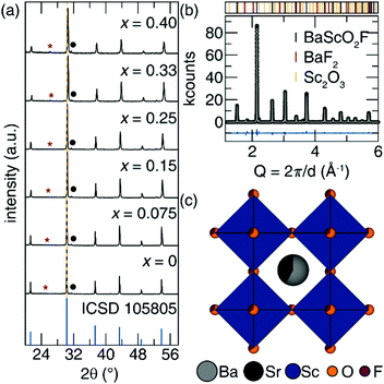
The structural flexibility of perovskites allows solid solutions to be formed with ease.33 The powder X-ray diffractograms of (Ba1−xSrx)0.98Eu0.02ScO2F (x = 0, 0.075, 0.15, 0.25, 0.33, 0.40), plotted in Fig. 1a, indicate the samples are isostructural with the parent phase, BaScO2F (ICSD no. 105805). The perovskite crystallizes with the average cubic space group Pm![[3 with combining macron]](https://www.rsc.org/images/entities/char_0033_0304.gif) m (No. 221).34 The powder X-ray diffractograms also reveal minor impurities of BaF2 and Sc2O3, marked with a red asterisk and a black dot, respectively. The incremental incorporation of Sr2+ in the host perovskite crystal structure is confirmed through the gradual shift of the diffraction peaks to larger angles (yellow dashed line, Fig. 1a) following the substitution of the smaller Sr2+ (r12-coord = 1.44 Å) for the larger Ba2+ (r12-coord = 1.61 Å).35 This shift in the diffraction peaks is corroborated by a slight, linear decrease in the refined lattice parameters, following Vegard's law of substitution (Fig. S1, ESI†).36 This confirms the ability of the oxyfluoride perovskite to accommodate Sr2+ in the cuboctahedral cavity without inducing large average structural changes. A solubility limit is reached when the Sr2+ concentration approaches 40%. All attempts to synthesize (Ba1−xSrx)0.98Eu0.02ScO2F with x > 0.40 resulted in major SrSc2O4 impurities, a known red-emitting phosphor upon Eu2+ substitution.37
m (No. 221).34 The powder X-ray diffractograms also reveal minor impurities of BaF2 and Sc2O3, marked with a red asterisk and a black dot, respectively. The incremental incorporation of Sr2+ in the host perovskite crystal structure is confirmed through the gradual shift of the diffraction peaks to larger angles (yellow dashed line, Fig. 1a) following the substitution of the smaller Sr2+ (r12-coord = 1.44 Å) for the larger Ba2+ (r12-coord = 1.61 Å).35 This shift in the diffraction peaks is corroborated by a slight, linear decrease in the refined lattice parameters, following Vegard's law of substitution (Fig. S1, ESI†).36 This confirms the ability of the oxyfluoride perovskite to accommodate Sr2+ in the cuboctahedral cavity without inducing large average structural changes. A solubility limit is reached when the Sr2+ concentration approaches 40%. All attempts to synthesize (Ba1−xSrx)0.98Eu0.02ScO2F with x > 0.40 resulted in major SrSc2O4 impurities, a known red-emitting phosphor upon Eu2+ substitution.37
Perovskites are famous for symmetry lowering distortions as a function of compositional change.38 Therefore, a Rietveld refinement of the solid solution end member, x = 0.40, was performed on high-resolution synchrotron X-ray powder diffraction data to confirm the average cubic crystal structure upon Sr2+ substitution. The Eu2+ ion was omitted from the refinement due to the low substitution concentration in the sample (2%). The refinement results, provided in Fig. 1b, indicate excellent agreement with the previously published crystal structure of BaScO2F, meaning the average cubic Pm![[3 with combining macron]](https://www.rsc.org/images/entities/char_0033_0304.gif) m perovskite structure following the A2+B3+[X2−]2[Y−] general structural formula is retained even upon maximum Sr2+ substitution. The resulting refined crystal structure data and atomic positions are listed in Table 1 and Table S1 (ESI†), respectively. The refined Sr2+ content was 43.3(8)%, agreeing with the nominally loaded 40% concentration. The resulting refined perovskite crystal structure of Ba0.566(2)Sr0.433(8)ScO2F (Fig. 1c) is composed of corner-connected [Sc(O/F)6] octahedra. The A2+ cation is in cuboctahedral coordination and sits in the cavity produced by the octahedral network. Both Eu2+ (r12-coord = 1.48 Å) and Sr2+ (r12-coord = 1.44 Å) are expected to occupy the cuboctahedral site since their ionic radii are similar to that of Ba2+ (r12-coord = 1.61 Å) and substantially larger than Sc3+ (r6-coord = 0.745 Å).35 The anion disorder has been previously confirmed through 19F-MAS NMR where a single 19F signal was observed, indicating a single fluoride site.39
m perovskite structure following the A2+B3+[X2−]2[Y−] general structural formula is retained even upon maximum Sr2+ substitution. The resulting refined crystal structure data and atomic positions are listed in Table 1 and Table S1 (ESI†), respectively. The refined Sr2+ content was 43.3(8)%, agreeing with the nominally loaded 40% concentration. The resulting refined perovskite crystal structure of Ba0.566(2)Sr0.433(8)ScO2F (Fig. 1c) is composed of corner-connected [Sc(O/F)6] octahedra. The A2+ cation is in cuboctahedral coordination and sits in the cavity produced by the octahedral network. Both Eu2+ (r12-coord = 1.48 Å) and Sr2+ (r12-coord = 1.44 Å) are expected to occupy the cuboctahedral site since their ionic radii are similar to that of Ba2+ (r12-coord = 1.61 Å) and substantially larger than Sc3+ (r6-coord = 0.745 Å).35 The anion disorder has been previously confirmed through 19F-MAS NMR where a single 19F signal was observed, indicating a single fluoride site.39
| Composition | Ba0.566(2)Sr0.433(8)ScO2F |
| Radiation type, λ (Å) | Synchrotron; 0.457868 |
| 2θ range (deg) | 0.5–50 |
| Temperature (K) | 100 |
| Crystal system | Cubic |
| Space group; Z |
Pm![[3 with combining macron]](https://www.rsc.org/images/entities/char_0033_0304.gif) m; 1 m; 1 |
| Lattice parameters (Å) | 4.1282(1) |
| Volume (Å3) | 70.352(2) |
| R p | 0.0948 |
| R wp | 0.1530 |
| χ 2 | 14.19 |
3.2 The effect of Sr2+ substitution on the electronic structure
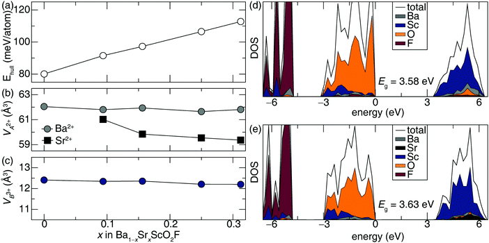
Density functional theory (DFT) calculations were performed to investigate the structural and electronic changes arising from the gradual substitution of Sr2+ in the perovskite crystal structure. To enumerate all possible O/F configurations, all irreducible representations of the Pm![[3 with combining macron]](https://www.rsc.org/images/entities/char_0033_0304.gif) m space group that satisfy the Lifshitz criterion were considered.40 The two lowest energy configurations adopt space groups P4/mmm and P42/mmc, with the latter favored by only 5 meV per atom (Fig. S2 and Table S2, ESI†). Therefore, all subsequent studies on compounds containing Sr2+ are based on the optimized structure with space group P42/mmc. The thermodynamic evolution of the computed Ba1−xSrxScO2F (x = 0, 0.09375, 0.15625, 0.25, 0.3125) was analyzed by calculating the energy above the convex hull (Ehull). Ehull measures the decomposition energy of an inorganic compound with respect to the thermodynamically stable phases in the DFT calculated (0 K) phase diagram. The compositions on the convex hull include BaScO2F, BaSc2O4, SrSc2O4 and BaF2. The gradual substitution of Sr2+ into Ba1−xSrxScO2F incrementally increases from 80 meV per atom (x = 0) to 113 meV per atom (x = 0.3125) above the Ehull, as plotted in Fig. 2a. The increasing Ehull indicates it becomes more energetically unfavorable to incorporate Sr2+ into the BaScO2F host crystal structure. This is consistent with the experimentally observed 40% solubility limit of Sr2+ and the formation of the SrSc2O4 impurity phase.
m space group that satisfy the Lifshitz criterion were considered.40 The two lowest energy configurations adopt space groups P4/mmm and P42/mmc, with the latter favored by only 5 meV per atom (Fig. S2 and Table S2, ESI†). Therefore, all subsequent studies on compounds containing Sr2+ are based on the optimized structure with space group P42/mmc. The thermodynamic evolution of the computed Ba1−xSrxScO2F (x = 0, 0.09375, 0.15625, 0.25, 0.3125) was analyzed by calculating the energy above the convex hull (Ehull). Ehull measures the decomposition energy of an inorganic compound with respect to the thermodynamically stable phases in the DFT calculated (0 K) phase diagram. The compositions on the convex hull include BaScO2F, BaSc2O4, SrSc2O4 and BaF2. The gradual substitution of Sr2+ into Ba1−xSrxScO2F incrementally increases from 80 meV per atom (x = 0) to 113 meV per atom (x = 0.3125) above the Ehull, as plotted in Fig. 2a. The increasing Ehull indicates it becomes more energetically unfavorable to incorporate Sr2+ into the BaScO2F host crystal structure. This is consistent with the experimentally observed 40% solubility limit of Sr2+ and the formation of the SrSc2O4 impurity phase.
Substituting Sr2+ for Ba2+ (and vice versa) is a well-known mechanism to shift the photoluminescence of phosphors. However, the structural mechanism to achieve a tunable emission can vary. For example, substituting the smaller Sr2+ for the larger Ba2+ decreases the average bond length and polyhedral volume of the (Ba/Sr) polyhedral units. This is observed in Ba2−xSrxSiO4:Eu2+, (Ba1−xSrx)8.46(Ce0.27Li0.27)Sc2Si6O24, and (Ba1−xSrx)Si3O4N2:Eu2+, among other examples, where the substitution of Sr2+ onto the rare-earth substitution site causes a red-shift in the observed emission spectrum.9,41,42 There are also instances, such as in SrY2O4:Eu2+, where the substitution of Ba2+ for Sr2+, red-shifts the observed emission. This is counterintuitive but it can be understood because Eu2+ preferentially substitutes onto the Y3+ sites in the host crystal structure. The mechanism for the red-shift involves the [SrO8] polyhedral volumes increasing upon Ba2+ substitution which contracts the surrounding [YO6] octahedra to increases the crystal field splitting and induce a red-shift in the emission.43 This phenomenon is also seen in the Sr1.8Ba0.2Sc0.5Ga1.5O5:Eu2+ phosphor.44 As a result, it is imperative to understand how the [BaO12], [SrO12], and [ScO6] polyhedral volumes evolve upon the substitution of Sr2+ in the BaScO2F host crystal structure. Structurally, the DFT optimized Ba1−xSrxScO2F (x = 0.0, 0.09375, 0.15625, 0.25, 0.3125) host crystal structure shows minor changes in the [Ba(O/F)12] and [Sc(O/F)6] polyhedral volumes, seen in Fig. 2b and c, respectively, whereas the polyhedral volume around Sr2+ (black square) continuously decreases with increasing x, as shown in Fig. 2b. This computed structural evolution agrees well with the trends in the experimentally measured lattice parameters. The computed decreasing [SrO12] and unchanging [ScO6] and [BaO12] polyhedral volumes suggest that the crystal field splitting will increase and red-shift the emission spectra of (Ba1−xSrx)0.98Eu0.02ScO2F upon increasing Sr2+ concentration.
Electronically, the gradual insertion of Sr2+ into Ba1−xSrxScO2F has a minor effect on the band structure. The PBE-level calculated density of states (DOS) shows relatively constant band gap (Eg) between BaScO2F (Eg = 3.58 eV) and Ba0.6875Sr0.3125ScO2F (Eg = 3.63 eV), plotted in Fig. 2d and e, respectively. The DOS of the other compounds analyzed (x = 0.09375, 0.15625, 0.25) are provided in Fig. S3 (ESI†). The element-projected DOS reveals that oxygen sets the valence band maximum (VBM) whereas Sc3+ 3d orbitals set the conduction band minimum (CBM). Consequently, the uniformity of the O2− and Sc3+ band positions arising from the uniformity of the [Sc(O/F)6] octahedra across the Ba1−xSrxScO2F solid solution yields a nearly constant Eg regardless of Sr2+ concentration.
Finally, the average structural rigidity across the solid solution was investigated through Debye temperature calculations. The DFT calculated Debye temperature, ΘD,DFT, was determined for Ba1−xSrxScO2F when x = 0 and 0.3125 to approximate the structural rigidity as a function of composition. Interestingly, the Debye temperature remains nearly constant across the solid solution, exhibiting only a 7 K difference between x = 0 (ΘD,DFT = 497 K) and x = 0.3125 (ΘD,DFT = 504 K). This indicates that the average structural rigidity is preserved upon Sr2+ substitution. Since the Debye temperature is considered a proxy for the photoluminescent quantum yield (PLQY) of rare-earth substituted phosphors, this suggests the 80% quantum yield of BaScO2F:Eu2+ should be retained.45,46
3.3 Photoluminescence
BaScO2F:Eu2+ is a phosphor that produces a bright cyan emission with a maximum PLQY of 80.3(5)% with 2% Eu2+ substitution.20 For the study here, the rare-earth substitution concentration was held constant as Sr2+ was incrementally introduced into the host crystal structure following the general formula (Ba1−xSrx)0.98Eu0.02ScO2F (x = 0.075, 0.15, 0.25, 0.33, and 0.40). The resulting excitation spectra are plotted in Fig. 3a. Each phosphor in the solid solution possesses a wide excitation band covering approximately 280 nm to 450 nm with three distinct maxima as provided in Table 2. The maxima of the excitation bands indicate excellent compatibility with UV LEDs.| λ ex (nm) | λ em,max (nm) | fwhm | |
|---|---|---|---|
| x = 0 | 282, 340, 365 | 487 | 103 nm, 4228 cm−1 |
| x = 0.075 | 279, 341, 369 | 496 | 103 nm, 4158 cm−1 |
| x = 0.15 | 276, 340, 371 | 504 | 100 nm, 4420 cm−1 |
| x = 0.25 | 272, 342, 372 | 509 | 105 nm, 4251 cm−1 |
| x = 0.33 | 267, 339, 372 | 512 | 102 nm, 4378 cm−1 |
| x = 0.40 | 265, 340, 373 | 516 | 107 nm, 4328 cm−1 |
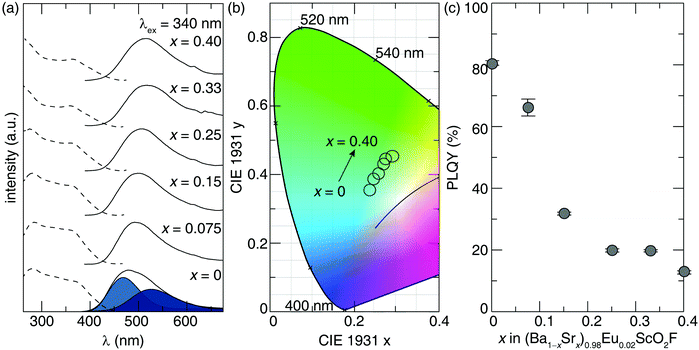
The emission spectra under 340 nm excitation, also shown in Fig. 3a, covers a broad region from 400 nm to 700 nm, where the emission peak maxima shifts from 481 nm (x = 0) to 516 nm (x = 0.40). This red-shift is reflected in the 1931 CIE XYZ coordinates of the solid solution, where the perceived emission color linearly shifts from cyan to green, indicating successful tunability of the observed emission color (Fig. 3b). Each phosphor's emission color is not saturated due to the broad full width at half maximum (fwhm) across the solid solution. Indeed, the fwhm of the emission peak falls between 100 nm and 107 nm (4158–4420 cm−1). Such a broad emission is inherent to the perovskite crystal structure of BaScO2F:Eu2+, which possesses two crystallographically independent Eu2+ atoms upon rare-earth substitution, as discussed previously.20 As a result, the emission spectrum of BaScO2F:Eu2+ (x = 0) is described by two distinct Gaussians to represent the emission from the independent Eu2+ atoms, illustrated in blue in Fig. 3a. The optical properties shown here indicate the gradual substitution of Sr2+ in BaScO2F:Eu2+ preserves the emission peak shape while red-shifting the emission. This suggests the local Eu2+ coordination environments across the solid solution are maintained, while Sr2+ substitution decreases the polyhedral volume (Fig. 2b), leading to the observed red-shift in the emission spectra. Additionally, there is little change between the fwhm of each emission peak, suggesting the magnitude of electron–phonon coupling is not affected by Sr2+ substitution (Table 2).47 This broad emission is ultimately advantageous in producing a full-spectrum white light by covering the blue, cyan, and green regions of the visible spectrum.
The observed red-shift in the emission spectrum is further supported by analyzing the crystallographic site preference for Eu2+ to substitute for Ba2+ or Sr2+ within the crystal structure. Rietveld refinements cannot accurately probe the Eu2+ location due to its low substitution concentration (2%). Instead, the site preference was determined by calculating the dopant formation energies of Ba1−xSrxScO2F (x = 0, 0.09375, 0.15625, 0.25, 0.3125). These models used 2 × 4 × 4 supercells and a constant Eu2+ concentration of 3% substituted onto either the Ba2+ site (EuBa) or the Sr2+ site (EuSr). The resulting dopant formation energy difference between EuBa and EuSr for x < 0.25 is relatively constant, ≈1.85 meV per atom favoring EuSr. This value decreases to ≈0.9 meV per atom when x = 0.3125 but still indicates that Eu2+ should preferentially substitute for Sr2+ (Fig. S4, ESI†). Analyzing the DFT optimized Ba1−xSrxScO2F:Eu2+ (x = 0, 0.09375, 0.15625, 0.25, 0.3125) models show the average bond length forming the Eu2+ coordination polyhedron as a function of Sr2+ gradually decreases (Table S3, ESI†), which increases the crystal field splitting around Eu2+ and inherently induces the observed red-shifted emission generating the green emission.
Even though a novel phosphor may emit in the desired color range, integration into LED light bulbs requires efficient photoluminescence. Therefore, the room temperature PLQY was measured to determine whether the high (80.3(5)%) quantum yield of Ba0.98Eu0.02ScO2F was maintained. The room temperature PLQY of (Ba1−xSrx)0.98Eu0.02ScO2F (x = 0.075, 0.15, 0.25, 0.33, 0.40) was determined using an excitation wavelength of 340 nm and plotted in Fig. 3c. Incorporating 7.5% Sr2+ causes the PLQY to decrease to 66.1(6)%. Doubling the Sr2+ concentration to 15% causes the efficiency to decrease by more than half (PLQY = 31.7(9)%), and substituting 40% Sr2+ into the structure results in a dramatic drop in the PLQY to 12.7(5)%. This result is surprising because the nearly constant Debye temperature suggests a minimal change in the PLQY should occur as a function of Sr2+ concentration.
The change in room temperature PLQY could stem from a variety of different sources. The minor presence of the Eu3+ 4f ↔ 4f emission around 605 nm at higher Sr2+ concentrations (Fig. 3a) can cause a drop in the PLQY. However, the Eu3+ as a luminescence quencher in the solid solution appears to be constant based on the intensity of these emission peaks. Thus, the presence of the trivalent rare-earth does not fully explain the dramatic drop in the PLQY with increasing Sr2+. Additionally, the decrease in the PLQY may be related to the temperature-dependence photoluminescence. The optical properties of rare-earth substituted phosphors can change considerably as a function of temperature. One of the most notable changes is a loss of photoluminescence emission intensity known as thermal quenching (TQ). Thus, the substitution of Sr2+ could cause an increase in TQ at room temperature that decreases the PLQY.
3.4 Thermal quenching and its relationship to local environment rigidity
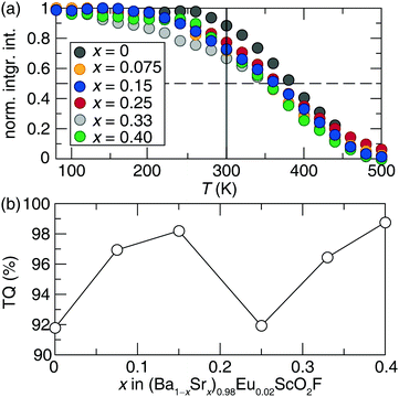
Measuring the photoluminescent emission spectrum of each phosphor from 80 K to 500 K and plotting the normalized, integrated intensity as a function of the temperature range in Fig. 4a shows BaScO2F:Eu2+ remains stable up to 260 K and begins to quench with increasing temperature. Adding Sr2+ in the phosphors causes thermal quenching to begin beyond 220 K, resulting in a ≈25% drop in the integrated emission intensity by room temperature (300 K), shown as the solid black line in Fig. 4a. This behavior is reflected in all of the phosphors across the solid solution. By room temperature, the Sr2+-substituted phosphors are already undergoing different rates of thermal quenching, causing a drop in photoluminescence intensity, which is responsible for the observed decrease in the PLQY.It is well understood that thermal quenching is accompanied by a decrease of the photoluminescence decay time, τ.48,49 Therefore, the time-resolved room temperature photoluminescent decay curve was measured under 330 nm excitation. The photoluminescent lifetime was determined by fitting the decay curve to a bi-exponential following eqn (4), where I is intensity, I0 is the initial intensity, A1, and A2 are the pre-exponential constants, τ1 and τ2 are decay times for exponential components in microseconds, and t is the measured time.
| I = I0 + A1e−t/τ1 + A1e−t/τ2 | (4) |
The thermal quenching of each phosphor can also be quantified by defining and plotting the TQ as the ratio between the integrated emission intensity at high temperature (500 K), which is above the operating temperature of most LED-based light bulbs, and room temperature (300 K). Fig. 4b plots the TQ values of (Ba1−xSrx)0.98Eu0.02ScO2F (x = 0, 0.075, 0.15, 0.25, 0.33, 0.40) and depicts an increase in TQ once Sr2+ is introduced into the system. This outcome is crucial as it reveals that the substitution of Sr2+ into the perovskite structure hinders the thermal stability of the emission. There are multiple TQ mechanisms responsible for the differences in temperature-dependent photoluminescence. One quenching mechanism is related to photoionization, where the excited state electron in the rare-earth 5d orbital transitions to the host conduction band and is subsequently lost. This process becomes a prominent quenching mechanism in phases when the host Eg is decreased as a function of composition.22 In this system, however, the magnitude of Eg remains relatively constant as a function of Sr2+ (Fig. 2d and e). Previous work has also shown that the thermal quenching of Eu2+ substituted phosphors correlates to the phosphor host's structural rigidity, which quantifies the cross-over quenching mechanism.50 Structural rigidity can be assessed based on the material's measured or calculated ΘD. Interestingly, the calculated Debye temperatures are nearly constant as a function of composition, ranging from 497 K (x = 0) to 504 K (x = 0.3125), or <2% different. The similar structural rigidity also does not provide any additional insight on the TQ response. These analyses in tandem show that these calculated average structure proxies for understanding a phosphor's optical properties are not sufficient for this system.
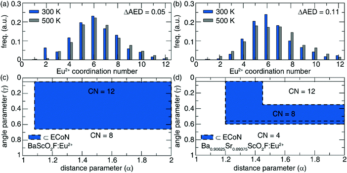
The local structure around the rare-earth ion must be considered instead. The Eu2+ local coordination environment can be investigated by computing the local activator environment distribution (AED) at two temperatures, room temperature and high temperature (300 K and 500 K), using AIMD calculations. Here, the AED is determined by extracting the number of simulations timesteps that the rare-earth forms a particular coordination number (CN). The change in the AED (ΔAED) between 300 K and 500 K is directly correlated with TQ.22 The calculated shift in AED in the pristine phosphor (x = 0) is small (ΔAED = 0.05), whereas the Sr2+ substituted phosphor (x = 0.09375) shows a larger shift in AED (ΔAED = 0.11) as shown in Fig. 5a and b, respectively. The larger ΔAED indicates that the rare-earth environment is more prone to local polyhedral distortions upon Sr2+ substitution. In addition, the larger ΔAED suggests increased TQ, which is experimentally observed in the onset of quenching occurring at lower temperatures yielding lower room temperature PLQY. The local environment rigidity can also be investigated via a two-dimensional projection of the Eu2+ coordination environment using the Voronoi tessellation representation. Here, a constant, normalized Voronoi area is assumed to be less sensitive to the thermally induced fluctuations and hence, less prone to thermal quenching. Plotting the Voronoi grid representation and highlighting the area of interest around Hoppe's effective rare-earth local environment for BaScO2F:Eu2+ (Fig. 5c) shows the CN is nearly always 12 whereas the CN in Ba0.90625Sr0.09375ScO2F:Eu2+ can be either 12, 8, or 4 (Fig. 5d). This reflects that substituting Sr2+ decreases the Eu2+ local rigidity indicated by the changing AED, consequently increasing the thermal quenching. These AIMD calculations are the only method that validates the decrease in TQ and PLQY of this new green-emitting phosphor that arises from the substitution of Sr2+ in Ba1−xSrxScO2F:Eu2+.
4. Conclusion
The development of novel green-emitting phosphors by substituting Sr2+ into the BaScO2F:Eu2+ perovskite was successful. The synthesis of the solid solution following (Ba1−xSrx)0.98Eu0.02ScO2F (x = 0, 0.075, 0.15, 0.25, 0.40) causes a 30 nm red-shift of the cyan emission of the parent phosphor to the green region of the visible spectrum. However, the high efficiency of the parent phosphor, Ba0.98Eu0.02ScO2F, is not retained across the solid solution, with a nearly 65% decrease in the PLQY when x = 0.40. In addition, there is severe thermal quenching observed in this solid solution. Average electronic structure calculations of the band gap and Debye temperature of Ba1−xSrxScO2F:Eu2+ (x = 0, 0.09375, 0.15625, 0.25, 0.3125) were inadequate to describe the trend in the observed optical properties. Instead, ab initio molecular dynamics simulations revealed that a decrease in the local activator environment rigidity is responsible for the increasing TQ and loss of PLQY. These results provide considerable support for using computational modeling, including standard DFT but more importantly, advanced AIMD calculations, to analyze the average structure and local rare-earth environment as a function of temperature to understand the optical properties of new luminescent materials.Conflicts of interest
The authors declare no competing financial interest.Acknowledgements
S. H., M. K., and J. B. would like to thank the National Science Foundation (CER-1911311) as well as the Welch Foundation (E-1981) for supporting this work. M. A. and S. P. O. also acknowledge funding from the National Science Foundation (CER-1911372), computing resources provided by the National Energy Research Scientific Computing Center (NERSC) under Contract No. DE-AC02-05CH11231 using NERSC award BES-ERCAP0018251, the Triton Shared Computing Cluster (TSCC) at the University of California, San Diego, and the Extreme Science and Engineering Discovery Environment (XSEDE) under grant ACI-1548562. This work used the resources available through the 11-BM beamline at the Advanced Photon Source, an Office of Science User Facility operated for the US Department of Energy (DOE) Office of Science by Argonne National Laboratory, under Contract No. DE-AC02-06CH11357.References
- U. S. Department of Energy, Energy Saver, https://www.energy.gov/energysaver/led-lighting, (accessed September, 2021).
- P. Pust, P. J. Schmidt and W. Schnick, Nat. Mater., 2015, 14, 454–458 CrossRef CAS PubMed
.
- J. Hye Oh, S. Ji Yang and Y. Rag Do, Light Sci. Appl., 2014, 3, e141 CrossRef
.
- M.-H. Fang, C. Ni, X. Zhang, Y.-T. Tsai, S. Mahlik, A. Lazarowska, M. Grinberg, H.-S. Sheu, J.-F. Lee, B.-M. Cheng and R.-S. Liu, ACS Appl. Mater. Interfaces, 2016, 8, 30677–30682 CrossRef CAS PubMed
.
- P. Pust, V. Weiler, C. Hecht, A. Tücks, A. S. Wochnik, A.-K. Henß, D. Wiechert, C. Scheu, P. J. Schmidt and W. Schnick, Nat. Mater., 2014, 13, 891–896 CrossRef CAS PubMed
.
- S. Li, Y. Xia, M. Amachraa, N. T. Hung, Z. Wang, S. P. Ong and R.-J. Xie, Chem. Mater., 2019, 31, 6286–6294 CrossRef CAS
.
- S. Li, L. Wang, D. Tang, Y. Cho, X. Liu, X. Zhou, L. Lu, L. Zhang, T. Takeda, N. Hirosaki and R.-J. Xie, Chem. Mater., 2018, 30, 494–505 CrossRef CAS
.
- K. Park, T. Kim, Y. Yu, K. Seo and J. Kim, J. Lumin., 2016, 173, 159–164 CrossRef CAS
.
- K. A. Denault, J. Brgoch, M. W. Gaultois, A. Mikhailovsky, R. Petry, H. Winkler, S. P. DenBaars and R. Seshadri, Chem. Mater., 2014, 26, 2275–2282 CrossRef CAS
.
- Y. Zhuo, S. Hariyani, J. Zhong and J. Brgoch, Chem. Mater., 2021, 33, 3304–3311 CrossRef CAS
.
- Y. Ohno, Color Rendering and Luminous Efficacy of White LED Spectra, SPIE, 2004.
- M. Zhao, Q. Zhang and Z. Xia, Acc. Mater. Res., 2020, 1, 137–145 CrossRef CAS
.
- P. D. Rack and P. H. Holloway, Mater. Sci. Eng., R, 1998, 21, 171–219 CrossRef
.
- Z. Xia, Q. Li and J. Sun, Mater. Lett., 2007, 61, 1885–1888 CrossRef CAS
.
- X. Zhang, X. Wang, J. Huang, J. Shi and M. Gong, Opt. Mater., 2009, 32, 75–78 CrossRef CAS
.
- K. A. Denault, N. C. George, S. R. Paden, S. Brinkley, A. A. Mikhailovsky, J. Neuefeind, S. P. DenBaars and R. Seshadri, J. Mater. Chem., 2012, 22, 18204–18213 RSC
.
- W. B. Im, S. Brinkley, J. Hu, A. Mikhailovsky, S. P. DenBaars and R. Seshadri, Chem. Mater., 2010, 22, 2842–2849 CrossRef CAS
.
- W. B. Im, N. George, J. Kurzman, S. Brinkley, A. Mikhailovsky, J. Hu, B. F. Chmelka, S. P. DenBaars and R. Seshadri, Adv. Mater., 2011, 23, 2300–2305 CrossRef CAS PubMed
.
- M. Hermus, P.-C. Phan, A. C. Duke and J. Brgoch, Chem. Mater., 2017, 29, 5267–5275 CrossRef CAS
.
- S. Hariyani and J. Brgoch, Chem. Mater., 2020, 32, 6640–6649 CrossRef CAS
.
- U. S. Department of Energy, Solid-State Lighting Research and Development Multi-Year Program Plan, https://www1.eere.energy.gov/buildings/publications/pdfs/ssl/ssl_mypp2014_web.pdf, (accessed October, 2021).
- M. Amachraa, Z. Wang, C. Chen, S. Hariyani, H. Tang, J. Brgoch and S. P. Ong, Chem. Mater., 2020, 32, 6256–6265 CrossRef CAS
.
- B. H. Toby, J. Appl. Crystallogr., 2001, 34, 210–213 CrossRef CAS
.
- K. Momma and F. Izumi, J. Appl. Crystallogr., 2011, 44, 1272–1276 CrossRef CAS
.
- J. C. de Mello, H. F. Wittmann and R. H. Friend, Adv. Mater., 1997, 9, 230–232 CrossRef CAS
.
- G. L. W. Hart and R. W. Forcade, Phys. Rev. B: Condens. Matter Mater. Phys., 2008, 77, 224115 CrossRef
.
- S. P. Ong, S. Cholia, A. Jain, M. Brafman, D. Gunter, G. Ceder and K. A. Persson, Comput. Mater. Sci., 2015, 97, 209–215 CrossRef
.
- G. Kresse and J. Furthmüller, Phys. Rev. B: Condens. Matter Mater. Phys., 1996, 54, 11169–11186 CrossRef CAS PubMed
.
- A. Chaudhry, R. Boutchko, S. Chourou, G. Zhang, N. Grønbech-Jensen and A. Canning, Phys. Rev. B: Condens. Matter Mater. Phys., 2014, 89, 155105 CrossRef
.
- S.-H. Wei and S. B. Zhang, Phys. Rev. B: Condens. Matter Mater. Phys., 2002, 66, 155211 CrossRef
.
- Z.-J. Wu, E.-J. Zhao, H.-P. Xiang, X.-F. Hao, X.-J. Liu and J. Meng, Phys. Rev. B: Condens. Matter Mater. Phys., 2007, 76, 054115 CrossRef
.
- W. G. Hoover, Phys. Rev. A: At., Mol., Opt. Phys., 1985, 31, 1695–1697 CrossRef PubMed
.
- A. S. Bhalla, R. Guo and R. Roy, Mater. Res. Innovations, 2000, 4, 3–26 CrossRef CAS
.
- R. L. Needs and M. T. Weller, J. Solid State Chem., 1998, 139, 422–423 CrossRef CAS
.
- R. D. Shannon and C. T. Prewitt, Acta Crystallogr., Sect. B: Struct. Sci., 1969, 25, 925–946 CrossRef CAS
.
- E.-a. Zen, Am. Mineral., 1956, 41, 523–524 CAS
.
- M. Müller, M.-F. Volhard and T. Jüstel, RSC Adv., 2016, 6, 8483–8488 RSC
.
- K. S. Aleksandrov and J. BartolomÉ, Phase Transitions, 2001, 74, 255–335 CrossRef CAS
.
- R. L. Needs and M. T. Weller, J. Solid State Chem., 1998, 139(2), 422–423 CrossRef CAS
.
- M. V. Talanov, V. B. Shirokov and V. M. Talanov, Acta Crystallogr., Sect. A: Found. Crystallogr., 2016, 72, 222–235 CrossRef CAS PubMed
.
- J. Brgoch, C. K. H. Borg, K. A. Denault, S. P. DenBaars and R. Seshadri, Solid State Sci., 2013, 18, 149–154 CrossRef CAS
.
- Z. Zhang and W. Yang, Opt. Mater. Express, 2019, 9, 1922–1932 CrossRef CAS
.
- Z. Yang, Y. Zhao, Y. Zhou, J. Qiao, Y.-C. Chuang, M. S. Molokeev and Z. Xia, Adv. Funct. Mater., 2022, 32, 2103927 CrossRef CAS
.
- Z. Yang, Y. Zhou, J. Qiao, M. S. Molokeev and Z. Xia, Adv. Opt. Mater., 2021, 9, 2100131 CrossRef CAS
.
- J. Brgoch, S. P. DenBaars and R. Seshadri, J. Phys. Chem. C, 2013, 117, 17955–17959 CrossRef CAS
.
- S. Hariyani, A. C. Duke, T. Krauskopf, W. G. Zeier and J. Brgoch, Appl. Phys. Lett., 2020, 116, 051901 CrossRef CAS
.
-
G. Blasse, Structure and Bonding, Springer, Berlin, Heidelberg, 1980, vol. 42 Search PubMed
.
- D. Van der Heggen, J. J. Joos and P. F. Smet, ACS Photonics, 2018, 5, 4529–4537 CrossRef CAS
.
- V. Bachmann, C. Ronda and A. Meijerink, Chem. Mater., 2009, 21, 2077–2084 CrossRef CAS
.
- Y. Zhuo, A. Mansouri Tehrani, A. O. Oliynyk, A. C. Duke and J. Brgoch, Nat. Commun., 2018, 9, 4377 CrossRef PubMed
.
Footnotes |
| † Electronic supplementary information (ESI) available. See DOI: 10.1039/d1tc05411b |
| ‡ These authors contributed equally to this work. |
| This journal is © The Royal Society of Chemistry 2022 |

