
Mussel-inspired silver-nanoparticle coating on porous titanium surfaces to promote mineralization
Jialong Chen abc,
May Lei Meia,
Quan-Li Li*b and
Chun-Hung Chu*a
abc,
May Lei Meia,
Quan-Li Li*b and
Chun-Hung Chu*a
aFaculty of Dentistry, The University of Hong Kong, 34 Hospital Road, Hong Kong, China. E-mail: chchu@hku.hk; Fax: +852-25599013; Tel: +852-28590287
bCollege & Hospital of Stomatology, Anhui Medical University, Key Laboratory of Oral Diseases Research of Anhui Province, Hefei, China. E-mail: ql-li@126.com; Fax: +86-551-65121527; Tel: +86-551-65118677
cCollege of Pharmacy, Anhui Medical University, Hefei, China
First published on 26th October 2016
Abstract
Biomaterials with high porosity for bone ingrowth facilitate the osseointegration of implants. However, this porosity structure is also favorable for bacterial colonization and biofilm formation, and hampers osseointegration on implant surfaces. The objective of the study was to establish a porous surface on titanium implants with antibacterial activity, enhanced mineralization and then good osteoblast-biocompatibility. A uniform, 3-dimensional, microporous structure was prepared by alkaline treatment on a titanium implant surface. Subsequently, the surface was coated with dopamine and silver nanoparticles using dopamine and silver nitrate solutions. The results of SEM, EDS, XPS, and water contact angle tests confirmed that the surface had been successfully coated with dopamine and silver nanoparticles with sizes of 30–50 nm. More mineralization happened on the surface with this coating than on the alkaline-treated surface after incubation with calcification solution for one week, which led to a rapid decrease of silver release. Antibacterial tests indicated that the coating inhibited bacterial colonization on them and growth around these samples, indicating that the coating eliminates the shortcoming of porous structure which renders the implant extremely susceptible to biomaterial-associated infection. Besides, this coating did not favor osteoblast attachment, proliferation and differentiation, however we found that the mineralized surface of this coating stimulated the proliferation of osteoblasts and enhanced the activity of alkaline phosphatase. Therefore, we conclude that the coating with dopamine and AgNPs can be effective against bacterial infections and facilitate mineralization during the early post-operative period and then can promote osseointegration due to the good osteoblast-biocompatibility of the mineralized surface.
Introduction
Titanium and its alloys have been used extensively as implants in orthopedic and dental applications because of the combination of outstanding properties, such as excellent biocompatibility, high strength, good fatigue and corrosion resistance.1 However, bacteria that cause prosthetic joint and dental implant infections grow in highly structured biofilms (i.e., sessile communities of microorganisms adhering to the biomaterial embedded in a matrix of an extracellular polymeric substance that they produced).2,3 This protective environment enables bacteria to escape the host's defenses and antibiotic attacks. Moreover, the increased competence suggested for biofilm-embedded bacteria, which results in a higher degree of horizontal transfer of genes, including antibiotic resistance markers and the occurrence of persister cells, might further enhance biofilm-related antibiotic resistance.4,5 As a consequence, antibiotic treatment is often insufficient to eradicate biofilm-related implant infections, leading to potentially life-threatening systemic infections, tissue injury, device malfunction, and ultimately, a need to remove the implant. Biomaterial surfaces that are less prone to bacterial adherence and colonization have helped researchers make serious progress in reducing infection rates over the last few decades.6 Surface modification with antibiotics and antibacterial agents is an efficient way to reduce the infection rate, such as with gentamicin, vancomycin, and chlorhexidine.7,8The use of silver as a promising alternative antibacterial agent therefore receives increasing attention due to a very broad spectrum against bacterial and fungal species in the physiological environment,9 and the surge of antibiotic-resistant bacterial strains which are becoming a major public health concern.10 However, in dental applications it stains dental tissue black due to the oxidation process of ionic silver11 in the application of silver diamine fluoride and amalgam. This undesired effect has hindered its widespread use. Silver nanoparticles (AgNPs) have recently drawn considerable attention due to the good color stability and large active surface areas.12–16 Biomaterials that contain AgNPs have been exhaustively investigated for the development of catheters, dental materials, orthopedic implants, and wound and burn dressings.17 To obtain satisfactory surfaces containing AgNPs, many methods have been developed to apply silver on surfaces, such as plasma immersion ion implantation,18 pulsed filtered cathodic vacuum arc deposition,19 physical vapor deposition,20 and so on. Nevertheless, the major drawbacks to these methods mentioned above contain poor AgNP/material adhesion and the difficulty in controlling AgNPs size.21–23 In addition, the need of special equipment and/or consumption of large amounts of energy and complicated multi-step procedures also limited further applications. Given these problems, a meaningful approach is to develop a simple and versatile strategy for surface modification with AgNPs. Smaller AgNPs with large surface areas can exhibit better antibacterial activity than larger AgNPs, but the agglomeration of the AgNPs with small sizes is an important consideration and could result in a quick loss of antibacterial activity.24,25 Many researchers proposed that the rough surface could decrease the aggregation of nanoparticles.26
Dopamine (DA), a mussel-inspired biomolecule, contains unusually high concentrations of catechol and amine groups. The catechol side chain of dopamine readily oxidizes to form reactive species that can further undergo Michael-type additions or Schiff-base formations with nucleophiles and radical coupling with other catechols or amines.27 Thus, dopamine offers a simple method of coating various organic and inorganic substrates.10,28,29 Another interesting feature is that dopamine, as a reducing and stabilizing agent, has been used to reduce Au(III) or Ag(I) metal ions in the solution to form noble metal nanoparticles via catechol oxidation without the need for any toxic components, leading to in situ formation of nanoparticles on the dopamine-modified surfaces.30,31 In addition, dopamine could promote cell adhesion, exhibit good biocompatibility,32 and induce mineralization,33 which are of particular interest in dental and orthopedic implantology for engineering surfaces with the ability to improve osseointegration.
Comparing with the traditional sample with flat and dense surface, the porous structures or coatings are of special interest because they could enhance protein adsorption, osteoblast proliferation, differentiation and then bone ingrowth into the porous structure, thus establishing a biological anchorage for the implant in the host bone.34,35 Osseointegration is strongly dependent on the structural characteristics of the surface, such as total open porosity and pore size. Porous structures with high porosity facilitate biological anchorage in the surrounding bone tissue via the ingrowth of mineralized tissue into the pores,36 but the resulting large surface area renders the implant extremely susceptible to bacterial colonization and subsequent biofilm formation. Therefore, there is particular interest in dental and orthopedic implantology to design surfaces that combine both porous structures to improve osseointegration and simultaneously improve the antibacterial property to reduce the infection risk.
The aim of the present study is to build a porous titanium surface carrying dopamine and uniformly distributed small AgNPs and then to evaluate whether this surface is able to exhibit antibacterial activity and enhanced mineralization and then good osteoblast-compatibility. For this, the porous titanium surface obtained by alkaline treatment was modified with dopamine using the dip-coating; then, AgNPs were coated onto the dopamine-modified surface in situ by reduction reaction between Ag+ and dopamine. Finally, the antibacterial and mineralization properties and osteoblast-compatibility of the modified titanium were evaluated in vitro. This method could have implications for dental- and orthopedic-related areas, because the efficient antibacterial activity and the high bioactivity of implant surfaces could be constructed by a simple method.
Materials and methods
Materials
Commercial pure titanium was purchased from Baoji Non-ferrous Metal Co., Ltd. (Shanxi Province, China). Dopamine–HCl, silver nitrate, Tris base, CaCl2·2H2O, KH2PO4, NaCl, Tris–HCl were purchased from Sigma-Aldrich. Nitric acid, ethanol and glutaraldehyde were purchased from Acros Organics. Fetal bovine serum, α-minimum Eagle's medium, trypsin–EDTA solution, penicillin and streptomycin were purchased from Gibco. Bacterial strains and mice MC3T3-E1 cell line were purchased from ATCC.Preparation and characterization of the coating with dopamine and silver nanoparticles
Titanium discs were polished into a reflective, mirror-like surface. The discs were ultrasonically cleaned first in a detergent solution, then in acetone, ethanol and finally deionized water. After soaking in a 5 M NaOH solution at 60 °C for 48 h, the cleaned specimens were soaked in deionized water at 80 °C for 8 h and were then denoted as TiOH. The specimens were immersed in a 2 mg mL−1 solution of dopamine (10 mM Tris buffer, pH 8.5) for about 24 h at room temperature in the dark. Then, the samples were sonicated for 10 min in deionized water (3 times) to remove the nonattached dopamine; these samples are denoted as Ti–O–DA. Then, 100 mg of silver nitrate (AgNO3) was dissolved in deionized water (10 mL, pH = 10). The dopamine-modified samples were then placed in a 24-well plate and incubated with a 600 μL AgNO3 solution in an orbital shaker incubator at 80 rpm and 37 °C for 24 h. Then, the samples were rinsed vigorously for 10 min in deionized water and dried in a vacuum for further use; the samples are denoted as Ti–O–DA–Ag.The surface topography of all of the samples was investigated using scanning electron microscopy (SEM, Hitachi S-4800). Energy-dispersive X-ray spectroscopy (EDS) analysis was also performed. The surface composition of the samples was analyzed by X-ray photoelectron spectroscopy (XPS, Thermo ESCALAB 250) with an Al Kα X-ray source (1486.6 eV photons). A wide-scan survey spectrum over a binding energy (BE) range of 0–1400 eV was recorded at a pass energy of 100 eV to estimate the chemical elemental composition and 30 eV for high-resolution detailed scans. The system was calibrated using the C1s peak at 284.8 eV. All spectra were recorded at a takeoff angle of 45 degrees. The maximum information depth of the XPS method was not more than 10 nm.
Water contact angle analysis was performed with a DSA100 drop-shape analysis system (DSA100, Krüss, Germany) using deionized water at room temperature. Five samples of each group were measured, and two separate measurements were made on each sample. All of the samples were sterilized with UV irradiation for 1 h prior to biological evaluation.
Mineralization of the coating in the calcification solution
The calcification solution (pH = 7.6) contained 2.58 mM calcium (CaCl2·2H2O), 1.55 mM phosphate (KH2PO4), and 180 mM NaCl and was buffered by 50 mM of Tris–HCl.33 The samples were placed in a 24-well plate and incubated with 1.5 mL of calcification solution in an orbital shaker incubator at 80 rpm and 37 °C. The calcification solution was replaced every day. The samples were taken out at 7 days, rinsed vigorously for 10 min with deionized water, and gradually dehydrated in a critical drying point prior to characterization.Silver release
Silver release was quantified for up to 30 days by Inductively Coupled Plasma Optical Emission Spectrometry (ICP-OES) (SPECTRO ARCOS; AMETEK, Inc., Kleve, Germany). The AgNP-coated samples were incubated in 6 mL of deionized water or calcification solution at 37 °C without agitation, respectively. The solutions were collected and then replaced with fresh solutions every two days. After 30 days, the remaining silver on the samples was dissolved in 6 mL of 3% nitric acid with ultrasonic washer for 60 min. The amounts of silver were determined through analysis of the resultant solutions by ICP-OES, with sample averages obtained from 3 separate tests.Antibacterial test
Gram-negative bacteria, Escherichia coli, and Gram-positive bacteria, Staphylococcus aureus and Streptococcus mutans, were used in the antibacterial tests. The numbers of both live and dead bacteria were used to indicate the antibacterial activity for the different materials. Samples were placed in a 24-well plate and incubated with different bacterial suspensions at a concentration of 107 CFU mL−1 at 37 °C for different periods of time. Then, the samples were taken out and gently washed with phosphate-buffered saline. The viability of the bacteria on the samples was assessed using LIVE/DEAD® BacLight™ Bacterial Viability kit (Invitrogen, Carlsbad, CA). Viable bacterial cells were stained green, whereas dead cells were stained red.The samples' antibacterial effect against the strains of Gram-positive and Gram-negative microorganisms was tested using zone of inhibition (ZOI) testing.37 The samples were placed face down on a solid lysogeny broth medium agar plate surface, which was spread evenly with 20 μL of the individual test-strain solutions (107 CFU mL−1). The inhibition zones were photographed after incubation for different times at 37 °C. The formation of a clear zone around the sample indicated antibacterial activity for the obtained surface.
The growth curve of the bacteria incubation with different samples was assayed to evaluate the samples' antibacterial properties. The samples were placed in a 24-well plate and incubated with 1.5 mL of different bacterial suspensions at a concentration of 107 CFU mL−1 at 37 °C. 100 μL of bacterial suspensions were taken out for optical density measurements at 660 nm (OD660) using a microplate reader at a different time. A growth control with no samples was employed for each parameter.
Cell attachment and proliferation
The mouse osteoblastic cell line (MC3T3-E1) was used. Cells were routinely cultured in α-minimum Eagle's medium (α-MEM) supplemented with 10% fetal bovine serum (FBS) and 1% penicillin/streptomycin in a humidified atmosphere of 5% CO2 at 37 °C. At 80–90% confluence, cells were trypsinizated and harvested by centrifugation, resuspended and diluted to the desired density in culture medium, and finally inoculated onto the as-sterilized specimens in 24-well plate at a density of 1 × 10⁵ cells per mL. 1 mL of cell suspension was added into the well, and then the plate was put into a cell incubator (at 37 °C and 5% CO2). After 12 and 24 h incubation, the samples were gently rinsed in PBS to remove loosely adherent cells from the surface and the adherent cells were fixed with 2.5% glutaraldehyde for 4 h. The expression of actin in cell was determined by Streptavidin Biotin FITC Complex (SABC) kit (Boster, Wuhan, China).38 The samples were immediately examined under a fluorescence microscope. Otherwise, after 1, 3, 5 or 7 days cultivation, the cell proliferation was determined by an MTT assay.Cell differentiation
Alkaline phosphatase (ALP) activity quantitative assay was performed using ALP assay kit (Nanjing Jiancheng Bioengineering Institute, China) at days 4, 7 and 10 after cell seeding on different samples. Briefly, the samples were rinsed with PBS three times to remove the medium. The adherent cells on each sample were lysed in 100 μL Ripa Lysis Buffer (Shanghai Sangon Biotech. Co., Ltd., China) for 30 min at 4 °C. The lysates and reagent were added to a 96-well plate according to the manufacturer's instruction, followed by incubation at 37 °C for 15 min and was measured at 520 nm using microplate reader. The total protein content was measured by Pierce BCA® Protein Assay Kits (Thermo Scientific, Rockford, IL), read at 562 nm and calculated according to a series of albumin standards. Finally, the relative ALP activity was normalized to the total protein content at the end of the experiment.Statistics
All of the experiments were performed at least 3 independent times. All of the data were compared with one-way ANOVA tests to evaluate their statistical significance using SPSS software. Tukey multiple comparisons tests were performed to find significant differences between the pairs. Probability values less than 0.05 were considered statistically significant. In the figures, statistically significant differences (p < 0.05) are denoted with an asterisk (*).Ethical statement
All experimental protocols used in this research were approved by the Ethical Committee of by Anhui Medical University (protocol number: 20131412).Results
Characterization of multifunctional coating
Fig. 1 shows the surface morphology of TiOH, Ti–O–DA, and Ti–O–DA–Ag, respectively. As shown in Fig. 1A and B, the surface of the NaOH-treated titanium is characterized by a uniform 3D microporous morphology with lots of struts. After dopamine functionalization (Fig. 1C and D), the surface was no different from the TiOH surface. Some studies reported that the surface morphology did not change significantly after coating with dopamine.21 The samples of Ti–O–DA were immersed in silver nitrate solution to obtain silver nanoparticles loaded on the surface as a hybrid. Fig. 1E and F show that the AgNPs are successfully fused in the top edges as well as the inner struts of the porous structure. The size of the AgNPs is about 30–50 nm and their shape is spherical.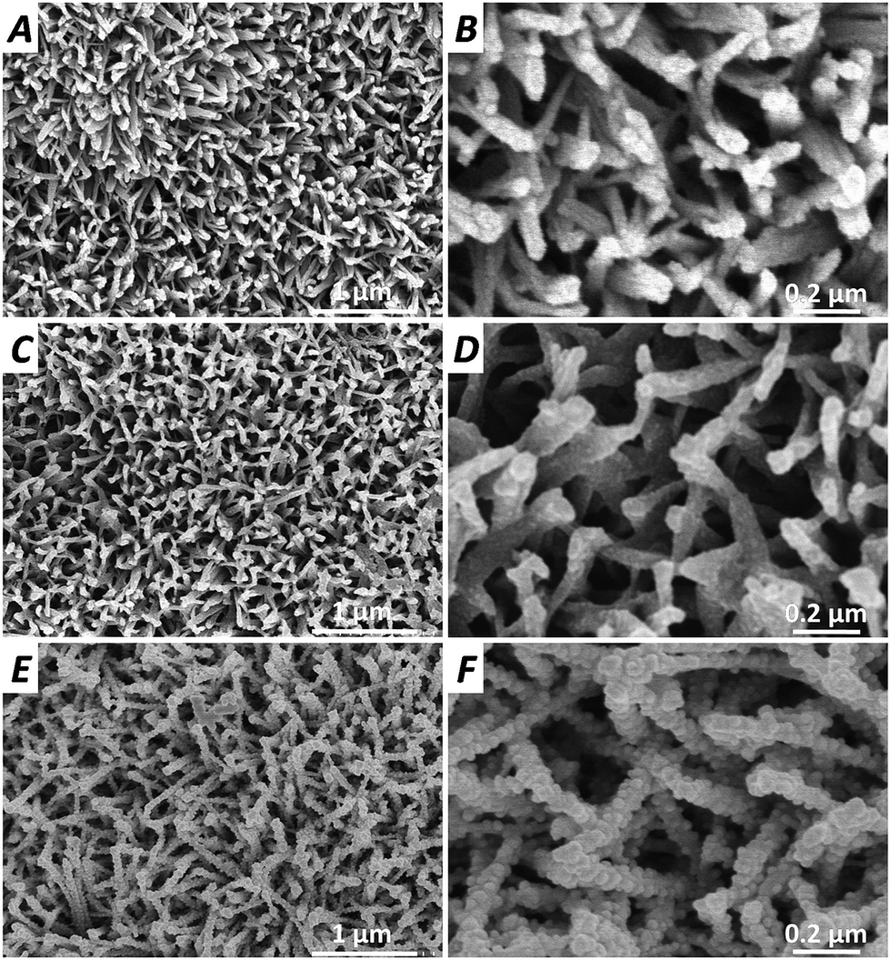 | ||
| Fig. 1 SEM images of TiOH (A and B), Ti–O–DA (C and D) and Ti–O–DA–Ag (E and F). | ||
The chemical composition of the surfaces at various stages of surface functionalization was determined using EDS. As shown in Fig. 2A, several types of peaks in the EDS spectrum were obtained from TiOH that corresponded to elemental titanium and oxygen. After dopamine functionalization (Fig. 2B), the presence of elemental carbon, which was not detected in the surface of TiOH, and the decrease in the atomic percentage of titanium indicate that dopamine was successfully immobilized onto the surface of TiOH. As shown in Fig. 2C, the atomic percentage of silver in the Ti–O–DA–Ag is 14.54%, indicating that a large amount of silver interlocked onto the dopamine-modified surface. The atomic percentages of titanium in the TiOH (A), Ti–O–DA (B), and Ti–O–DA–Ag (C) were 73.04%, 51.13%, and 34.04%, respectively, in turn, this further supports the conclusion that dopamine and AgNPs were successfully immobilized onto the surface of TiOH step by step.
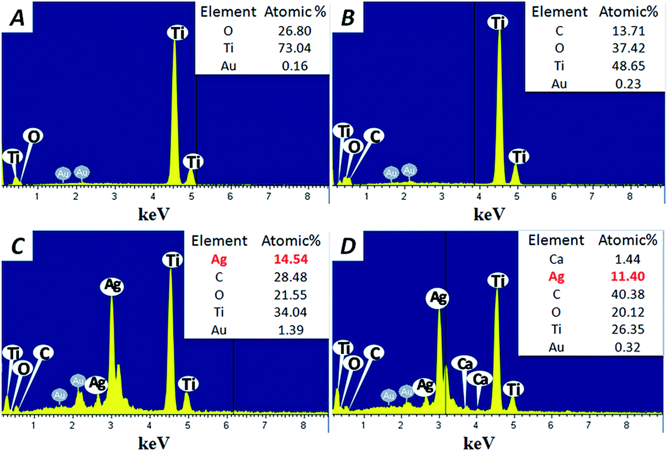 | ||
| Fig. 2 EDS pattern of DS pattern of TiOH (A), Ti–O–DA (B), and Ti–O–DA–Ag before (C) and after (D) incubation for one week in the calcification solution, the elemental atoms results in the inset table. | ||
The chemical composition of the surfaces at various stages of surface functionalization was determined by XPS. The XPS wide-scan spectra and the high-resolution spectra of Ag3d of the TiOH, Ti–O–DA, and Ti–O–DA–Ag are shown in Fig. 3. After dopamine functionalization (Ti–O–DA), the presence of N1s peak (∼399 eV) indicates that dopamine was successfully immobilized onto the surface of TiOH, because dopamine contains the large amount of nitrogen. Meanwhile, the Ti2p peak disappeared, indicating that dopamine had covered the substrate materials. Ag3d peak was clearly present in the Ti–O–DA–Ag, while two specific peaks with binding energies of 368.45 eV and 374.45 eV (shown in Fig. 3, right), were attributed to Ag3d5/2 and Ag3d3/2 electrons of Ag0, respectively. The spin energy separation was identified as 6.0 eV, indicating that the silver on the dopamine-modified surface is metallic Ag0 in nature;31 in turn, this further supported the conclusion that AgNPs have been successfully loaded on the surface.
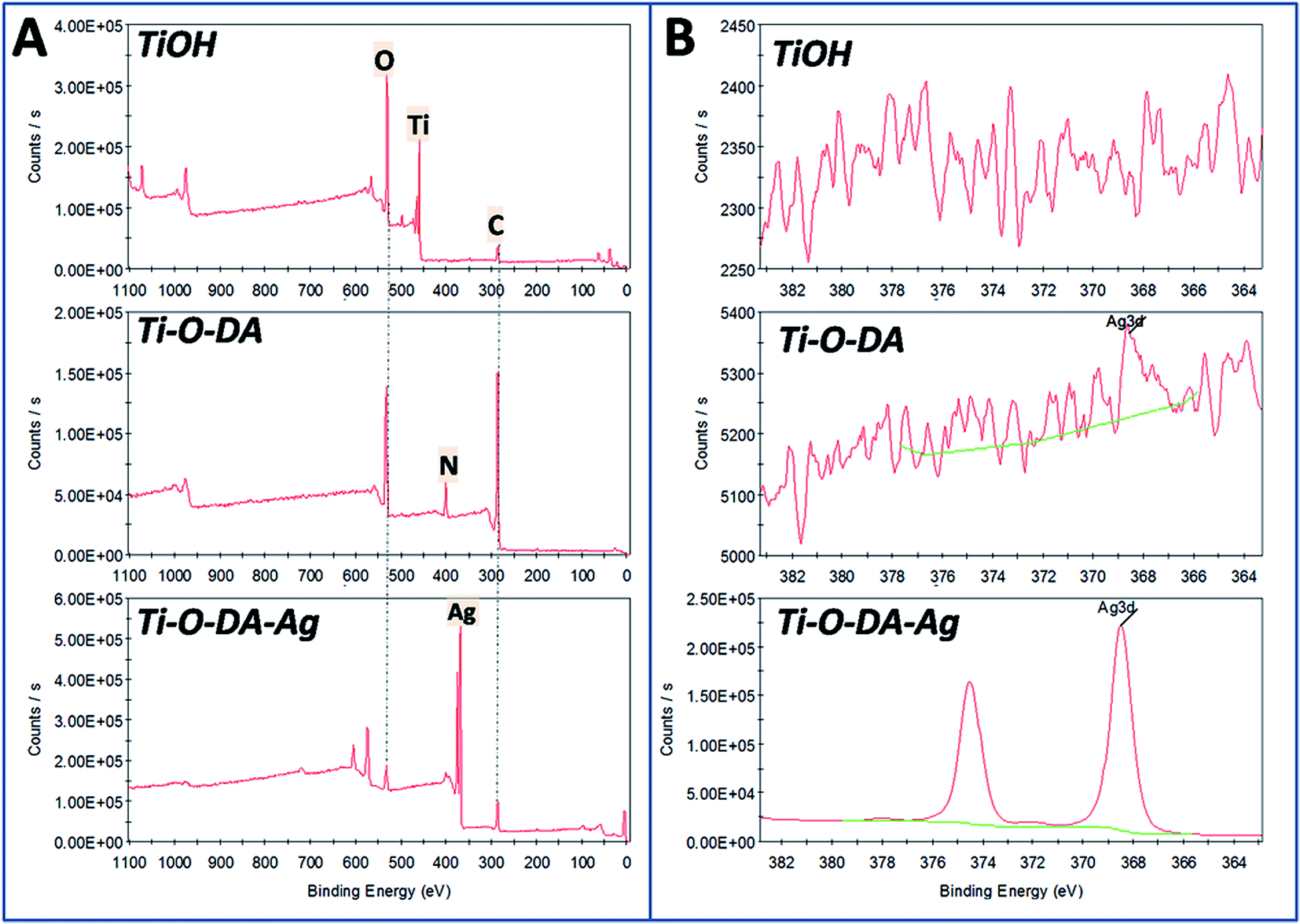 | ||
| Fig. 3 The XPS wide-scan spectra (A) and the high-resolution spectra of Ag3d (B) of TiOH, Ti–O–DA and Ti–O–DA–Ag. | ||
The measurement of the water contact angle (WCA) is well known as a useful technique to investigate surface characteristics. The WCA value of the different surfaces is shown in Fig. 4. Compared to the original Ti (79.0 ± 6.4°), the WCA value of TiOH decreased significantly to 25.3 ± 4.1°. After dopamine functionalization (Ti–O–DA), the WCA value increased significantly to 50.2 ± 5.4°. After AgNPs coating (Ti–O–DA–Ag), the WCA value further increased to 76.5 ± 6.7°. These results indirectly indicated that dopamine and AgNPs were successfully immobilized onto porous titanium surface.
 | ||
| Fig. 4 Water contact angle of Ti, TiOH, Ti–O–DA and Ti–O–DA–Ag. | ||
Mineralization of the multifunctional coating
Fig. 5 shows SEM images of the different surfaces obtained by incubation with calcification solution to assess the material's mineralization. After being soaked in the calcification solution for one week, the surface morphology of these samples was changed by mineralized products. It can be seen that the mineralized products filled into pore space and made the struts of TiOH grow thicker after incubation with calcification solution (Fig. 5A and B). Compared to TiOH, there were more mineralized products on the surfaces of Ti–O–DA (Fig. 5C and D) and Ti–O–DA–Ag (Fig. 5E and F), which almost covered these pores. Compared to spherical mineralized products on Ti–O–DA, the mineralized products on Ti–O–DA–Ag were irregular shape. These results indicate that the surfaces of Ti–O–DA and Ti–O–DA–Ag promote mineralization stronger than TiOH, one explanation for this is that the exposed dopamine on these surfaces promote mineralization in vitro.33 Further evidence is given by EDS in Fig. 2D. EDS reveals the presence of calcium element on the surface of Ti–O–DA–Ag after incubation with calcification solution for one week. This result also indicate that mineralization happened on the surface of Ti–O–DA–Ag, which is in good agreement with the SEM images (Fig. 5E and F). Because calcium phosphates can facilitate de novo bone formation at the bone–implant interface,39 we speculate that the surface of Ti–O–DA–Ag could adsorb mineral elements from body fluid to achieve mineralization during the early post-operative period and then improve the bone–implant fixation.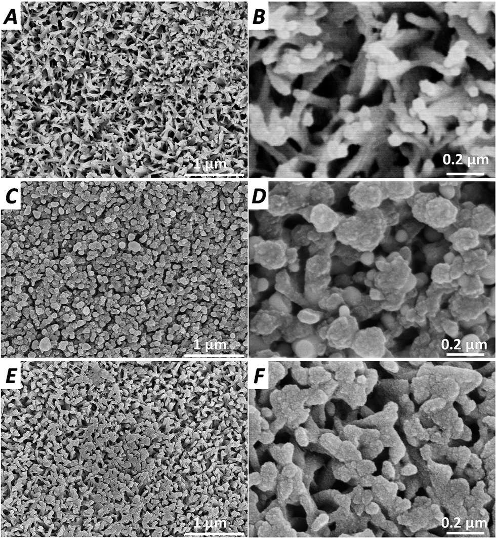 | ||
| Fig. 5 SEM images of TiOH (A and B), Ti–O–DA (C and D) and Ti–O–DA–Ag (E and F) after incubation for one week in the calcification solution. | ||
Silver release
Time-dependent silver release from AgNP-coated substrates was measured by ICP-OES and the silver release profiles are shown in Fig. 6. ICP-OES measurements revealed that the AgNP-coated surface in deionized water (Ti–O–DA–Ag) showed a slight rise in the first 2 days (3.95 ± 0.42 μg) and then a strongly sustained release silver release (>1.5 μg, inset, red spots), the cumulative release of silver into deionized water increase linearly with increased release time within 30 days. Meanwhile, a lot of silver sharply released into the calcification solution during first 10 days, after which little additional silver release was observed. The inset shows that the silver release every two days in the calcification solution decreased rapidly from 9.25 ± 0.42 μg to 1.21 ± 0.12 μg within the first 10 days. After 30 days of silver release, the remaining silver on the samples was 8.94 ± 0.62 μg (23.1% of total) for deionized water and 15.66 ± 0.49 μg (41.0% of total) for calcification solution, showing good durability. In other studies, the silver release from the AgNPs-modified titanium in phosphate buffered saline solution (PBS)21,40 or 0.85% NaCl solution41 is similar to it in calcification solution. The situation is somewhat different in deionized water, because ion exchange happened between silver on the samples and cation (e.g. Ca2+ and Na+) in the saline solution. With ion exchange, parts of surfaces were coated with cation (e.g. Ca2+ and Na+), which slowed down the silver release. Besides, the mineralized layer of Ti–O–DA–Ag (Fig. 5E and F) reduced the contact areas between AgNPs on the inner struts of porous structures and saline solution, and then the ion exchange at the inner sites of the porous structure was suppressed and the silver release decreased rapidly. | ||
| Fig. 6 Cumulative release of silver from AgNP-coated surfaces into 6 mL deionized water or calcification solution is sustained for 30 days. Solutions were changed every two day and silver concentration measured via ICP-OES. The inset is the silver mass released into solution every two days. | ||
Antibacterial activity
The viability of the attached bacterial cells was evaluated using a confocal laser scanning micrograph via staining with a combination of dyes. As shown in Fig. 7, the surface of the TiOH and Ti–O–DA supported rapid and extensive attachment of Escherichia coli (E. coli), Staphylococcus aureus (S. aureus), and Streptococcus mutans (S. mutans); however, attachment onto the AgNP-coated surface was suppressed by more than 95% compared to TiOH or Ti–O–DA over the same time period. Most of the bacterial cells on the surfaces of the TiOH and Ti–O–DA were viable (stained green) throughout the immersion period, while the dead bacterial cells (stained red) observed on these surfaces were mainly attributed to cell death during the bacterial growth process rather than antibacterial activity. For the AgNP-modified surface (Ti–O–DA–Ag), the number of bacterial cells decreased very significantly, and the percentage of dead cells (stained red) was higher than on the TiOH. Only a few sparsely distributed, single viable cells were observed, indicative of the high efficiency of AgNP conjugates in destroying the bacteria. This result indicated that the AgNP-modified surface inhibit the attachment and growth of bacteria. | ||
| Fig. 7 Representative confocal laser scanning micrographs of E. coli, S. aureus, and S. mutans on the TiOH, Ti–O–DA and Ti–O–DA–Ag surfaces, visualizing live (green) cells, dead (red) cells. | ||
The antibacterial activity of the different surfaces was investigated by measuring the ability to inhibit E. coli, S. aureus, and S. mutans growth around samples on agar culture plates, as shown in Fig. 8. Bacterial colonies were clearly observed in contact with TiOH and Ti–O–DA, while clear transparent rings (zone of inhibition) were obtained around Ti–O–DA–Ag, showing the inhibiting effect on bacterial activity. There results indicate that Ti–O–DA–Ag did inhibit growth or kill bacteria around them.
 | ||
| Fig. 8 Zone of inhibition (ZOI) testing of the TiOH, Ti–O–DA and Ti–O–DA–Ag surfaces against E. coli, S. aureus, and S. mutans. | ||
Previous studies revealed that AgNPs may lose their antibacterial activity in air within 5 days.42 To evaluate the samples' stability in air, all of the samples were stored in air for at least one week and then used in the experiment below. The bacterial growth in the solution with different samples was monitored by measuring the optical density at 660 nm (OD660). The higher the OD, the greater the opacity based on the turbidity of the cell suspension. As shown in Fig. 9, the surfaces without AgNPs (i.e., TiOH and Ti–O–DA) did not show noticeable antibacterial activity against E. coli, S. aureus, or S. mutans growth, as the curve was similar to that of the control bacteria. The growth of the three types of bacteria was completely inhibited when the samples of Ti–O–DA–Ag were immersed, confirming that AgNP-coated samples keep their antibacterial activity in air and inhibit bacterial growth in their local environment. It is reasonable to conclude that the surface of Ti–O–DA–Ag possess high and long-term antibacterial activity due to the high stability of AgNPs.
 | ||
| Fig. 9 Bacterial growth curves of E. coli, S. aureus, and S. mutans in LB media with different samples. Comparison of the antibacterial effect of the samples with and without AgNPs. The controls contain bacterial suspensions without samples (control). | ||
Osteoblast-compatibility
The mouse osteoblastic cell line (MC3T3-E1) was used to evaluate the osteoblast-compatibility of the different surfaces. Fig. 10 shows the typical fluorescence microscopic images of cytoskeletal actin of cells adherent on the different surfaces after 12 h and 24 h of culture, respectively. After 12 h of culture, the size of adherent cells on the TiOH (Fig. 10A) was bigger than it on other surfaces (Fig. 10B and C). Meanwhile, almost all adherent cells on the Ti–O–DA–Ag (Fig. 10B) were round and didn't spread, which indicated that this surface wasn't favorable for the osteoblast attachment. After incubation for one week in the calcification solution, the mineralized surface of Ti–O–DA–Ag was incubated with MC3T3-E1. Compared with pure Ti–O–DA–Ag, there were more cells adherent on the surface after mineralization (Fig. 10C), indicating that the mineralized surfaces of Ti–O–DA–Ag had better cytocompatibility. After 24 h of culture, cells on the TiOH (Fig. 10D) formed a confluent layer, but cells on the Ti–O–DA–Ag (Fig. 10E) were still detached and the cell morphology was thinner than on the TiOH, indicating that the surface of Ti–O–DA–Ag wasn't favorable for the osteoblast attachment and growth. The number of cell adherent on Ti–O–DA–Ag-mineralization (Fig. 10F) was higher than on the pure Ti–O–DA–Ag, while the cell morphology on the mineralized surface was similar to that on TiOH, indicating that the mineralized surface of Ti–O–DA–Ag improve osteoblast-compatibility. | ||
| Fig. 10 Immunofluorescent images of cytoskeletal actin for osteoblast (MC3T3-E1) on TiOH (A and D) and Ti–O–DA–Ag before (B and E) and after (C and F) incubation for one week in the calcification solution. | ||
The quantitative analysis of cells adhered on the different surfaces was performed by MTT assay and shown in Fig. 11A. The OD values on the TiOH were significantly higher than on the Ti–O–DA–Ag, indicating that the AgNP-coated surface showed some cytotoxicity. With the release of silver and the surface mineralization, the osteoblast-compatibility of this surface was significantly improved. After 5 d of culture, the MTT metabolism on Ti–O–DA–Ag-mineralization was higher than on TiOH, indicating that the mineralized surface of Ti–O–DA–Ag had good osteoblast-compatibility.
 | ||
| Fig. 11 (A) The quantitative analysis of osteoblast (MC3T3-E1) cultured on the different surfaces at days 1, 3, 5 and 7, measured by MTT assay; (B) ALP quantitative assay of MC3T3-E1 on different samples at days 4, 7 and 10. (n = 5) data presented as mean ± SEM and analyzed using a one-way ANOVA, *p < 0.01. | ||
Cell differentiation
Besides the initial cellular attachment and proliferation, the subsequent ALP activity is critical factor for enhanced osseointegration. ALP activity was measured after the cells were cultured for 4, 7 and 10 days on the different samples. The results of quantitative assay showed that ALP activity for cells cultured on all samples increased over time throughout the assay period (Fig. 11B). There were no significant differences between different samples at day 4, but Ti–O–DA–Ag-mineralization presented higher ALP activity than other samples at day 7 d and 10 d, indicating that the mineralized surface of Ti–O–DA–Ag could induce osteoblast differentiation.Discussion
It is well known that titanium, with its porous structure, has the merits of high bioactivity and lower elastic modulus.43,44 Also, a 3D porous structure, which is a characteristic feature of native bone tissue, could increase the specific surface area to improve the osseointegration of orthopedic implants.45 Dopamine could induce mineralization, which also can improve osseointegration.33 Here, compared to TiOH, more mineralized products presented on the surfaces of Ti–O–DA and Ti–O–DA–Ag (Fig. 6), indicating that dopamine on the Ti–O–DA–Ag could also enhance mineralization. Meanwhile, the mineralization weaken the silver release (Fig. 6). We speculate that the surface of Ti–O–DA–Ag could adsorb mineral elements from body fluid to achieve mineralization during the early post-operative period.All 3D porous structures with high porosity allow more bone ingrowth and therefore support improved anchorage in the surrounding bone,36 but such structures render the implant extremely susceptible to bacterial colonization and subsequent biofilm formation. After metal implants were introduced into human body, bacterial infections should be prevented in the first place.4,5 Silver nanoparticles were introduced to endow the 3D porous surface with antibacterial activity. To load AgNPs onto the surface, dopamine was used on the Ti–O–DA to reduce the Ag+ in the silver nitrate solution to AgNPs, due to the catechol groups in the dopamine.46 After reduction, the AgNPs were tightly bound to the dopamine-modified surface without aggregation (Fig. 1F). Compared to other reduction processes,47 no additional reductant or heating is needed. Thus, this strategy of obtaining the AgNP-modified surface is simple, facile, and environmentally friendly. Compared to TiO2 nanotubes (3D structure),48 such a porous structure could permit more AgNPs to be uniformly deposited not only on the top edges but also the inner porous structures, leading to a large number of AgNPs being carried on the surface, as shown in the EDS result (Fig. 2C). Good distribution and high-cover density of AgNPs on the samples are important for surface-based applications,49 so this strategy endows surfaces with excellent antibacterial activity, not only by inhibiting bacterial colonization on Ti–O–DA–Ag (Fig. 7) but also by inhibiting the growth of a wide antibacterial spectrum containing Gram-negative bacterium and Gram-positive bacteria around Ti–O–DA–Ag (Fig. 8 and 9), speculating that this surface could prevent bacterial infections during the early post-operative period. Dopamine, as a reducing and stabilizing agent,30,31 could hinder the oxidation and/or aggregation of AgNPs in air, leading to a significant reduction of antibacterial activity.50 Thus, AgNPs on Ti–O–DA–Ag could sustain their high antibacterial activity after exposure to air for at least one week (Fig. 9).
Cellular activities which indicate enhanced osteoblast functions include cell spreading, proliferation, and differentiation (e.g. ALP activity).32 The prerequisite for successful osseointegration of the implant in vivo is the attachment of osteoblasts to the implant surface. Though some studies reported that the AgNP-modified surfaces had no significant cytotoxicity,17,37,40 here, the surface of Ti–O–DA–Ag wasn't favorable for the osteoblast attachment (Fig. 10), proliferation (Fig. 11A), and increased enzyme production (Fig. 11B). After incubation with the calcification solution for one week, surface mineralization of Ti–O–DA–Ag happened and the osteoblast-compatibility of this surface was significantly improved. Osteoblast on Ti–O–DA–Ag-mineralization presented higher proliferation activity at 5 d (Fig. 11A) and higher ALP activity at 7 d (Fig. 11B) than on TiOH. Xu et al.51 also reported that a mussel-inspired Ca, P-modified polydopamine layer accelerated osteogenesis. We speculate that the surface of Ti–O–DA–Ag could adsorb mineral elements from body fluid to achieve mineralization during the early post-operative period and then the mineralized surface could be favorable for the osteoblast attachment, proliferation, and differentiation.
Conclusion
The coating with dopamine and silver nanoparticles (AgNPs) was successfully prepared on porous titanium surface by alkaline treatment immersion in a dopamine solution, followed by immersion in a silver nitrate solution. AgNPs with small size (30–50 nm) were fused in the top edges as well as the inner struts of the porous structure. More mineralization occurred on this coating than on TiOH, which led to a more rapid decrease of silver release from this coating in the calcification solution. The coating did inhibit bacterial colonization and therefore is suggested to eliminate the shortcoming of porous structure which render an implant extremely susceptible to bacterial colonization. The coating wasn't favorable for the osteoblast attachment, proliferation and differentiation, but this could be improved after mineralization. Therefore, the coating with dopamine and AgNPs on the surface with porous structure may not only reduce the risk of infection but also facilitate mineralization for osseointegration of titanium implants. This simple, facile, and environmentally friendly technique is therefore believed to have great potential for clinical application.Acknowledgements
This work was supported by the Natural Science Foundation of China (no. 31300792 and 31670967), China Postdoctoral Science Foundation (no. 2014M561813), the Natural Science Foundation of Anhui Province (no. 1408085QH149) and the Hong Kong Scholars Program.References
- L. Z. Zhao, P. K. Chu, Y. M. Zhang and Z. F. Wu, J. Biomed. Mater. Res., Part B, 2009, 91B, 470–480 CrossRef CAS PubMed.
- J. W. Costerton, Clin. Orthop. Relat. Res., 2005, 437, 7–11 CrossRef.
- D. W. Paquette, N. Brodala and R. C. Williams, Dent. Clin. North Am., 2006, 50, 361–374 CrossRef PubMed.
- E. M. Hetrick and M. H. Schoenfisch, Chem. Soc. Rev., 2006, 35, 780–789 RSC.
- R. O. Darouiche, N. Engl. J. Med., 2004, 350, 1422–1429 CrossRef CAS PubMed.
- C. R. Arciola, F. I. Alvi, Y. H. An, D. Campoccia and L. Montanaro, Int. J. Artif. Organs, 2005, 28, 1119–1125 CAS.
- K. C. Popat, M. Eltgroth, T. J. LaTempa, C. A. Grimes and T. A. Desai, Biomaterials, 2007, 28, 4880–4888 CrossRef CAS PubMed.
- V. Antoci, C. S. Adams, J. Parvizi, H. M. Davidson, R. J. Composto, T. A. Freeman, E. Wickstrom, P. Ducheyne, D. Jungkind, I. M. Shapiro and N. J. Hickok, Biomaterials, 2008, 29, 4684–4690 CrossRef CAS PubMed.
- C. Marambio-Jones and E. M. V. Hoek, J. Nanopart. Res., 2010, 12, 1531–1551 CrossRef CAS.
- D. E. Fullenkamp, J. G. Rivera, Y. K. Gong, K. H. Lau, L. He, R. Varshney and P. B. Messersmith, Biomaterials, 2012, 33, 3783–3791 CrossRef CAS PubMed.
- A. Rosenblatt, T. C. Stamford and R. Niederman, J. Dent. Res., 2009, 88, 116–125 CrossRef CAS PubMed.
- M. Fischer, M. Vahdatzadeh, R. Konradi, J. Friedrichs, M. F. Maitz, U. Freudenberg and C. Werner, Biomaterials, 2015, 56, 198–205 CrossRef CAS PubMed.
- Y. H. Zheng, J. B. Li, X. Y. Liu and J. Sun, Int. J. Nanomed., 2012, 7, 875–884 CAS.
- H. L. Cao, X. Y. Liu, F. H. Meng and P. K. Chu, Biomaterials, 2011, 32, 693–705 CrossRef CAS PubMed.
- V. E. Santos Jr, A. Vasconcelos Filho, A. G. Targino, M. A. Flores, A. Galembeck, A. F. Caldas Jr and A. Rosenblatt, J. Dent., 2014, 42, 945–951 CrossRef PubMed.
- T. Furuzono, T. Iwamoto, Y. Azuma, M. Okada and Y. Sawa, J. Artif. Organs, 2013, 16, 451–457 CrossRef CAS PubMed.
- B. S. Necula, J. P. T. M. van Leeuwen, L. E. Fratila-Apachitei, S. A. J. Zaat, I. Apachitei and J. Duszczyk, Acta Biomater., 2012, 8, 4191–4197 CrossRef CAS PubMed.
- W. Zhang, Y. J. Luo, H. Y. Wang, J. Jiang, S. H. Pu and P. K. Chu, Acta Biomater., 2008, 4, 2028–2036 CrossRef CAS PubMed.
- A. Ewald, S. K. Gluckermann, R. Thull and U. Gbureck, BioMedical Engineering Online, 2006, 5, 22 CrossRef PubMed.
- V. Antad, L. Simonot and D. Babonneau, J. Nanopart. Res., 2014, 16, 1–13 CrossRef.
- J. J. Wang, Z. Y. Li, Y. Q. Liang, S. L. Zhu, Z. D. Cui, H. J. Bao, Y. D. Liu and X. J. Yang, Mater. Express, 2015, 5, 191–200 CrossRef CAS.
- C. M. Xie, X. Lu, K. F. Wang, F. Z. Meng, O. Jiang, H. P. Zhang, W. Zhi and L. M. Fang, ACS Appl. Mater. Interfaces, 2014, 6, 8580–8589 CAS.
- N. Esfandiari, A. Simchi and R. Bagheri, J. Biomed. Mater. Res., Part A, 2014, 102, 2625–2635 CrossRef CAS PubMed.
- C. Baker, A. Pradhan, L. Pakstis, D. J. Pochan and S. I. Shah, J. Nanosci. Nanotechnol., 2005, 5, 244–249 CrossRef CAS PubMed.
- A. Panáček, L. Kvítek, R. Prucek, M. Kolář, R. Večeřová, N. Pizúrová, V. K. Sharma, T. j. Nevěčná and R. Zbořil, J. Phys. Chem. B, 2006, 110, 16248–16253 CrossRef PubMed.
- A. M. Mohammad, A. I. Abdelrahman, M. S. El-Deab, T. Okajima and T. Ohsaka, Colloids Surf., A, 2008, 318, 78–83 CrossRef CAS.
- J. H. Waite, Int. J. Adhes. Adhes., 1987, 7, 9–14 CrossRef CAS.
- A. GhavamiNejad, A. R. K. Sasikala, A. R. Unnithan, R. G. Thomas, Y. Y. Jeong, M. Vatankhah-Varnoosfaderani, F. J. Stadler, C. H. Park and C. S. Kim, Adv. Funct. Mater., 2015, 25, 2867–2875 CrossRef CAS.
- J. Chen, Q. Li, J. Xu, L. Zhang, M. F. Maitz and J. Li, J. Mater. Chem. B, 2015, 3, 2615–2623 RSC.
- J. B. Fei, J. Zhao, C. L. Du, A. H. Wang, H. Zhang, L. R. Dai and J. B. Li, ACS Nano, 2014, 8, 8529–8536 CrossRef CAS PubMed.
- H. Y. Luo, C. W. Gu, W. H. Zheng, F. Dai, X. L. Wang and Z. Zheng, RSC Adv., 2015, 5, 13470–13477 RSC.
- X. Hu, K. G. Neoh, Z. Shi, E. T. Kang, C. Poh and W. Wang, Biomaterials, 2010, 31, 8854–8863 CrossRef CAS PubMed.
- Y. Z. Zhou, Y. Cao, W. Liu, C. H. Chu and Q. L. Li, ACS Appl. Mater. Interfaces, 2012, 4, 6901–6910 CAS.
- K. L. Lin, L. G. Xia, J. B. Gan, Z. Y. Zhang, H. Chen, X. Q. Jiang and J. Chang, ACS Appl. Mater. Interfaces, 2013, 5, 8008–8017 CAS.
- L. G. Xia, K. L. Lin, X. Q. Jiang, Y. J. Xu, M. L. Zhang, J. Chang and Z. Y. Zhang, J. Mater. Chem. B, 2013, 1, 5403–5416 RSC.
- G. Ryan, A. Pandit and D. P. Apatsidis, Biomaterials, 2006, 27, 2651–2670 CrossRef CAS PubMed.
- X. M. Zhang, Z. Y. Li, X. B. Yuan, Z. D. Cui, H. J. Bao, X. Li, Y. D. Liu and X. J. Yang, Mater. Sci. Eng., C, 2013, 33, 2816–2820 CrossRef CAS PubMed.
- J. L. Chen, C. Chen, Z. Y. Chen, J. Y. Chen, Q. L. Li and N. Huang, J. Biomed. Mater. Res., Part A, 2010, 95A, 341–349 CrossRef CAS PubMed.
- M. Svehla, P. Morberg, B. Zicat, W. Bruce, D. Sonnabend and W. R. Walsh, J. Biomed. Mater. Res., 2000, 51, 15–22 CrossRef CAS PubMed.
- Z. J. Jia, P. Xiu, M. Li, X. C. Xu, Y. Y. Shi, Y. Cheng, S. C. Wei, Y. F. Zheng, T. F. Xi, H. Cai and Z. J. Liu, Biomaterials, 2016, 75, 203–222 CrossRef CAS PubMed.
- T. S. Sileika, H. D. Kim, P. Maniak and P. B. Messersmith, ACS Appl. Mater. Interfaces, 2011, 3, 4602–4610 CAS.
- Y. G. Wang, L. N. Cao, S. W. Guan, G. N. Shi, Q. Luo, L. Miao, I. Thistlethwaite, Z. P. Huang, J. Y. Xu and J. Q. Liu, J. Mater. Chem., 2012, 22, 2575–2581 RSC.
- G. A. Crawford, N. Chawla, K. Das, S. Bose and A. Bandyopadhyay, Acta Biomater., 2007, 3, 359–367 CrossRef CAS PubMed.
- J. L. Chen, Q. L. Li, J. Y. Chen, C. Chen and N. Huang, Appl. Surf. Sci., 2009, 255, 6894–6900 CrossRef CAS.
- S. Soumya, P. R. Sreerekha, D. Menon, S. V. Nair and K. P. Chennazhi, J. Mater. Chem., 2012, 22, 1904–1915 RSC.
- Z. Q. Shi, J. T. Tang, L. Chen, C. R. Yan, S. Tanvir, W. A. Anderson, R. M. Berry and K. C. Tam, J. Mater. Chem., 2015, 3, 603–611 RSC.
- V. K. Sharma, K. M. Siskova, R. Zboril and J. L. Gardea-Torresdey, Adv. Colloid Interface Sci., 2014, 204, 15–34 CrossRef CAS PubMed.
- Z. J. Guo, C. Chen, Q. Gao, Y. B. Li and L. Zhang, Mater. Lett., 2014, 137, 464–467 CrossRef CAS.
- S. K. Li, Y. X. Yan, J. L. Wang and S. H. Yu, Nanoscale, 2013, 5, 12616–12623 RSC.
- M. Lv, S. Su, Y. He, Q. Huang, W. B. Hu, D. Li, C. H. Fan and S. T. Lee, Adv. Mater., 2010, 22, 5463–5467 CrossRef CAS PubMed.
- M. C. Xu, D. Zhai, L. G. Xia, H. Li, S. Y. Chen, B. Fang, J. Chang and C. T. Wu, Nanoscale, 2016, 8, 13790–13803 RSC.
| This journal is © The Royal Society of Chemistry 2016 |