
 Open Access Article
 This Open Access Article is licensed under a
 Creative Commons Attribution 3.0 Unported Licence
Orthogonal halogen and hydrogen bonds involving a peptide bond model†
Vera
Vasylyeva
a,
Susanta K.
Nayak
b,
Giancarlo
Terraneo
*ab,
Gabriella
Cavallo
ab,
Pierangelo
Metrangolo
*abc and
Giuseppe
Resnati
ab
aNFMLab, D.C.M.I.C. “Giulio Natta”, Politecnico di Milano, Via Mancinelli 7, 20131 Milan, Italy. E-mail: pierangelo.metrangolo@polimi.it; giancarlo.terraneo@polimi.it; Fax: (+39) 02 2399 3180; Tel: (+39) 02 2399 3041
bCenter for Nano Science and Technology@Polimi, Istituto Italiano di Tecnologia, Via Pascoli 70/3, 20133 Milan, Italy
cVTT-Technical Research Centre of Finland, P.O. Box 1000, FI-02044 VTT, Finland
First published on 24th July 2014
Abstract
The peptide bond model N-methylacetamide self-assembles with a range of dihalotetrafluorobenzenes forming co-crystals that all show the occurrence of orthogonal hydrogen and halogen bonds.
Orthogonal self-assembly relies on the use of multiple interaction motifs applied in the same system to drive the assembly of different components. The specific and highly controllable interactions that are used do not influence each other's assembly profile and can be manipulated independently and simultaneously.1
In 2009, P. S. Ho and co-workers2 demonstrated, through a Protein Data Bank (PDB) survey, that in protein–ligand complexes hydrogen (HB) and halogen (XB) bonds3 occur orthogonally, in terms of both their geometric features and their chemical behaviour, when sharing the carbonyl oxygen atom of the peptide bond as a common bond acceptor. Lately, we have demonstrated that HB and XB can successfully be combined in an orthogonal manner to drive the self-assembly of complex and functional supramolecular networks.4 In this latter case, the two interactions did not share the same acceptor partner. Very recently, Bruce et al. successfully reproduced Ho's orthogonal motifs by using N-methylacetamide and N-methylbenzamide as peptide bond models with a selected number of iodinated XB donors.5 However, in their hands N-methylacetamide decomposed to methylacetamide and any attempt to get analogous motifs involving brominated donors failed.
In this communication, we demonstrate in small-molecule self-assembly that HB and XB occur orthogonally on the same acceptor site in a very reliable and consistent manner also when brominated XB donors are used. We chose N-methylacetamide (NMA, 1) as the smallest molecule that mimics the peptide bond (–NH–C![[double bond, length as m-dash]](https://www.rsc.org/images/entities/char_e001.gif) O) and thus the protein backbone.6 NMA effectively self-assembles with a range of dihalotetrafluorobenzenes resulting in co-crystals characterized by XBs occurring orthogonal to the classical HB pattern that characterizes the homomeric assembly of NMA.
O) and thus the protein backbone.6 NMA effectively self-assembles with a range of dihalotetrafluorobenzenes resulting in co-crystals characterized by XBs occurring orthogonal to the classical HB pattern that characterizes the homomeric assembly of NMA.
Very few examples of ‘engineered’ orthogonal XB and HB in the context of crystal engineering can be found in the Cambridge Structural Database (CSD),7 one exception being the isonicotinamide–chloroacetic acid co-crystal8 (details about the CSD search are given in the ESI†).
Our goal was to rationally design systems based on the orthogonal HB/XB supramolecular synthon and verify its robustness for use in crystal engineering. For this reason, NMA 1, affording the carbonyl oxygen acceptor site, was co-crystallized with several dihalotetrafluorobenzenes (DXTFBs), the XB donors, 1,2-dibromotetrafluorobenzene (12DBrTFB, 2a), 1,3-dibromotetrafluorobenzene (13DBrTFB, 2b), 1,4-dibromotetrafluorobenzene (14DBrTFB, 2c), 1,2-diiodotetrafluorobenzene (12DITFB, 2d), 1,3-diiodotetrafluorobenzene (13DITFB, 2e), and 1,4-diiodotetrafluorobenzene (14DITFB, 2f). The corresponding complexes 3a–f were obtained (Scheme 1).
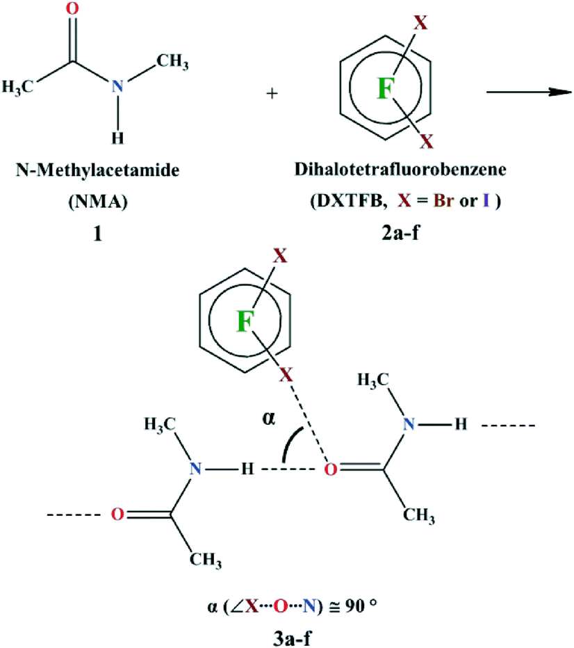 | ||
| Scheme 1 Synthesis of N-methylacetamide (NMA, 1) co-crystals with various dihalotetrafluorobenzenes (DXTFB, X = Br or I). The orthogonal angle (α) is defined as the angle between XB (C–X⋯O, X = Br or I) and HB (N–H⋯O), i.e., ∠X⋯O⋯N. | ||
We first re-determined the crystal structure of 1 at low temperature by in situ cryo-crystallization using a zone-melting procedure with an optical heating and crystallization device (OHCD).9 This was intended to get rid of static disorder that is found in the previously reported structures of NMA.10 A fully ordered crystal structure of 1 resulted in the Pnma space group. Classical N–H⋯O (1.878(2) Å)11 HBs link NMA molecules into infinite 1D chains running along the a crystallographic axis (Fig. 1). The two interacting groups N–H and CO are in the trans conformation, mimicking the molecular arrangement of the polypeptide backbone observed in β-sheet structures.12
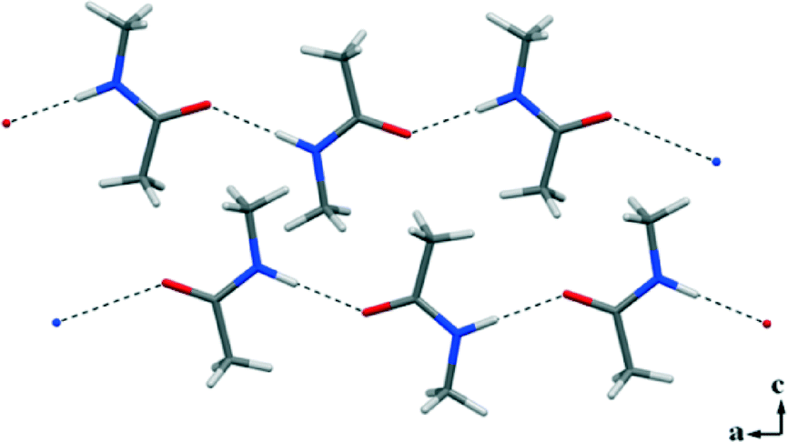 | ||
| Fig. 1 N–H⋯O HBs drive the formation of 1D infinite chains in the crystal packing of the homocrystal 1. | ||
DSC thermograms of 1![[thin space (1/6-em)]](https://www.rsc.org/images/entities/char_2009.gif) :1 mixtures of 1 and 2a–f revealed that both on cooling and on heating, the peaks for crystallization/melting of the starting compounds were not observed, confirming quantitative co-crystal formations with the adopted tectons' ratio. New melting endotherms appeared at temperatures higher than the melting point of pure NMA (12–20 °C), except for 3a that melted at 18 °C, giving a quite sharp peak (Table 1). Interestingly, the melting points of 3a, b, d, and e are higher than, and of 3c and f lower than, those of the corresponding XB donor tectons.‡
:1 mixtures of 1 and 2a–f revealed that both on cooling and on heating, the peaks for crystallization/melting of the starting compounds were not observed, confirming quantitative co-crystal formations with the adopted tectons' ratio. New melting endotherms appeared at temperatures higher than the melting point of pure NMA (12–20 °C), except for 3a that melted at 18 °C, giving a quite sharp peak (Table 1). Interestingly, the melting points of 3a, b, d, and e are higher than, and of 3c and f lower than, those of the corresponding XB donor tectons.‡
O and N–H stretching modes, melting points, and orthogonal angle α (∠X⋯O⋯N, with X = Br, I) of 1 and co-crystals 3a–f
Good-quality single crystals 3a–f were reproducibly obtained from 1:1 ratios of the starting compounds and successfully analysed by X-ray diffraction (3a and 3b are low-melting solids and required the OHCD method). All adducts show remarkable similarities in their supramolecular arrangements. The classical N–H⋯O HBs observed in the crystal structure of pure NMA are preserved in co-crystals 3a–f and organize the NMA molecules into infinite 1D chains as in pure 1. N–H⋯O contacts span the range 1.831(4)–2.182(1) Å that do not differ much from the values seen in pure NMA, although in all of the adducts the NMA molecule is disordered. Moreover, in all of the structures 3a–f the carbonyl O atom is involved in an additional XB involving at least one of the two XB donor sites of the dihalotetrafluorobenzenes used.
A distinctive feature of the XB is its directionality. As expected, it has been observed in the C–X⋯O angles of 3a–f, which span the range 168.5°–177.7°. X⋯O interaction distances are in the range 2.800(2)–2.871(4) Å for X = Br (3a–c) and 2.706(2)–3.001(1) Å for X = I (3d–f), which correlate well to the calculated interaction energy curves of different complexes between halophenyl derivatives and NMA.13
Only one of the XB donor sites of the DXTFBs in 3a–e is involved in the XB with the carbonyl oxygen atom. Differently, in 3f, both of the iodine atoms of 14DITFB are halogen-bonded to the carbonyl oxygens of two NMA molecules belonging to two different hydrogen-bonded chains. In all of the six co-crystals, the oxygen atom of NMA is simultaneously involved in short HBs. Special attention during the analysis of the crystal structures has been given to the angle α, which has been defined as the angle between XB and HB, ∠X–O–N as shown in Scheme 1. These angles in 3a–f vary in the range 77.9–98.5°, thus demonstrating the intrinsic tendency of HB and XB to occur orthogonal to each other when sharing the same sp2 O atom (Fig. 2).
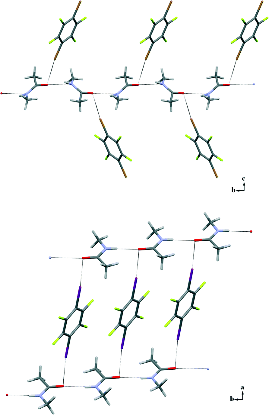 | ||
| Fig. 2 Partial crystal packing showing how infinite 1D chains (horizontally positioned) formed by NMA via N–H⋯O HBs further interact with 14DITFB via Br⋯O XBs in 3c (top) and I⋯O XBs in 3f (bottom). The disorder on the NMA molecule is omitted for clarity. | ||
Interestingly, while in 1 and 3f the N–H⋯O HB occurs perfectly in the carbonyl plane, in the structures of 3a–e it assumes an out-of-plane arrangement deviating from the carbonyl plane by angles in the range 36.19°–72.57°. The orthogonal XB occurs in an out-of-plane arrangement in all of the structures 3a–f, with the largest deviations in 3c and 3f where the positive σ-holes13 of bromine and iodine atoms enter the oxygen atom approximately in the equatorial region; the corresponding angles are 62.91° in 3c and 99.41° in 3f (see the ESI,† Fig. S4). Similar out-of-plane X⋯O interactions have been recently noted in protein–ligand complexes.14
The IR spectra of the co-crystals 3a–f essentially contain modified vibrations of both the corresponding starting compounds and the observed band shifts may give an indication of how much the orthogonal HB and XB perturb the electron density of the carbonyl group. Pure NMA shows a broad νCO centred at 1634 cm−1. This band is consistently red-shifted in all of the co-crystals 3a–f, the largest shift (27 cm−1) being observed in 3f. These red shifts suggest that the simultaneous formation of HB and XB results in a reduced electron density on the carbonyl group with respect to the pure NMA.15 Interestingly, iodinated co-crystals show larger shifts than brominated co-crystals, and para derivatives show larger shifts than ortho and meta derivatives. The shifts of the N–H stretching modes of the amide group (at 3290 cm−1 in pure 1) do not show any clear trend. Moreover, no correlation has been found between the N–H stretching values relative to the increasing energies of the XBs, as reflected in CO stretching, demonstrating that the interactions are truly independent (see plot in the ESI†).
In conclusion, we have reported the first halogen-bonded co-crystals formed by differently substituted (o, m, and p) dihalotetrafluorobenzenes (halogen = I and Br), functioning as halogen-bond donor modules, and N-methylacetamide, a well-known peptide bond model. Six co-crystals were obtained and they all show geometrically orthogonal hydrogen and halogen bonds involving simultaneously the carbonyl oxygen atom. This demonstrates the great robustness of this orthogonal synthon, which occurs in 100% supramolecular yield16 of the attempted co-crystals. The two interactions are also chemically orthogonal as the XB formation does not alter the 1D and hydrogen-bonded β-sheet mimetic chains typical of the homomeric assembly of N-methylacetamide. A PDB survey performed by Ho et al. showed that hydrogen and halogen bonds are orthogonal, both in terms of their geometric alignments and their chemical behaviour, when sharing the carbonyl oxygen atom of a peptide bond. Our results demonstrate that this feature is not a peculiarity occurring only when biomacromolecules are involved,3 but it is a general feature of the two interactions and is probably inherent to their intrinsic chemical nature.
The reported results pave the way to a new design concept in orthogonal self-assembly and crystal engineering and may also have important implications in other fields such as materials processing. As far as this field is concerned, amyloid and silk fibers are examples of ordered nanomaterials and they both feature robust β-sheet elements. The manipulation of the self-assembly and structural complexity of these nanomaterials during processing is still far from being completely understood.17 An approach based on the orthogonal interaction of β-sheets with halogen-bond donor mesogens may be particularly valuable.18 Current studies in our laboratory are addressing this issue and the results will be reported elsewhere.
Acknowledgements
This research was supported by the MIUR (FIRB project “FLUORIMAGING” no. RBAP1183B5) and the ERC (starting grant ERC-2012-StG_20111012 FOLDHALO, grant agreement number 307108).Notes and references
- M. D. Yilmaz and J. Huskens, Soft Matter, 2012, 8, 11768 RSC.
- A. R. Voth, P. Khuu, K. Oishi and P. S. Ho, Nat. Chem., 2009, 1, 74 CrossRef CAS PubMed.
- G. R. Desiraju, P. S. Ho, L. Kloo, A. C. Legon, R. Marquardt, P. Metrangolo, P. Politzer, G. Resnati and K. Rissanen, Pure Appl. Chem., 2013, 85, 1711 CrossRef CAS.
- J. Martí-Rujas, L. Colombo, J. Lü, A. Dey, G. Terraneo, P. Metrangolo, T. Pilati and G. Resnati, Chem. Commun., 2012, 48, 8207 RSC.
- A. Takemura, L. J. McAllister, S. Hart, N. E. Pridmore, P. B. Karadakov, A. C. Whitwood and D. W. Bruce, Chem. – Eur. J., 2014, 20, 6721 CrossRef CAS PubMed.
- NMA has been widely used as a model for peptide and protein folding studies, which resulted in various spectroscopic and theoretical investigations. In spite of its biological relevance, however, very few structural studies of NMA are known. The CSD, in fact, contains only three co-crystal structures of NMA with relevant organic compounds forming different hydrogen-bonded motifs (refcodes: IBOXOO, ANOPUP, ITYRMA10).
- CSD search (ConQuest Version 1.15, 2013).
- C. B. Aakeroy, A. M. Beatty and B. A. Helfrich, J. Am. Chem. Soc., 2002, 124, 14425 CrossRef PubMed.
- R. Boese and M. Nussbaumer, in Organic Crystal Chemistry, ed. J. B. Garbarczyk and D. W. Jones, Oxford University Press, Oxford, 1994, p. 20 Search PubMed.
- J. L. Katz and B. Post, Acta Crystallogr., 1960, 13, 624 CrossRef CAS; F. Hamzaoui and F. Baert, Acta Crystallogr., Sect. C: Cryst. Struct. Commun., 1994, 50, 757 CrossRef.
- T. S. Takhur, Y. Azim, T. Srinu and G. R. Desiraju, Curr. Sci., 2010, 98, 793 Search PubMed.
- S.-I. Mizushima, T. Simanouti, S. Nagakura, K. Kuratani, M. Tsuboi, H. Baba and O. Fujioka, J. Am. Chem. Soc., 1950, 72, 3490 CrossRef CAS.
- T. Clark, M. Hennemann, J. S. Murray and P. Politzer, J. Mol. Model., 2007, 13, 291 CrossRef CAS PubMed.
- L. A. Hardegger, B. Kuhn, B. Spinnler, L. Anselm, R. Ecabert, M. Stihle, B. Gsell, R. Thoma, J. Diez, J. Benz, J.-M. Plancher, G. Hartmann, D. W. Banner, W. Haap and F. Diederich, Angew. Chem., Int. Ed., 2011, 50, 314 CrossRef CAS PubMed.
- L. Russo, S. Biella, M. Lahtinen, R. Liantonio, P. Metrangolo, G. Resnati and K. Rissanen, CrystEngComm, 2007, 9, 341 RSC.
- C. B. Aakeröy, D. J. Salmon, M. M. Smith and J. Desper, Cryst. Growth Des., 2006, 6, 1033 Search PubMed.
- S. Ling, C. Li, J. Adamcik, S. Wang, Z. Shao, X. Chen and R. Mezzenga, ACS Macro Lett., 2014, 3, 146 CrossRef CAS.
- N. Houbenov, R. Milani, M. Poutanen, J. Haataja, V. Dichiarante, J. Sainio, J. Ruokolainen, G. Resnati, P. Metrangolo and O. Ikkala, Nat. Commun., 2014, 5, 4043, DOI:10.1038/ncomms5043.
- G. M. Sheldrick, Acta Crystallogr., Sect. A: Found. Crystallogr., 2008, 64, 112 CrossRef CAS PubMed.
- Mercury 3.3 is a software program for crystal structure visualization, exploration, and analysis, which is a copyright work of the Cambridge Crystallographic Data Centre (CCDC) and its licensors. Also see C. F. Macrae, I. J. Bruno, J. A. Chisholm, P. R. Edgington, P. McCabe, E. Pidcock, L. Rodriguez-Monge, R. Taylor, J. van de Streek and P. A. Wood, J. Appl. Crystallogr., 2008, 41, 466 CrossRef CAS.
Footnotes |
| † Electronic supplementary information (ESI) available: Experimental part, DSC, IR spectroscopic and crystallographic data. CCDC 899779–899785. For ESI and crystallographic data in CIF or other electronic format see DOI: 10.1039/c4ce01514b |
| ‡ Single-crystal X-ray diffraction data for 1 and 3a–f were recorded using Mo-Kα radiation on a Bruker KAPPA APEX II diffractometer. Data were collected in ω and φ scan with a scan width of 0.5. The data were reduced with empirical absorption corrections. Structures were solved by direct methods using SHELXL97.19 The molecular diagrams shown were generated using Mercury 3.3.20 The non-hydrogen atoms are refined anisotropically and hydrogen atoms were positioned geometrically for 3a–d. All crystallographic details are listed in Table S2 and intermolecular interactions are listed in Table S3 in the ESI.† |
| This journal is © The Royal Society of Chemistry 2014 |