AGuIX nanoparticle-nanobody bioconjugates to target immune checkpoint receptors†
Léna
Carmès‡
 ab,
Guillaume
Bort‡
 acd,
François
Lux
 *ae,
Léa
Seban
 f,
Paul
Rocchi
ab,
Zeinaf
Muradova
f,
Agnès
Hagège
f,
Paul
Rocchi
ab,
Zeinaf
Muradova
f,
Agnès
Hagège
 g,
Laurence
Heinrich-Balard
h,
Frédéric
Delolme
i,
Virginie
Gueguen-Chaignon
i,
Charles
Truillet
 j,
Stephanie
Crowley
k,
Elisa
Bello
k,
Tristan
Doussineau
b,
Michael
Dougan
k,
Olivier
Tillement
a,
Jonathan D.
Schoenfeld
f,
Needa
Brown
*fl and
Ross
Berbeco
*f
aInstitut Lumière-Matière, UMR 5306, Université Lyon1-CNRS, Université de Lyon, Villeurbanne Cedex 69100, France. E-mail: francois.lux@univ-lyon1.fr
bNH TherAguix SA, Meylan 38240, France
cInstitut Curie, PSL Research University, CNRS, UMR9187, INSERM, U1196, Chemistry and Modeling for the Biology of Cancer, F-91400, Orsay, France
dUniversité Paris-Saclay, CNRS, UMR9187, INSERM, U1196, Chemistry and Modeling for the Biology of Cancer, F-91400, Orsay, France
eInstitut Universitaire de France (IUF), Paris, France
fDepartment of Radiation Oncology, Brigham and Women's Hospital, Dana-Farber Cancer Institute, and Harvard Medical School, Boston 02115, USA. E-mail: ross_berbeco@dfci.harvard.edu
gUniversité de Lyon, CNRS, Université Claude Bernard Lyon 1, Institut des Sciences Analytiques, UMR 5280, 69100, Villeurbanne, France
hUniversité Lyon 1, CNRS, MATEIS, UMR5510, Univ Lyon, Université Claude Bernard Lyon 1, Villeurbanne 69100, France
iUniversité Lyon, Université Claude Bernard Lyon 1, ENS de Lyon, CNRS UAR3444, Inserm US8, SFR Biosciences, F-69007 Lyon, France
jUniversité Paris-Saclay, CEA, CNRS, Inserm, BioMaps, SHFJ, Orsay 91400, France
kDivision of Gastroenterology, Massachusetts General Hospital, Boston, MA 02114, USA
lDepartment of Physics, Northeastern University, Boston 02115, USA. E-mail: ne.brown@northeastern.edu
First published on 6th December 2023
Abstract
This article presents bioconjugates combining nanoparticles (AGuIX) with nanobodies (VHH) targeting Programmed Death-Ligand 1 (PD-L1, A12 VHH) and Cluster of Differentiation 47 (CD47, A4 VHH) for active tumor targeting. AGuIX nanoparticles offer theranostic capabilities and an efficient biodistribution/pharmacokinetic profile (BD/PK), while VHH's reduced size (15 kDa) allows efficient tumor penetration. Site-selective sortagging and click chemistry were compared for bioconjugation. While both methods yielded bioconjugates with similar functionality, click chemistry demonstrated higher yield and could be used for the conjugation of various VHH. The specific targeting of AGuIX@VHH has been demonstrated in both in vitro and ex vivo settings, paving the way for combined targeted immunotherapies, radiotherapy, and cancer imaging.
1. Introduction
In recent decades, inorganic nanoparticles (NPs) have demonstrated significant promise for oncologic indications. One noteworthy example is the AGuIX (activation and guidance of irradiation X) NPs. AGuIX are ultra-small NPs composed of gadolinium (Gd) chelates on a polysiloxane core, displaying a mean hydrodynamic diameter of 4 ± 2 nm. The corresponding mean molecular weight (MW) has been estimated at around 20 kDa. These NPs are currently being investigated in several clinical trials targeting different types of cancer, including brain metastasis (phase II: NCT03818386 and NCT04899908), cervical cancer (phase I: NCT03308604), glioblastoma (phase I/II: NCT04881032), and pancreatic and lung cancers (phase I/II: NCT04789486).1,2 AGuIX NPs possess theranostic properties, providing dual functionality for imaging and radiotherapy. This is achieved through the presence of paramagnetic gadolinium ions (Gd3+) embedded within the NPs. These gadolinium ions contribute to positive contrast effects in T1 magnetic resonance imaging (MRI) and enhance the effectiveness of radiotherapy due to their high atomic number (Z = 64).3,4 The small size of AGuIX NP enables preferential passive uptake in tumors while facilitating rapid elimination through the kidneys (in humans t1/2 = 1.29 ± 0.27 h), thus minimizing the risk of toxicity. Additionally, AGuIX NPs demonstrate tumor uptake comparable to that of other organic or inorganic NPs, achieving approximately 1.19 ± 0.87% ID per g through passive targeting after 24 hours in large variety of animal model.5,6 Nevertheless, to further enhance the efficacy of AGuIX NP, improving specificity and tumor retention time via active tumor targeting is an important next step for AGuIX translation and future generations.Preclinical studies employing bioconjugation of AGuIX with antibodies and peptides have showcased the effectiveness of active targeting, resulting in a minimum 1.43-fold increase in tumor accumulation.5,7–9 Peptides have gained significant popularity as ligands for targeting cancer cells due to their compact size and ease of production. However, they often exhibit low affinity for the target compared to antibodies.10 Conversely, antibodies have demonstrated remarkable efficacy as targeting molecules, but their larger size hampers tissue penetration and prevents access to certain areas in tumoral tissues.11 For these reasons, our work focuses on bioconjugates based on nanobodies, otherwise known as variable heavy domain of heavy chain (VHH). These targeting biomolecules have a molecular weight of approximatively 15 kDa and are roughly ten times smaller than monoclonal antibodies, and therefore offer enhanced efficiency in targeting.12 Their reduced size enables them to readily penetrate tumor tissues and bind to a greater number of receptors with enhanced affinity and specificity. Furthermore, VHH are swiftly eliminated from the bloodstream, minimizing the risk of toxic accumulation. They possess a remarkable folding capacity and robust physicochemical properties, endowing them with superior stability and excellent solubility.12–16 These distinctive features enable VHH to overcome some limitations associated with monoclonal antibody therapies.17,18 It is worth highlighting that both AGuIX NPs (nanoparticles) and VHHs (single-domain antibodies) exhibit a similar compact size, approximately 15 to 20 kDa, endowing them with similar in vivo properties, particularly in terms of tissue penetration, circulation, and clearance. By bioconjugating them, it is possible to combine these benefits in a single product. Herein, we design bioconjugates that target Programmed Death Ligand 1 (PD-L1) and the Cluster of Differentiation 47 (CD47) receptors by using the A12 and A4 VHH respectively.19–21 A12 exhibits high affinity in the low nanomolar range for human and murine PD-L1, making it suitable for targeting its expression. Similarly, A4 exhibits a binding affinity of approximately 10 pM for murine CD47.20,21
The PD-L1 ligand and the innate immune regulator CD47 are relevant immunotherapy targets. PD-L1 can be expressed on tumor cells and binds to PD-1 on the surface of T-cells, triggering immune cell exhaustion and inhibiting anti-tumor immunity. Interfering with this negative immune checkpoint, PD-L1 inhibitors have demonstrated efficacy across many different cancer types. Specifically, atezolizumab, an anti-PD-L1 therapy, was approved for treatment of lung cancer in the U.S. in 2016 after showing a 25.4% overall survival rate after 3 years in the treatment of metastatic non-squamous or squamous non-small cell lung cancer (NSCLC).22 Four years later, in 2020, duvarlumab, a second anti-PD-L1 antibody, was also approved for lung cancer treatment, demonstrating a 5-year overall survival rate of 42.9%, when administered following definitive chemoradiation therapy.23 These two antibodies have been approved as a second treatment after one or two doses of chemotherapy. However, despite these promising results, the majority of patients with solid tumors do not respond to PD-L1 therapy, highlighting the need for combination approaches and better patient stratification.24
CD47 is an important receptor expressed on tumor infiltrating macrophages that interacts with signal regulatory protein alpha (SIRPα). This regulator is overexpressed in various malignancies and prevents phagocytosis by providing a “don't eat me” signal to immune cells. Targeting and blocking CD47 on tumors restores this phagocytic response which can then upregulate secondary, adaptive responses.20,25–27 Antibodies targeting the CD47 receptor are currently being evaluated in multiple clinical trials.28–30
The limited efficacy of existing immunotherapy in treating most solid tumors has highlighted the need to better define the tumor immune microenvironment and develop combination treatment approaches. PD-L1/CD47 AGuIX combinations could help achieve both aims. Three of the approved immune checkpoint inhibitors (ICI) that target the PD-1/PD-L1 axis require biomarker confirmation of PD-L1, highlighting the need for PD-L1 in vivo diagnostic tools.31 Similarly, visualization of CD47 expression may help identify tumors amenable to macrophage based therapies. Both PD-L1 and CD47 inhibitors have been demonstrated to synergize with radiotherapy in preclinical models;32–35 enhancing the effectiveness of radiotherapy with AGuIX could further increase the potency of this combination. Therefore, our primary focus was on developing AGuIX NPs that could effectively target and delineate crucial immunomarkers, specifically PD-L1 and CD47. This research serves as a foundational demonstration of the chemical synthesis approach, aimed at creating a platform that harnesses both the diagnostic and therapeutic potential of AGuIX, in conjunction with the immunotherapeutic capabilities of immunocheckpoint targeting.
To achieve this objective, we grafted AGuIX NPs with two different VHHs for targeted applications using two highly specific techniques: (1) sortagging36–38 and (2) click chemistry.39–41 The sortagging is based on an enzyme specific reaction and click on an azide–alkyne reaction.40,42–45 A12 and A4-modified AGuIX were prepared to compare both conjugation techniques and to assess receptor targeting using various in vitro assays. For the selected coupling synthesis, the ex vivo approach was studied using A12 nanobody in a highly aggressive murine model of melanoma.
2. Materials and methods
2.1. AGuIX NPs
The Gd-chelated polysiloxane NPs (AGuIX) were provided by NH TherAguix (Meylan, France) as a lyophilized powder. Their synthesis has been extensively documented in the scientific literature.46,47 They contain roughly between 10 and 20 Gd atoms per particle, which can be quantified using ICP-MS (inductively coupled plasma-mass spectrometry).3,48 The presence of the gadolinium atoms confers to AGuIX radiation dose amplification and MRI contrast properties. Throughout the paper, each AGuIX NPs concentration is stated in g L−1 of AGuIX NP or M of Gd element. Within the polysiloxane structure AGuIX contain primary amines in the (3-aminopropyl)triethoxysilane (APTES) function.49,50 Each NP is estimated to possess a similar number of amine functions as Gd chelates.51,52 These amines will be utilized as the main functional groups for all the biofunctionalizations. The AGuIX NPs used in this study have all been pre-grafted on the free amino function present on polysiloxane matrix with the cyanine 5.5 fluorescent dye (detailed synthesis in ESI†).2.2. VHHs
The A12 and A4 VHHs were synthesized at the Massachusetts General Hospital (USA), as described a previously published protocol.21 A12 and A4 sequences were sub-cloned into the WK6 E. coli periplasmic expression vector pHEN6 to enable Gibson cloning and the inclusion of a C-terminal sortase motif and 6xHis tag. E. coli containing the plasmid were grown to mid-log phase at 37 °C and VHH expression induced with 1 mM IPTG at 30 °C overnight. Centrifugation (5000g, 15 min, 4 °C) was used to harvest the cells and resuspend them in 25 mL 1× TES buffer. Cells were submitted to osmotic shock in 1![[thin space (1/6-em)]](https://www.rsc.org/images/entities/char_2009.gif) :
:![[thin space (1/6-em)]](https://www.rsc.org/images/entities/char_2009.gif) 4 0.25× TES buffer overnight at 4 °C. The periplasmic fraction was separated by centrifugation (8000 rpm, 30 min, 4 °C) and loaded onto Ni-NTA beads (Qiagen) and eluted in 50 mM Tris, pH 8, 150 mM NaCl, 500 mM imidazole. Eluted protein was loaded onto a Superdex 75 10/300 column. Recombinant VHH purity was assessed by SDS-PAGE and concentrated with an Amicon 10
4 0.25× TES buffer overnight at 4 °C. The periplasmic fraction was separated by centrifugation (8000 rpm, 30 min, 4 °C) and loaded onto Ni-NTA beads (Qiagen) and eluted in 50 mM Tris, pH 8, 150 mM NaCl, 500 mM imidazole. Eluted protein was loaded onto a Superdex 75 10/300 column. Recombinant VHH purity was assessed by SDS-PAGE and concentrated with an Amicon 10![[thin space (1/6-em)]](https://www.rsc.org/images/entities/char_2009.gif) 000 kDa filtration unit (Millipore). VHH were stored at −80 °C.
000 kDa filtration unit (Millipore). VHH were stored at −80 °C.
2.3. Synthesize of AGuIX@VHH by sortagging
AGuIX-Cy5.5-C(W/T)GGG (600 μM GGG(W/T)C final concentration) and VHH (30 or 40 μM final concentration) were introduced in buffer (50 mM Tris-HCl, pH 7.5, 150 mM NaCl). Ligation was triggered (t0) by the addition of StrA7 m (2.5 μM final concentration). The mixture was stirred (orbital) at room temperature for 3 h. After reaction, the crude was negatively selected using Ni-NTA bead (400 μL slurry for 1 mg VHH, spin at 800 G for 1 min) to remove 6xHis-tag starting materials (StrA7 m, VHH and released VHH residue after ligation), and filtered (MWCO 50 kDa vivaspin, dilution ×100![[thin space (1/6-em)]](https://www.rsc.org/images/entities/char_2009.gif) 000). AGuIX@VHH was purified by SEC (size exclusion chromatography, method B detailed in ESI†) and stored at −20 °C. AGuIX-Cy5.5-C(W/T)GGG preparations and synthesis optimizations are detailed in ESI.†
000). AGuIX@VHH was purified by SEC (size exclusion chromatography, method B detailed in ESI†) and stored at −20 °C. AGuIX-Cy5.5-C(W/T)GGG preparations and synthesis optimizations are detailed in ESI.†
2.4. Synthesize AGuIX@VHH through click chemistry
A12-azide (50 μM, final concentration in reaction) was added to AGuIX-DBCO at (100 AGuIX mg mL−1 final concentration in reaction) in 10 mM phosphate-buffered saline (PBS) solution. The solution was stirred for 24 h at 4 °C. A temporal follow-up of the click reaction is monitored by SEC using method A (detailed in ESI†). After reaction, AGuIX@VHH was purified by SEC (method B) to remove sub-products (NHS-terminated, azide-PEG4-NHS and AGuIX-DBCO excess) and stored at −20 °C. AGuIX-Cy5.5-DBCO and A12-azide preparations and synthesis optimizations are detailed in ESI.†2.5. Material characterizations
2.6. Competition ELISA
High-affinity 96-well plates were coated with either 2 μg mL−1 PD-L1 or 1.25 μg mL−1 CD47 and allowed to adhere overnight at 4 °C. Plates were washed and blocked with a 10% FBS solution. Plates were incubated with sample VHH (A12 or A4) or NPs (AGuIX, AGuIX@A12, or AGuIX@A4) followed by incubation with 6.25 nM biotin-anti-PD-L1 or 50 nM biotin-anti-CD47. Plates were then incubated with avidin-HRP (Abcam ab7403, 1![[thin space (1/6-em)]](https://www.rsc.org/images/entities/char_2009.gif) :
:![[thin space (1/6-em)]](https://www.rsc.org/images/entities/char_2009.gif) 40
40![[thin space (1/6-em)]](https://www.rsc.org/images/entities/char_2009.gif) 000 dilution) followed by TMB (ThermoFisher N301) to detect colorimetric changes. TMB conversation was stopped at 40 min and absorbance was read at 450 nm. The equilibrium inhibitory dissociation constant (Ki) was curve fitted using Graphpad 8.0.1 Top and Bottom are the plateaus in the units of the y-axis. Log
000 dilution) followed by TMB (ThermoFisher N301) to detect colorimetric changes. TMB conversation was stopped at 40 min and absorbance was read at 450 nm. The equilibrium inhibitory dissociation constant (Ki) was curve fitted using Graphpad 8.0.1 Top and Bottom are the plateaus in the units of the y-axis. Log![[thin space (1/6-em)]](https://www.rsc.org/images/entities/char_2009.gif) Ki is the log of the molar equilibrium dissociation constant of the unlabeled ligand (AGuIX, AGuIX@A12, or AGuIX@A4). RadioligandNM is the concentration of the labeled ligand (biotin-anti-PD-L1 or biotin-anti-CD47) and HotKdNM is the equilibrium dissociation constant of the labeled ligand (biotin-anti-PD-L1 or biotin-anti-CD47).
Ki is the log of the molar equilibrium dissociation constant of the unlabeled ligand (AGuIX, AGuIX@A12, or AGuIX@A4). RadioligandNM is the concentration of the labeled ligand (biotin-anti-PD-L1 or biotin-anti-CD47) and HotKdNM is the equilibrium dissociation constant of the labeled ligand (biotin-anti-PD-L1 or biotin-anti-CD47).
2.7. Biacore analyses
Biomolecular interactions between immobilized receptor PD-L1 and CD47 and analyte AGuIX@A12 or AGuIX@A4 were assessed by Surface Plasmon Resonance (SPR) on Biacore 2000 instrument (Cytiva).53 For the kinetic assays, the A12 was injected up to a concentration of 61.2 nM, the A4 up to 67 nM, the AGuIX NPs at 0–915.7 nM in Gd3+, the AGuIX@A12 VHH at 0–61.2 nM, (0–915.7 nM equivalent in Gd3+) and the AGuIX@A4 VHH at 0–16.7 nM (0–240 nM equivalent in Gd3+). The chip was prepared as described in ESI.† All characteristic interaction constants (equilibrium dissociation (KD) and kinetic rate of association and dissociation (kA and kD)) were determined by curve fitting using the Langmuir 1![[thin space (1/6-em)]](https://www.rsc.org/images/entities/char_2009.gif) :
:![[thin space (1/6-em)]](https://www.rsc.org/images/entities/char_2009.gif) 1 binding model implemented in Biaevaluation software 4.1.1.54 The detailed method is described in ESI.†
1 binding model implemented in Biaevaluation software 4.1.1.54 The detailed method is described in ESI.†
2.8. Cell culture
Murine melanoma (wild-type B16F10 or hiPDL1-B16F10) cells were culture in 10% fetal bovine serum (FBS; Invitrogen, USA) and 1% pen/strep (10![[thin space (1/6-em)]](https://www.rsc.org/images/entities/char_2009.gif) 000 U mL−1 penicillin and 10
000 U mL−1 penicillin and 10![[thin space (1/6-em)]](https://www.rsc.org/images/entities/char_2009.gif) 000 μg mL−1 streptomycin; Invitrogen, US) supplemented Roswell Park Memorial Institute medium (RPMI 1640; Gibco, Invitrogen, USA) at 37 °C, 5% CO2 and optimal humidity.
000 μg mL−1 streptomycin; Invitrogen, US) supplemented Roswell Park Memorial Institute medium (RPMI 1640; Gibco, Invitrogen, USA) at 37 °C, 5% CO2 and optimal humidity.
2.9. Fluorescence microscopy
Cover slips were placed in 48-well plates and seeded with either hiPDL1-B16 or wild-type B16 cells (50![[thin space (1/6-em)]](https://www.rsc.org/images/entities/char_2009.gif) 000 cells per well) and allowed to attached overnight at 37 °C. Cells were incubated with Cy5.5 (683/703 nm) tagged NPs (0.2 mg mL−1) for 1 h. Cells were fixed, blocked, and permeabilizing with a solution of 10% FBS and 0.3% Triton X-100. Cells were stained with CellMask Orange Plasma Membrane stain (ThermoFisher, 554/567 nm) and mounted with DAPI (350/470 nm) and Flouromount-G. Images were taken using a Zeiss AxioObserver microscope 63x. Fluorescence images were analyzed using ImageJ (version 2.14.0/1.54f) and corrected total cell fluorescence was calculated: (CTCF = integrated densitycell – (areacell × mean fluorescencebackground)).
000 cells per well) and allowed to attached overnight at 37 °C. Cells were incubated with Cy5.5 (683/703 nm) tagged NPs (0.2 mg mL−1) for 1 h. Cells were fixed, blocked, and permeabilizing with a solution of 10% FBS and 0.3% Triton X-100. Cells were stained with CellMask Orange Plasma Membrane stain (ThermoFisher, 554/567 nm) and mounted with DAPI (350/470 nm) and Flouromount-G. Images were taken using a Zeiss AxioObserver microscope 63x. Fluorescence images were analyzed using ImageJ (version 2.14.0/1.54f) and corrected total cell fluorescence was calculated: (CTCF = integrated densitycell – (areacell × mean fluorescencebackground)).
2.10. Animal tumor model
Animal experiments were performed according to the European Directive 2010/63/EU and its application in French law (Decree No. 2013-118). Research was approved by a local ethics committee (APAFIS #34522-2022010412087915 v1). The Comité d'Ethique en Expérimentation Animale (CEtEA) at the Commissariat à l’énergie atomique et aux énergies alternatives (CEA) at Ile De France (IdF) is registered by the French Ministry of Research (CEEA No. 44). This committee examines applications for authorization of projects using animals, and issues opinions to the French Ministry of Higher Education, Research and Innovation (MESRI), authorized to issue authorizations for animal experimentation. Experiments were conducted at the imaging facility CEA-SHFJ (authorization D91-471-105/ethics committee no. 44). C57BL/6J mice (Janvier-Labs, France) were housed by 6 mice in each cage (bedding material: aspen wood) at room temperature 22 °C, humidity 40%, under a regular 12 h dark/light cycle. Food and water were available ad libitum. 6 weeks old female C57BL/6 mice were purchased from Janvier laboratory. B16F10 cells (ATCC, USA) were cultured in DMEM media (Life Technologies, France) supplemented with 10% FBS (Life Technologies, France) and 1% penicillin/streptomycin (100 mg mL−1), and 1% l-glutamine, and maintained at 37 °C with 5% CO2 until confluence. Mice were subcutaneously injected with 1 × 10⁶ B16F10 cells suspended in DPBS (1 × 100 μL) into both flanks while anesthetized with 2% isoflurane.2.11. Ex vivo biodistribution
The animals were anaesthetized with isoflurane (induction: 3%, maintenance: 1.5–2.0%) in a mixture of 100% O2 (flow rate = 1.0–1.5 L min−1). At day 8 post-inoculation, 1.92 ± 0.09 MBq (mean ± SD), corresponding to the dose of 7.12 ± 0.35 and 2.29 ± 0.07 μg of AGuIX (mean ± SD) for AGuIX-[89Zr]- and for AGuIX-[89Zr]@A12 respectively,55 were intravenously injected (i.v.) into tumor-bearing mice (n = 8 for both AGuIX and AGuIX-A12, 22.0 ± 1.9 g per mouse). The mice were euthanized at 4 h and 24 h post-injection (n = 4 per time points) and tissue activity was determined for several organs of interest after harvesting (blood pool, intestines, kidneys, spleen, pancreas, liver, muscle, bone, brain, tumor). Activity in various organs of interest is represented in percentage of injected dose per gram (%ID per g) (Table S2†).2.12. Statistical analysis
The results are reported as mean ± SEM, as stated in the figure captions. Statistical analyses were conducted using GraphPad Prism (version 8.0.1). For comparisons involving three or more means, a one-way ANOVA followed by the Kruskal–Wallis nonparametric test (for non-Gaussian populations) was employed for in vitro internalization assays. All in vitro experiments were performed in triplicate. Mann–Whitney test for multiple comparisons was used to analyze ex vivo biodistribution data. Statistical significance was considered at p < 0.05.3. Results and discussion
To evaluate the best approach for biofunctionalization of AGuIX NPs with VHH, we compared sortagging and click chemistry. The sortagging bioconjugation approach permits for the selection of specific sites for modification, enabling more accurate predictions regarding the biological impact of the chemical alteration. Modifying a protein terminus is expected to have a smaller effect on the protein's folding and functionality as well as the added advantage of defining the orientation of the immobilized protein, which is anticipated to better preserve its function compared to other immobilization techniques. This enzymatic reaction relies on the enzyme Sortase A (SrtA), a transpeptidase produced by Gram-positive bacteria, which catalyzes the formation of a peptide bond between two peptides to attach specific proteins to the cell wall or pili assembly.56–58 SrtA specifically recognizes the LPXTG sequence (with X representing any amino acid) and by nucleophilic attack makes it reactive to an N-terminal oligoglycine, forming a peptide bond between the threonine of LPXT and the oligoglycine. This natural ligation system has been repurposed for protein modification and has gained popularity as a research tool due to its high ligation selectivity, simplicity, robustness, and the availability of various SrtA variants (expressed in Escherichia coli or commercially available), as well as other required materials.37,59 Sortagging reactions have been used to graft specific biomolecules, such as peptides or proteins, onto a protein or other biological targets of interest and has already proved efficacious in NP conjugation.60–62Click chemistry describes chemical reactions aimed at achieving high yield and high selectivity in the formation of carbon-heteroatom bond. Click chemistry reactions are based on the 1,3-dipolar cycloaddition of an azide and an alkyne to form a 1,2,3-triazole. This reaction has been widely utilized in various applications due to its simplicity in terms of preparation and purification steps, enabling rapid generation of new products with high reaction rates, such as bioconjugates.60,63–65 The formation of the triazole linkage is irreversible and quantitative, providing excellent reaction stability. Azide and dibenzocyclooctyne (DBCO) are selected as strain-promoted azide–alkyne cycloaddition reactions (SPAAC) tools due to their relatively small functional groups with a favorable rate constant (k2 = 0.2–0.5 M−1 s−1)39 which have been shown to be effective. The crucial challenge of click chemistry is to preserve the integrity and functionality of the biomolecule after grafting.39,66,67 Given the advantages and challenges associated with each method, we have synthesized AGuIX and VHH conjugates using both approaches to compare them.
3.1. Synthesis of AGuIX-VHH by sortagging
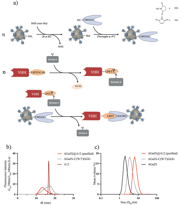
The sortagging reaction is based on enzymatic synthesis involving three primary steps: (1) introduction of maleimide (Mal) moieties on AGuIX NPs, (2) introduction of the peptide GGG(W/T)C using the Mal moieties, and (3) introduction of VHH through the transpeptidation enzymatic reaction (Fig. 1a).AGuIX NPs were modified to mimic the functionality of the N-terminal oligoglycine residue typically employed in sortagging. Initially, a Mal functional group was introduced onto the surface of AGuIX NPs using a bifunctional NHS/Mal linker. AGuIX-Mal was further modified with a short peptide linker that incorporates a cysteine amino acid at the C-terminal and a triglycine residue (GGG(W/T)C) at the N-terminal. To enable the use of sortagging, the VHH was engineered by incorporating a C-terminal StrA motif LPETGG and a 6xHis tag. The addition of the 6xHis tag motif facilitated protein purification and removal of the C-terminal residue released during sortagging.20 The transpeptidation reaction between A12 and AGuIX-C(W/T)GGG was verified using SEC monitoring. After purification, the final product exhibited a retention time (tR) of 13.2 minutes, corresponding to a product size ranging from 29 to 44 kDa (Fig. 1b and Fig. S1c†). The equilibrium for the reaction was reached within 2–3 h at room temperature. Following isolation using Ni-beads to capture and remove the 6xHis-containing reagents and by-products (StrA7 m, residual peptides from VHH C-terminal, and unreacted VHH), AGuIX@A12 was purified via filtration and dispersed in PBS (Fig. S1c†). A VHH conversion of 32–33% was achieved during the sortagging process, resulting in an isolated yield of AGuIX@A12 of 5.3% (relative to A12) with a Gd/A12 ratio of 20 (Fig. 1b and Fig. S1e†). The hydrodynamic diameter (DH) of AGuIX@A12, as assessed by DLS, was 6.1 nm, slightly larger than that of AGuIX-C(W/T)GGG (4.1 nm) (Fig. 1c). This outcome confirms the modification of the particle surface and hints at the possibility of VHH grafting. After optimization, sortagging proved successful in grafting VHH onto the surface of AGuIX NPs. The key parameter to increase the grafting yield of VHH was to increase the equivalence of the oligoglycine in the reaction. However, this strategy also resulted in a higher NP/A12 ratio, which ideally should be around 1 for imaging applications. The NP/A12 ratio of 1 could be achieved after optimization of several parameters (Fig. S1a–e†), while VHH conversion decreased to 15%. The binding affinity, evaluated using the inhibitory constant (Ki) through a competitive enzyme-linked immunosorbent assay (ELISA), confirmed that the addition of AGuIX to A12 did not impact the competitive binding affinities (Fig. S2†). Furthermore, AGuIX's lack of interaction with PD-L1 proteins also confirmed that the high affinity is solely attributed to the presence of the nanobody on AGuIX's surface. Moreover, the similar log![[thin space (1/6-em)]](https://www.rsc.org/images/entities/char_2009.gif) Ki values of 12.8 ± 1.1 nM, and 5.5 ± 0.6 nM for A12 and AGuIX@A12 obtained, respectively, strongly reinforced this result (Fig. S2†).
Ki values of 12.8 ± 1.1 nM, and 5.5 ± 0.6 nM for A12 and AGuIX@A12 obtained, respectively, strongly reinforced this result (Fig. S2†).
3.2. Synthesis of AGuIX@VHH by click chemistry
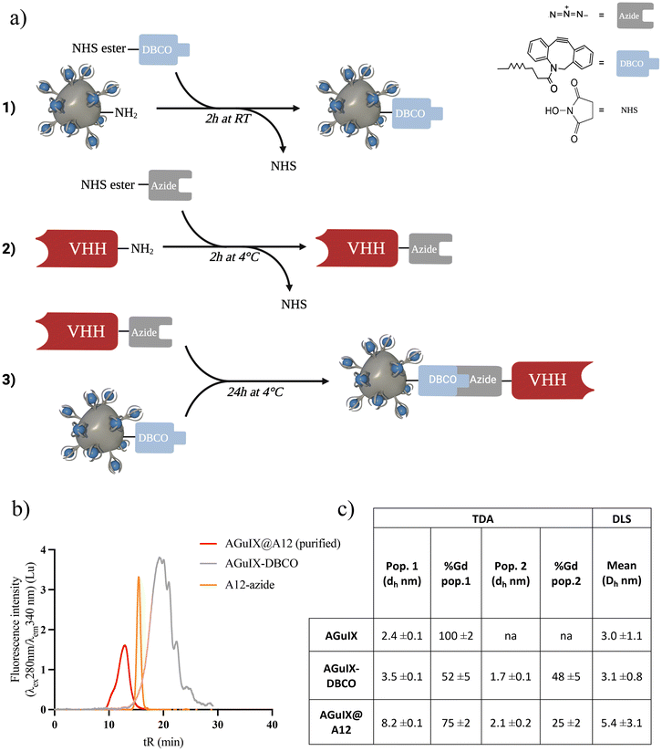
Click chemistry based on strain-promoted azide–alkyne cycloaddition involves three primary steps: (1) introduction of the azide group on the VHH, (2) introduction of the DBCO group on the AGuIX NP, and (3) introduction of the VHH through the click chemistry reaction (Fig. 2a). The first step of the synthesis involves functionalizing the VHH with the azide group based on NHS-ester reaction with lysine residues.68–70 Functionalization was confirmed by SEC (Fig. S3a†) and matrix-assisted laser desorption/ionization-time of flight (MALDI-TOF) mass spectrometry (Fig. S3b†).68The AGuIX-DBCO was prepared by the same reaction consisting in NHS reaction with the primary amines on the NP surface. After synthesis and purification, the modified NP presented a ratio of 10 Gd/DBCO, i.e. 1 to 2 DBCO functions per NP (Fig. S3c and d†). AGuIX-DBCO high excess ratio (450Gd/A12) was chosen for the click chemistry to minimize any reactant rate limitations and ensure complete reaction (Fig. 2b). At least 95% of the VHH in solution successfully reacted with AGuIX, resulting in the formation of AGuIX@A12 with a size ranging from approximately 29 to 44 kDa, as determined by Superdex 75 protein calibration (Fig. 2d and Fig. S3e, f†). Purification by preparative SEC was effective in removing the unreacted AGuIX-DBCO and other by-products, isolating the AGuIX@A12 bioconjugate. Dynamic light scattering (DLS) and Taylor Dispersion Analysis (TDA) size measurements of the bioconjugate were consistent (Fig. 3c and Fig. S3i†). The analyses of hydrodynamic diameters using DLS and TDA exhibit coherence and complementarity. DLS offers a standard resolution, providing an average value, but it is limited in its ability to differentiate between species as such ultrasmall sizes. On the other hand, application of TDA to nanoparticles is a recent method that has proven to be effective in distinguishing various populations of ultrasmall size.71
An increase in hydrodynamic diameter was observed following biofunctionalization (in DLS, DH, AGuIX = 3.0 ± 1.1 nm vs. DH, AGuIX@A12 = 5.4 ± 3.1 nm). TDA is a highly accurate and absolute method based on deconvolution methods, enabling the hydrodynamic diameters of AGuIX NPs to be determined via their diffusion coefficients.53 This analysis revealed the presence of two populations within the AGuIX@A12 sample in which 75 ± 2% of AGuIX NPs were effectively functionalized with the VHH, while 25 ± 2% remained unfunctionalized. The minority population (25 ± 2%) are the remaining AGuIX-DBCO (Fig. S3j†). In summary, AGuIX@A12 was obtained with a 20Gd/A12 (approx. 1 NP/A12) and a reaction yield close to 20% (based on VHH). Just as with the sortagging product, the affinity of AGuIX@A12 was assessed using ELISA, resulting in a log![[thin space (1/6-em)]](https://www.rsc.org/images/entities/char_2009.gif) Ki value of 13.0 ± 0.4 nM, similar to A12. The addition of AGuIX to the A12 nanobody through click chemistry did not influence competitive binding affinities; therefore, click chemistry did not compromise functional targeting properties (Fig. S2†).
Ki value of 13.0 ± 0.4 nM, similar to A12. The addition of AGuIX to the A12 nanobody through click chemistry did not influence competitive binding affinities; therefore, click chemistry did not compromise functional targeting properties (Fig. S2†).
3.3. Methods comparison
First, we observed that both sortagging and click chemistry methods yielded AGuIX@VHH bioconjugates with relatively similar characteristics. The hydrodynamic size measurements (DH, sortagging: 6.1 ± 3.7 nm and DH, click: 5.4 ± 3.1 nm) and SEC chromatograms (tR![[thin space (1/6-em)]](https://www.rsc.org/images/entities/char_2009.gif) sortagging: 13.2 min and tR
sortagging: 13.2 min and tR![[thin space (1/6-em)]](https://www.rsc.org/images/entities/char_2009.gif) click: 12.9 min) were consistent between the two methods. Furthermore, a crucial factor determining the reliability of the synthesis is the maintenance of the VHH's affinity with the PD-L1 after its grafting to AGuIX. Evaluation of this affinity through a competitive ELISA demonstrated that, in both synthesis approaches, the binding affinity remained robust and similar to the reference VHH (log
click: 12.9 min) were consistent between the two methods. Furthermore, a crucial factor determining the reliability of the synthesis is the maintenance of the VHH's affinity with the PD-L1 after its grafting to AGuIX. Evaluation of this affinity through a competitive ELISA demonstrated that, in both synthesis approaches, the binding affinity remained robust and similar to the reference VHH (log![[thin space (1/6-em)]](https://www.rsc.org/images/entities/char_2009.gif) Ki
Ki![[thin space (1/6-em)]](https://www.rsc.org/images/entities/char_2009.gif) sortagging: 5.5 ± 0.6 nM and log
sortagging: 5.5 ± 0.6 nM and log![[thin space (1/6-em)]](https://www.rsc.org/images/entities/char_2009.gif) Ki
Ki![[thin space (1/6-em)]](https://www.rsc.org/images/entities/char_2009.gif) click: 13.0 ± 0.4 nM). Although both conjugation products maintained similar binding affinities for the PD-L1 ligand, the reactivity of VHH upon click reaction conditions was higher, with a grafting yield before purification ηclick > 95% (vs. ηsortagging < 50%). This observation aligns with existing literature, where sortagging reactivity efficacy may be limited due to equilibrium parameters (Fig. S1e†).58,66,72 Moreover, the AGuIX-DBCO created during the click chemistry process could be separated from the final AGuIX@VHH bioconjugate, unlike the AGuIX-C(W/T)GGG used for sortagging. This separation provided a larger amount of purified AGuIX NP to be used for the click chemistry, reaching reaction equilibrium. Moreover, economic considerations are crucial when strategizing the scale-up of a process. It is noteworthy that biologics as used in the sortagging reaction are more expensive and less stable compared to chemical compounds. In conclusion, both sortagging and click chemistry methods afforded the AGuIX@VHH bioconjugates, however, the higher yield and lower cost associated with click chemistry have encouraged its adoption for further investigations (Table S1†).
click: 13.0 ± 0.4 nM). Although both conjugation products maintained similar binding affinities for the PD-L1 ligand, the reactivity of VHH upon click reaction conditions was higher, with a grafting yield before purification ηclick > 95% (vs. ηsortagging < 50%). This observation aligns with existing literature, where sortagging reactivity efficacy may be limited due to equilibrium parameters (Fig. S1e†).58,66,72 Moreover, the AGuIX-DBCO created during the click chemistry process could be separated from the final AGuIX@VHH bioconjugate, unlike the AGuIX-C(W/T)GGG used for sortagging. This separation provided a larger amount of purified AGuIX NP to be used for the click chemistry, reaching reaction equilibrium. Moreover, economic considerations are crucial when strategizing the scale-up of a process. It is noteworthy that biologics as used in the sortagging reaction are more expensive and less stable compared to chemical compounds. In conclusion, both sortagging and click chemistry methods afforded the AGuIX@VHH bioconjugates, however, the higher yield and lower cost associated with click chemistry have encouraged its adoption for further investigations (Table S1†).
3.4. Proof of concept on A4 VHH
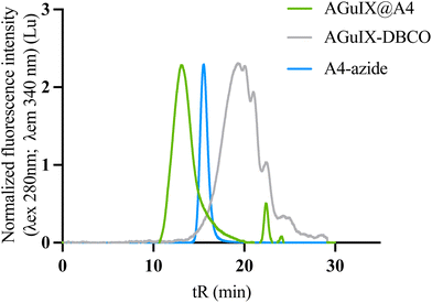
To demonstrate the reproducibility and robustness of the click chemistry approach, we conjugated a second VHH to AGuIX, this one specific to the CD47 receptor (A4. MW = 14.8 kDa). The first functionalization of the VHH with azide-PEG4-NHS demonstrated comparable efficacy to that of A12 (mean size of MWA12-azide: 15.33 kDa (2 azide grafted) and MWA4-azide:15.5 kDa (2–3 azide grafted)) (Fig. S6†). The SEC indicate that the AGuIX@A4 (tR: 13 min) product has a size comparable to AGuIX@A12 (tR: 12.9 min) (Fig. 3). AGuIX@A4 was synthesized with a >95% conversion yield, similar to AGuIX@A12. After purification, the final purified product showed a ratio of 17Gd/VHH ratio, with a final yield of around 26%.3.5. In vitro characterizations of AGuIX@VHH
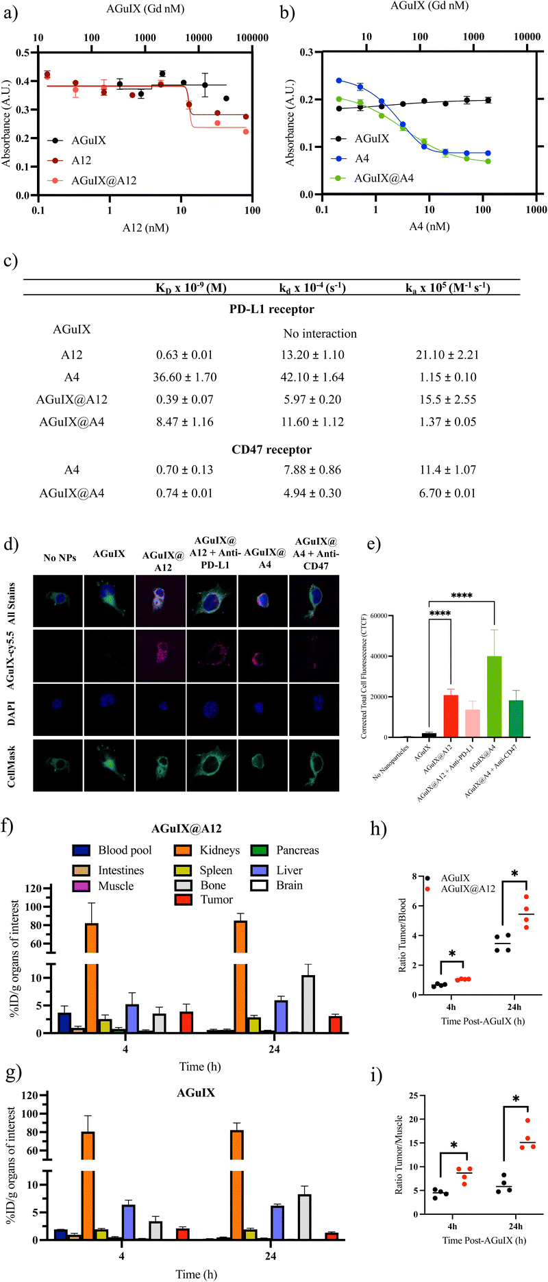
A competitive ELISA confirmed that the addition of AGuIX to either VHH, A12 (Fig. 4a) or A4 (Fig. 4b), did not impact competition binding affinities. We demonstrated similar logKi values of 12.8 ± 1.1 nM and 13.0 ± 0.4 nM for A12 and AGuIX@A12, respectively, and 2.9 ± 0.2 nM and 3.3 ± 0.4 nM for A4 and AGuIX-A4, respectively. Moreover, AGuIX on its own exhibited no interaction with PD-L1 or CD47 ligands, providing additional support for the notion that the robust affinity observed primarily results from the incorporation of functional VHH on the AGuIX surface. These results were further validated using a second method: biacore analysis which is based on a surface plasmon resonance (SPR) technique that which quantifies the association and dissociation phenomena.The affinity analyses conducted on the VHHs, and their associated receptors confirmed a strong binding affinity between these VHHs and their respective receptors, as shown by the equilibrium dissociation constants (KD) (0.63 ± 0.01 nM for A12 on PD-L1 and 0.70 ± 0.13 nM for A4 on CD47) (Fig. 4c and Fig. S4, S5†). These results were supported by the literature, particularly in the case of A4, which has been extensively documented.73 Comparing the KD values of AGuIX@VHH with their unmodified counterparts (0.39 ± 0.07 nM for AGuIX@A12 and 0.74 ± 0.01 nM for AGuIX@A4), we observed similar binding affinities further confirming that conjugation of AGuIX did not impact VHH binding. This similarity in KD values between AGuIX@VHH and unmodified VHH provided confirmation of the relevance of click chemistry in preserving the function of the VHHs. The A4, specific for the CD47 receptor, was selected to verify the specificity of the AGuIX@A12 to the PD-L1 receptor. The characteristic constants (equilibrium dissociation (KD) and kinetic rate of association and dissociation (kA and kD)) indicated a remarkable 10-fold stronger and faster binding affinity for A12 towards PD-L1 compared to A4, regardless of the bioconjugation status. These results confirm the specificity of the interaction between the nanobodies and their respective receptors (detailed in ESI†).
To confirm ligand–receptor interaction on cells, internalization and receptor blocking assays on melanoma B16F10 tumor cells, modified for high PD-L1 expression, were conducted to further validate the functionality of VHH bound to AGuIX (Fig. 4d and e). Melanoma hiPDL1-B16F10 murine cells are highly metastatic, aggressive models mimicking patient phenotypes, expressing both PD-L1 and CD47.51 The conjugation of A12 and A4 significantly increased internalization of AGuIX as seen in the representative images (Fig. 4d) and corrected total cell fluorescence (CTCF, Fig. 4e). Blocking with either anti-PD-L1 or anti-CD47 decreased internalization confirming specificity and functionality of the AGuIX@VHH. Studies were repeated in wild-type B16F10 cells (lower PD-L1 expression)74 (Fig. S7†) with similar increase in internalization with the conjugation of A12 and A4 to AGuIX. Although complete loss of internalization was not observed, this may be an indication of only partial blocking of PD-L1 and CD47 receptors allowing for some receptor mediated internalization.
3.6. Ex vivo AGuIX@A12 targeting
Given the widespread use of PD-1/PD-L1 inhibitors in clinical practice, we further evaluated the AGuIX@A12 product in proof-of-concept ex vivo studies. Targeting of PD-L1 was investigated in a murine melanoma model to confirm that AGuIX@A12 binding increases tumor accumulation. An ex vivo biodistribution study using zirconium-89 (89Zr) radiolabeling was performed for accurate quantification of AGuIX and AGuIX@A12 in different organs at 4 h and 24 h post-intravenous injection (Fig. 4f–i). A short and long time point was selected based on previous preclinical biodistribution studies and current clinical trials involving AGuIX.Currently, in clinical trials, radiotherapy is delivered 4 hours after AGuIX administration, justifying the clinical relevance of a 4-hour biodistribution time point and multiple preclinical trials showed that tumor accumulation was increased up to 4 h, while the 24-hour time point was chosen to study the expected longer persistence of targeted nanoparticles in the tumor.1,5,75–77 As expected from previous work, both NPs accumulate within a few hours into the kidneys, leading to fast washout from the blood pool due to their ultrasmall size; lower accumulation of NPs in the liver can be detected.76,78 Accumulation in the bone was attributed to the free [89Zr] (non-coupled on the NPs).79 Both NPs can also accumulate in the tumor area due to the enhanced permeability and retention (EPR) effect, previously shown in several rodent models and in human studies.6 However, a significantly higher accumulation of AGuIX-[89Zr]@A12 was present in the tumor area compared to untargeted AGuIX-[89Zr] (At 4 h, AGuIX: 2.10 ± 0.15% ID per g vs. AGuIX@A12: 3.90 ± 0.67% ID per g) (Fig. 4f–i). This difference was observed 4 h post-injection and was more pronounced 24 h post-injection (at 24 h, AGuIX: 1.34 ± 0.06% ID per g vs. AGuIX@A12: 3.10 ± 0.15% ID per g). This higher accumulation and retention of AGuIX@VHH suggests targeting of the PD-L1 receptors at the surface of cancer cells, that is expected to increase active accumulation and slow the AGuIX-VHH washout within the tumor area. This ex vivo validation of the higher targeting of AGuIX@A12 vs. AGuIX is evidence of the in vivo targeting of PD-L1 by AGuIX@A12. AGuIX@A12 is thus a promising candidate for future work involving MRI-detection of PD-L1 or for more targeted radiosensitization of PD-L1 expressing tumors.
4. Conclusion
We report the preparation of AGuIX@VHH bioconjugates using two distinct methods, sortagging and click chemistry. The two methods resulted in NPs with similar physicochemical characteristics using the A12 VHH. However, click chemistry exhibited higher VHH conversion while using lower-cost reagents, making it a more favourable approach for further investigations. Interestingly, the two bioconjugation methods yielded AGuIX@VHH with strong binding affinities, suggesting that site-specific VHH modification, such as sortagging, may not be required when coupling to relatively small NP such as AGuIX NP. PD-L1 targeting of AGuIX-A12 prepared by click chemistry was validated by ex vivo autoradiograpy demonstrating substantially greater uptake and persistence than untargeted NP. The A4 nanobody was used to the reliability and reproducibility of click synthesis for these constructs. Overall, click chemistry emerges as a favourable and promising approach for preparing a broad array of potential AGuIX@VHH bioconjugates that can be applied to multiple nanobodies, with diverse applications in targeted therapeutic interventions and imaging. Further comprehensive evaluation and validation in larger preclinical studies and clinical trials are necessary to fully explore the imaging and therapeutic potential of AGuIX@VHH and their role in enhancing cancer treatment.Author contributions
L. C., G. B. and P. R. designed and performed the chemical experiments and product characterization. L. S., Z. M. and N. B. conducted in vitro ELISA experiments and microscopy imaging. L. H. performed the Biacore experiments. A. H. conducted the TDA-ICP-MS experiments. V. G.-C. carried out SEC preparative purifications. F. D. conducted the MALDI-TOF experiments. C. T. performed the ex vivo experiments. T. D. helped with NP formulation and characterization. L. C., G. B. and N. B. wrote the manuscript. F. L., O. T., M. D., J. S., G. B., N. B. and R. B. conceptualized the idea, supervised the study, and revised the manuscript. All authors discussed the results and approved the final version of the manuscript.Conflicts of interest
L. C. is an employee of NH TherAGuIX that develops AGuIX NPs. O. T. and F. L. possess shares in this company. J. S. reports research support paid to the institution: Merck, BMS, Regeneron, Debiopharm, EMD, Seronoand consulting/Scientific Advisory Board/Travel fees: Castle Biosciences, Genentech, Immunitas, Debiopharm, BMS, LEK, Catenion, ACI Clinical, Astellas, Stimit, Merck KGA, SIRPant, EMD Serono. Stock options: Immunitas.Acknowledgements
Special thanks to the international IRP Harvard-Lyon Radioboost project, which aims to boost radiotherapy with ultra-small nanoparticles. Support for this project was provided by Viewray, Inc (agreement #2019A017789).References
- C. Verry, S. Dufort, J. Villa, M. Gavard, C. Iriart, S. Grand, J. Charles, B. Chovelon, J.-L. Cracowski, J.-L. Quesada, C. Mendoza, L. Sancey, A. Lehmann, F. Jover, J.-Y. Giraud, F. Lux, Y. Crémillieux, S. McMahon, P. J. Pauwels, D. Cagney, R. Berbeco, A. Aizer, E. Deutsch, M. Loeffler, G. Le Duc, O. Tillement and J. Balosso, Radiother. Oncol., 2021, 160, 159–165 CrossRef CAS PubMed.
- E. Thivat, M. Casile, J. Moreau, I. Molnar, S. Dufort, K. Seddik, G. Le Duc, O. De Beaumont, M. Loeffler, X. Durando and J. Biau, BMC Cancer, 2023, 23, 344 CrossRef CAS PubMed.
- F. Lux, V. L. Tran, E. Thomas, S. Dufort, F. Rossetti, M. Martini, C. Truillet, T. Doussineau, G. Bort, F. Denat, F. Boschetti, G. Angelovski, A. Detappe, Y. Crémillieux, N. Mignet, B.-T. Doan, B. Larrat, S. Meriaux, E. Barbier, S. Roux, P. Fries, A. Müller, M.-C. Abadjian, C. Anderson, E. Canet-Soulas, P. Bouziotis, M. Barberi-Heyob, C. Frochot, C. Verry, J. Balosso, M. Evans, J. Sidi-Boumedine, M. Janier, K. Butterworth, S. McMahon, K. Prise, M.-T. Aloy, D. Ardail, C. Rodriguez-Lafrasse, E. Porcel, S. Lacombe, R. Berbeco, A. Allouch, J.-L. Perfettini, C. Chargari, E. Deutsch, G. Le Duc and O. Tillement, Br. J. Radiol., 2019, 92, 20180365 Search PubMed.
- F. Lux, L. Sancey, A. Bianchi, Y. Crémillieux, S. Roux and O. Tillement, Nanomedicine, 2015, 10, 1801–1815 CrossRef CAS PubMed.
- L. Carmès, M. Banerjee, P. Coliat, S. Harlepp, X. Pivot, O. Tillement, F. Lux and A. Detappe, Adv. Ther., 2023, 2300019 CrossRef.
- G. Bort, F. Lux, S. Dufort, Y. Crémillieux, C. Verry and O. Tillement, Theranostics, 2020, 10, 1319–1331 CrossRef CAS PubMed.
- A. Detappe, C. Mathieu, C. Jin, M. P. Agius, M.-C. Diringer, V.-L. Tran, X. Pivot, F. Lux, O. Tillement, D. Kufe and P. P. Ghoroghchian, Int. J. Radiat. Oncol., 2020, 108, 1380–1389 CrossRef PubMed.
- A. Detappe, M. Reidy, Y. Yu, C. Mathieu, H. V.-T. Nguyen, T. P. Coroller, F. Lam, P. Jarolim, P. Harvey, A. Protti, Q.-D. Nguyen, J. A. Johnson, Y. Cremillieux, O. Tillement, I. M. Ghobrial and P. P. Ghoroghchian, Nanoscale, 2019, 11, 20485–20496 RSC.
- E. Thomas, C. Mathieu, P. Moreno-Gaona, V. Mittelheisser, F. Lux, O. Tillement, X. Pivot, P. P. Ghoroghchian and A. Detappe, Adv. Healthcare Mater., 2022, 11, 2101565 CrossRef CAS PubMed.
- M. M. Gomari, S. Abkhiz, T. G. Pour, E. Lotfi, N. Rostami, F. N. Monfared, B. Ghobari, M. Mosavi, B. Alipour and N. V. Dokholyan, Mol. Med., 2022, 28, 146 CAS.
- P. Chames, M. Van Regenmortel, E. Weiss and D. Baty, Br. J. Pharmacol., 2009, 157, 220–233 CrossRef CAS PubMed.
- S. Sun, Z. Ding, X. Yang, X. Zhao, M. Zhao, L. Gao, Q. Chen, S. Xie, A. Liu, S. Yin, Z. Xu and X. Lu, Int. J. Nanomed., 2021, 16, 2337–2356 CrossRef PubMed.
- A. Muruganandam, J. Tanha, S. Narang and D. Stanimirovic, FASEB J., 2002, 16, 240–242 CrossRef CAS PubMed.
- J. Wesolowski, V. Alzogaray, J. Reyelt, M. Unger, K. Juarez, M. Urrutia, A. Cauerhff, W. Danquah, B. Rissiek, F. Scheuplein, N. Schwarz, S. Adriouch, O. Boyer, M. Seman, A. Licea, D. V. Serreze, F. A. Goldbaum, F. Haag and F. Koch-Nolte, Med. Microbiol. Immunol., 2009, 198, 157–174 CrossRef CAS PubMed.
- M. Liu, Y. Zhu, T. Wu, J. Cheng and Y. Liu, Chem. – Eur. J., 2020, 26, 7442–7450 CrossRef CAS PubMed.
- E. Y. Yang and K. Shah, Front. Oncol., 2020, 10, 1182 CrossRef PubMed.
- T. De Meyer, S. Muyldermans and A. Depicker, Trends Biotechnol., 2014, 32, 263–270 CrossRef CAS PubMed.
- P. Bannas, J. Hambach and F. Koch-Nolte, Front. Immunol, 2017, 8, 1603 CrossRef PubMed.
- M. Dougan, J. R. Ingram, H.-J. Jeong, M. M. Mosaheb, P. T. Bruck, L. Ali, N. Pishesha, O. Blomberg, P. M. Tyler, M. M. Servos, M. Rashidian, Q.-D. Nguyen, U. H. von Andrian, H. L. Ploegh and S. K. Dougan, Cancer Immunol. Res., 2018, 6, 389–401 CrossRef CAS PubMed.
- J. R. Ingram, O. S. Blomberg, J. T. Sockolosky, L. Ali, F. I. Schmidt, N. Pishesha, C. Espinosa, S. K. Dougan, K. C. Garcia, H. L. Ploegh and M. Dougan, Proc. Natl. Acad. Sci. U. S. A., 2017, 114, 10184–10189 CrossRef CAS PubMed.
- J. R. Ingram, M. Dougan, M. Rashidian, M. Knoll, E. J. Keliher, S. Garrett, S. Garforth, O. S. Blomberg, C. Espinosa, A. Bhan, S. C. Almo, R. Weissleder, H. Lodish, S. K. Dougan and H. L. Ploegh, Nat. Commun., 2017, 8, 647 CrossRef PubMed.
- L. Fehrenbacher, A. Spira, M. Ballinger, M. Kowanetz, J. Vansteenkiste, J. Mazieres, K. Park, D. Smith, A. Artal-Cortes, C. Lewanski, F. Braiteh, D. Waterkamp, P. He, W. Zou, D. S. Chen, J. Yi, A. Sandler and A. Rittmeyer, Lancet, 2016, 387, 1837–1846 CrossRef CAS PubMed.
- D. R. Spigel, C. Faivre-Finn, J. E. Gray, D. Vicente, D. Planchard, L. Paz-Ares, J. F. Vansteenkiste, M. C. Garassino, R. Hui, X. Quantin, A. Rimner, Y.-L. Wu, M. Özgüroğlu, K. H. Lee, T. Kato, M. de Wit, T. Kurata, M. Reck, B. C. Cho, S. Senan, J. Naidoo, H. Mann, M. Newton, P. Thiyagarajah and S. J. Antonia, J. Clin. Oncol., 2022, 40, 1301–1311 CrossRef CAS PubMed.
- A. Bouleau, V. Lebon and C. Truillet, Pharmacol. Ther., 2021, 222, 107786 CrossRef CAS PubMed.
- Y.-C. Chen, W. Shi, J.-J. Shi and J.-J. Lu, J. Cancer Res. Clin. Oncol., 2022, 148, 1–14 CrossRef CAS PubMed.
- E. C. Piccione, S. Juarez, J. Liu, S. Tseng, C. E. Ryan, C. Narayanan, L. Wang, K. Weiskopf and R. Majeti, mAbs, 2015, 7, 946–956 CrossRef CAS PubMed.
- M. P. Chao, C. H. Takimoto, D. D. Feng, K. McKenna, P. Gip, J. Liu, J.-P. Volkmer, I. L. Weissman and R. Majeti, Front. Oncol., 2019, 9, 1380 CrossRef PubMed.
- H. A. Burris III, A. I. Spira, M. H. Taylor, O. O. Yeku, J. F. Liu, P. N. Munster, E. P. Hamilton, J. S. Thomas, F. Gatlin, R. T. Penson, T. A. Abrams, M. S. Dhawan, J. M. Walling, J. W. Frye, K. Romanko, V. Sung, C. Brachmann and A. B. El-Khoueiry, J. Clin. Oncol., 2021, 39, 2516–2516 Search PubMed.
- Z. Jiang, H. Sun, J. Yu, W. Tian and Y. Song, J. Hematol. Oncol., 2021, 14, 180 CrossRef CAS PubMed.
- I. E. Krop, N. Masuda, T. Mukohara, S. Takahashi, T. Nakayama, K. Inoue, H. Iwata, T. Toyama, Y. Yamamoto, D. M. Hansra, M. Takahashi, A. Osaki, K. Koyama, T. Inoue, T. Yonekura, J. Mostillo, S. Ohwada, Y. Tanaka, D. W. Sternberg and K. Yonemori, J. Clin. Oncol., 2022, 40, 1002–1002 CrossRef.
- R. K. Vaddepally, P. Kharel, R. Pandey, R. Garje and A. B. Chandra, Cancers, 2020, 12, 738 CrossRef CAS PubMed.
- H. Sato, N. Okonogi and T. Nakano, Int. J. Clin. Oncol., 2020, 25, 801–809 CrossRef CAS PubMed.
- L. Wen, F. Tong, R. Zhang, L. Chen, Y. Huang and X. Dong, Front. Oncol, 2021, 11, 799957 CrossRef CAS PubMed.
- Y. Nishiga, A. P. Drainas, M. Baron, D. Bhattacharya, A. A. Barkal, Y. Ahrari, R. Mancusi, J. B. Ross, N. Takahashi, A. Thomas, M. Diehn, I. L. Weissman, E. E. Graves and J. Sage, Nat. Cancer, 2022, 3, 1351–1366 CrossRef CAS PubMed.
- E. Rostami, M. Bakhshandeh, H. Ghaffari-Nazari, M. Alinezhad, M. Alimohammadi, R. Alimohammadi, G. Mahmoodi Chalbatani, E. Hejazi, T. J. Webster, J. Tavakkol-Afshari and S. A. Jalali, PLoS One, 2022, 17, e0273547 CrossRef CAS PubMed.
- Y. Zhang, K.-Y. Park, K. F. Suazo and M. D. Distefano, Chem. Soc. Rev., 2018, 47, 9106–9136 RSC.
- X. Dai, A. Böker and U. Glebe, RSC Adv., 2019, 9, 4700–4721 RSC.
- M. Ritzefeld, Chem. – Eur. J., 2014, 20, 8516–8529 CrossRef CAS PubMed.
- L. Taiariol, C. Chaix, C. Farre and E. Moreau, Chem. Rev., 2022, 122, 340–384 CrossRef CAS PubMed.
- J. M. Eeftens, J. van der Torre, D. R. Burnham and C. Dekker, BMC Biophys., 2015, 8, 9 CrossRef PubMed.
- T. I. Chio and S. L. Bane, Methods Mol. Biol., 2020, 2078, 83–97 CrossRef CAS PubMed.
- J. M. Baskin, J. A. Prescher, S. T. Laughlin, N. J. Agard, P. V. Chang, I. A. Miller, A. Lo, J. A. Codelli and C. R. Bertozzi, Proc. Natl. Acad. Sci. U. S. A, 2007, 104, 16793–16797 CrossRef CAS PubMed.
- E. Kim and H. Koo, Chem. Sci., 2019, 10, 7835–7851 RSC.
- B. Stump, ChemBioChem, 2022, 23, e202200016 CrossRef CAS PubMed.
- Explaining Nobel Prize-winning chemistry techniques, https://www.buffalo.edu/ubnow/stories/2022/10/nobel-prize-chemistry.html, (accessed March 9, 2023).
- G. Le Duc, S. Roux, A. Paruta-Tuarez, S. Dufort, E. Brauer, A. Marais, C. Truillet, L. Sancey, P. Perriat, F. Lux and O. Tillement, Cancer Nanotechnol., 2014, 5, 4 CrossRef PubMed.
- A. Mignot, C. Truillet, F. Lux, L. Sancey, C. Louis, F. Denat, F. Boschetti, L. Bocher, A. Gloter, O. Stéphan, R. Antoine, P. Dugourd, D. Luneau, G. Novitchi, L. C. Figueiredo, P. C. de
![[thin space (1/6-em)]](https://www.rsc.org/images/entities/char_2009.gif) Morais, L. Bonneviot, B. Albela, F. Ribot, L. Van
Morais, L. Bonneviot, B. Albela, F. Ribot, L. Van![[thin space (1/6-em)]](https://www.rsc.org/images/entities/char_2009.gif) Lokeren, I. Déchamps-Olivier, F. Chuburu, G. Lemercier, C. Villiers, P. N. Marche, G. Le
Lokeren, I. Déchamps-Olivier, F. Chuburu, G. Lemercier, C. Villiers, P. N. Marche, G. Le![[thin space (1/6-em)]](https://www.rsc.org/images/entities/char_2009.gif) Duc, S. Roux, O. Tillement and P. Perriat, Chem. – Eur. J., 2013, 19, 6122–6136 CrossRef CAS PubMed.
Duc, S. Roux, O. Tillement and P. Perriat, Chem. – Eur. J., 2013, 19, 6122–6136 CrossRef CAS PubMed. - N. Brown, P. Rocchi, L. Carmès, R. Guthier, M. Iyer, L. Seban, T. Morris, S. Bennett, M. Lavelle, J. Penailillo, R. Carrasco, C. Williams, E. Huynh, Z. Han, E. Kaza, T. Doussineau, S. M. Toprani, X. Qin, Z. D. Nagel, K. A. Sarosiek, A. Hagège, S. Dufort, G. Bort, F. Lux, O. Tillement and R. Berbeco, Theranostics, 2023, 13, 4711–4729 CrossRef CAS PubMed.
- E. Thomas, L. Colombeau, M. Gries, T. Peterlini, C. Mathieu, N. Thomas, C. Boura, C. Frochot, R. Vanderesse, F. Lux, M. Barberi-Heyob and O. Tillement, Int. J. Nanomed., 2017, 12, 7075–7088 CrossRef CAS PubMed.
- C. Truillet, E. Thomas, F. Lux, L. T. Huynh, O. Tillement and M. J. Evans, Mol. Pharm., 2016, 13, 2596–2601 CrossRef CAS PubMed.
- E. Thomas, C. Mathieu, P. Moreno-Gaona, V. Mittelheisser, F. Lux, O. Tillement, X. Pivot, P. P. Ghoroghchian and A. Detappe, Adv. Healthcare Mater., 2022, 11, 2101565 CrossRef CAS PubMed.
- E. Thomas, Cancer, Université de Lyon, 2017 Search PubMed.
- L. Labied, P. Rocchi, T. Doussineau, J. Randon, O. Tillement, H. Cottet, F. Lux and A. Hagège, Anal. Chim. Acta, 2021, 1185, 339081 CrossRef CAS PubMed.
- L. Heinrich, N. Tissot, D. J. Hartmann and R. Cohen, J. Immunol. Methods, 2010, 352, 13–22 CrossRef CAS PubMed.
- V.-L. Tran, F. Lux, N. Tournier, B. Jego, X. Maître, M. Anisorac, C. Comtat, S. Jan, K. Selmeczi, M. J. Evans, O. Tillement, B. Kuhnast and C. Truillet, Adv. Healthcare Mater., 2021, 10, e2100656 CrossRef PubMed.
- T. Spirig, E. M. Weiner and R. T. Clubb, Mol. Microbiol., 2011, 82, 1044–1059 CrossRef CAS PubMed.
- H. E. Morgan, W. B. Turnbull and M. E. Webb, Chem. Soc. Rev., 2022, 51, 4121–4145 RSC.
- J. M. Antos, M. C. Truttmann and H. L. Ploegh, Curr. Opin. Struct. Biol., 2016, 38, 111–118 CrossRef CAS PubMed.
- J. E. Glasgow, M. L. Salit and J. R. Cochran, J. Am. Chem. Soc., 2016, 138, 7496–7499 CrossRef CAS PubMed.
- S. A. M. van Lith, S. M. J. van Duijnhoven, A. C. Navis, W. P. J. Leenders, E. Dolk, J. W. H. Wennink, C. F. van Nostrum and J. C. M. van Hest, Bioconjugate Chem., 2017, 28, 539–548 CrossRef CAS PubMed.
- L. Liu, J. L. Gray, E. W. Tate and A. Yang, Trends Biotechnol., 2023, 41, 1385–1399 CrossRef CAS PubMed.
- M. W. Popp, J. M. Antos, G. M. Grotenbreg, E. Spooner and H. L. Ploegh, Nat. Chem. Biol., 2007, 3, 707–708 CrossRef CAS PubMed.
- N. Kotagiri, Z. Li, X. Xu, S. Mondal, A. Nehorai and S. Achilefu, Bioconjugate Chem., 2014, 25, 1272–1281 CrossRef CAS PubMed.
- V. Aragon-Sanabria, A. Aditya, L. Zhang, F. Chen, B. Yoo, T. Cao, B. Madajewski, R. Lee, M. Z. Turker, K. Ma, S. Monette, P. Chen, J. Wu, S. Ruan, M. Overholtzer, P. Zanzonico, C. M. Rudin, C. Brennan, U. Wiesner and M. S. Bradbury, Clin. Cancer Res., 2022, 28, 2938–2952 CrossRef CAS PubMed.
- L. Williams, L. Li, P. J. Yazaki, P. Wong, A. Miller, T. Hong, E. K. Poku, S. Bhattacharya, J. E. Shively and M. Kujawski, Biotechnol. J., 2023, e2300115 CrossRef PubMed.
- A. Debon, E. Siirola and R. Snajdrova, JACS Au, 2023, 3, 1267–1283 CrossRef CAS PubMed.
- C. J. Pickens, S. N. Johnson, M. M. Pressnall, M. A. Leon and C. J. Berkland, Bioconjugate Chem., 2018, 29, 686–701 CrossRef CAS PubMed.
- V. Solntceva, M. Kostrzewa and G. Larrouy-Maumus, Front. Cell. Infect. Microbiol., 2021, 10, 621452 CrossRef PubMed.
- S. Mädler, C. Bich, D. Touboul and R. Zenobi, J. Mass Spectrom., 2009, 44, 694–706 CrossRef PubMed.
- J. S. Nanda and J. R. Lorsch, Methods in Enzymology, ed. J. Lorsch, Academic Press, 2014, vol. 536, pp. 87–94 Search PubMed.
- L. Labied, P. Rocchi, T. Doussineau, J. Randon, O. Tillement, H. Cottet, F. Lux and A. Hagège, Anal. Chim. Acta, 2021, 1185, 339081 CrossRef CAS PubMed.
- Q. Wu, H. L. Ploegh and M. C. Truttmann, ACS Chem. Biol., 2017, 12, 664–673 CrossRef CAS PubMed.
- J. T. Sockolosky, M. Dougan, J. R. Ingram, C. C. M. Ho, M. J. Kauke, S. C. Almo, H. L. Ploegh and K. C. Garcia, Proc. Natl. Acad. Sci. U. S. A., 2016, 113, E2646–E2654 CrossRef CAS PubMed.
- A. Quijano-Rubio, A. M. Bhuiyan, H. Yang, I. Leung, E. Bello, L. R. Ali, K. Zhangxu, J. Perkins, J.-H. Chun, W. Wang, M. J. Lajoie, R. Ravichandran, Y.-H. Kuo, S. K. Dougan, S. R. Riddell, J. B. Spangler, M. Dougan, D.-A. Silva and D. Baker, Nat. Biotechnol., 2023, 41, 532–540 CrossRef CAS PubMed.
- S. Kotb, A. Detappe, F. Lux, F. Appaix, E. L. Barbier, V.-L. Tran, M. Plissonneau, H. Gehan, F. Lefranc, C. Rodriguez-Lafrasse, C. Verry, R. Berbeco, O. Tillement and L. Sancey, Theranostics, 2016, 6, 418–427 CrossRef CAS PubMed.
- E. Thivat, M. Casile, J. Moreau, I. Molnar, S. Dufort, K. Seddik, G. Le Duc, O. De Beaumont, M. Loeffler, X. Durando and J. Biau, BMC Cancer, 2023, 23, 344 CrossRef CAS PubMed.
- C. Verry, L. Sancey, S. Dufort, G. Le Duc, C. Mendoza, F. Lux, S. Grand, J. Arnaud, J. L. Quesada, J. Villa, O. Tillement and J. Balosso, BMJ Open, 2019, 9, e023591 CrossRef PubMed.
- F. Lux, V. L. Tran, E. Thomas, S. Dufort, F. Rossetti, M. Martini, C. Truillet, T. Doussineau, G. Bort, F. Denat, F. Boschetti, G. Angelovski, A. Detappe, Y. Crémillieux, N. Mignet, B.-T. Doan, B. Larrat, S. Meriaux, E. Barbier, S. Roux, P. Fries, A. Müller, M.-C. Abadjian, C. Anderson, E. Canet-Soulas, P. Bouziotis, M. Barberi-Heyob, C. Frochot, C. Verry, J. Balosso, M. Evans, J. Sidi-Boumedine, M. Janier, K. Butterworth, S. McMahon, K. Prise, M.-T. Aloy, D. Ardail, C. Rodriguez-Lafrasse, E. Porcel, S. Lacombe, R. Berbeco, A. Allouch, J.-L. Perfettini, C. Chargari, E. Deutsch, G. Le Duc and O. Tillement, Br. J. Radiol., 2019, 92, 20180365 Search PubMed.
- D. S. Abou, T. Ku and P. M. Smith-Jones, Nucl. Med. Biol., 2011, 38, 675–681 CrossRef CAS PubMed.
Footnotes |
| † Electronic supplementary information (ESI) available. See DOI: https://doi.org/10.1039/d3nr04777f |
| ‡ These authors contributed equally to this work. |
| This journal is © The Royal Society of Chemistry 2024 |


