 Open Access Article
Creative Commons Attribution 3.0 Unported Licence
Thioamide-based fluorescent sensors for dipeptidyl peptidase 4†
Hoang Anh T.
Phan
 a,
Yanan
Chang
 a,
Samuel A.
Eaton
 a and
E. James
Petersson
 *ab
*ab
aDepartment of Chemistry, School of Arts and Sciences, University of Pennsylvania, 231 South 34th Street, Philadelphia, Pennsylvania 19104, USA. E-mail: ejpetersson@sas.upenn.edu
bDepartment of Biochemistry and Biophysics, Perelman School of Medicine, University of Pennsylvania, 421 Curie Boulevard, Philadelphia, PA 19104, USA
First published on 23rd October 2024
Abstract
Dipeptidyl peptidase 4 (DPP-4) is a promising biomarker for cancer and metabolic diseases. We demonstrate the design of novel fluorescent DPP-4 probes based on the protease's native substrates using a thioamide as a quencher for measuring in vitro kinetics, inhibition with sitagliptin, and DPP-4 activity in saliva samples.
Dipeptidyl peptidase 4 (DPP-4) is a serine protease that preferably cleaves at a proline or an alanine at the second amino acid of its substrates, liberating dipeptides.1 Interestingly, DPP-4 is a multifunctional protein also known as CD26, which serves as T cell costimulatory receptor in addition to its enzymatic activity.2 When DPP-4 was first discovered by Hopsu-Havu and Glenner in 1966, it was initially called glycyl-proline naphthylamidase due to the fact that it could cleave the substrate glycyl-DL-prolyl-β-naphthylamide (GP-βNA).3 DPP-4 can inactivate peptides whose N-termini are important for biological function or cleave peptides to generate new ones.4,5
Among the natural substrates of DPP-4 are the peptides from the pituitary adenylate cyclase-activating polypeptide (PACAP)/glucagon family such as glucagon-like polypeptide 1 (GLP-1) and gastric inhibitory polypeptide (GIP), which are important in regulating glucose homeostasis.4,6 GLP-1 stimulates insulin release, but it can be rapidly cleaved by DPP-4 with a half-life of less than 2 minutes. There have been major advances in DPP-4-based therapeutics for treatment of diabetes, especially in creating stabilized GLP-1 analogs and DPP-4 inhibitors such as sitagliptin (Januvia).7–10 Beyond GLP-1, DPP-4 cleaves a diverse repertoire of bioactive peptides, such as vasoactive intestinal peptide (VIP), substance P, pancreatic polypeptide (PP), peptide YY (PYY), and neuropeptide Y (NPY). DPP-4 is expressed in different tissues as either an anchored transmembrane protease or a soluble circulating one, and its anomalous activity has also been linked to cancer (solid tumors), hematological malignancies, immune diseases, and infectious diseases.4,5 Studies have identified DPP-4 as a promising biomarker for early diagnosis and treatment of diseases such as lung diseases, diabetic kidney disease,11 rheumatoid arthritis,12 post-kidney transplant tubulitis,12 and liver disease.13 There is thus a need to develop probes to monitor DPP-4 activity real-time.
Common chromogenic substrates for DPP-4 include GP-βNA,3 Ala-Pro-7-amino-4-trifluoromethyl-coumarin (AP-AFC), and Gly-Pro-7-amido-4-methylcoumarin (GP-AMC). Many DPP-4 probes utilize a pro-fluorophore Förster resonance energy transfer (FRET), photoinduced electron transfer (PeT), or intramolecular charge transfer (ICT) approach where the fluorophore is quenched when conjugated to a short recognition dipeptide (e.g., Gly-Pro; Ala-Pro), and can return to its highly emissive state upon cleavage by the protease.14–17 These strategies can perturb and compromise the recognition sequences, thereby preventing accurate reporting of proteolysis. For instance, in ICT-based probes GP-βNA, AP-AFC, and GP-AMC, the chromophore is attached directly to the cleavage site so no information can be gained on the C terminal side (called the prime side) of the cleavage site. Even the N-terminal (non-prime) side information can be compromised. A study showed that P1 substitution in the P2–P1-AMC probes did not reflect mutation effects observed in the full-length peptide substrates of DPP-4.18 Additionally, an approach utilizing FRET pairs, that require labeling on either side of the scissile bond, is not compatible with monitoring aminopeptidases or carboxypeptidases where recognition of the amino acid at the substrate terminus is essential.
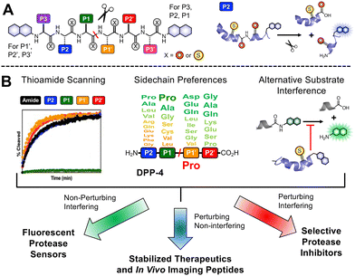
Our laboratory has previously shown that thioamides can quench donor fluorophores such as tryptophan, tyrosine, fluorescein, acridonylalanine, and 7-methoxycoumarin, serving as a minimally perturbing probe for protein dynamics and proteolysis.19–23 For the general design, we typically incorporate a thioamide quencher on the opposite of the scissile bond from the fluorophore; upon cleavage by proteolysis, these two will be separated, resulting in a turn-on of fluorescence that can be monitored in real time (Fig. 1A). We have systematically studied thioamide positional effects on our fluorescence sensor probes for cysteine proteases (papain, cathepsins L, V, K, B, and S) and serine proteases (trypsin, chymotrypsin, and kallikrein), identifying trends in perturbing and non-perturbing sites for thioamide labeling.24 We have also tested thioamide peptides to determine whether they interfere with the cleavage of other substrates (showing that they bind at the active site).21–23,25 This initial screening allows us to use thioamide in one of three ways at a given site (Fig. 1B): (1) non-perturbing, interfering sites can be used for fluorescence protease sensors,20 (2) perturbing, non-interfering sites can be used for in vivo therapeutic or imaging peptides (e.g., thioamide-stabilized therapeutic GLP-1 or NPY-based cancer imaging probes),21,25 (3) perturbing, interfering sites can be used for selective protease inhibitors (e.g., thioamide cathepsin L inhibitors).23 Herein, we follow this workflow to develop thioamide peptide probes for DPP-4 based on its native substrates. Our probes, containing 7-methoxycoumarin-4-yl-alanine (Mcm or μ, Fig. 2A) as a fluorophore, can effectively report real-time DPP-4 activity in in vitro steady-state assays, inhibition assays, and saliva samples. We thus highlight the value of thioamides for the minimally perturbing design of substrate-based protease probes.
We based our first peptide probe design on NPY since it is known to be an excellent substrate of DPP-4.4,26 NPY is a natural 36-amino-acid neuromodulator that is abundant in the brain as well as in the peripheral nervous system.27 NPY can modulate NPY receptors, which have been implicated in many diseases such as metabolic diseases, obesity, pain, cancer, and cardiovascular regulation. DPP-4 typically cleaves full-length NPY at Pro2 at its N-terminus, resulting in NPY3-36.4,5,26 DPP-4 cleavage modulates NPY receptor selectivity since the NPY3-36 fragment is no longer active toward the NPY Y1 receptor (with vasoconstrictive properties), yet has a strong affinity toward NPY Y2 and Y5 receptors (as a vascular growth factor).4,5
Since DPP-4 cleaves the NPY peptides at Pro2, we have only two options for thioamide placement, at the first tyrosine residue Y1 (P2 position) or at the second proline P2 (P1 position). As a preliminary assessment, we synthesized the all-amide full-length NPY peptide, along with its P1 thioamide analog (NPY-PS2) and its P2 thioamide analog (NPY-YS1), to evaluate which thioamide position on NPY would significantly perturb recognition by DPP-4. In these assays, cleavage of the peptides was monitored high-performance liquid chromatography (HPLC), and fragment identities were confirmed by matrix-assisted laser desorption ionization mass spectrometry (MALDI MS) (Fig. 2B, primary data are shown in Fig. S11–S13, ESI†). The P2 thioamide analog, NPY-YS1, cleaved 6.5 times slower than the all-amide counterpart, while the P1 thioamide, NPY-PS2, cleaved 372 times slower. This is in contrast to our results with GLP-1 and GIP thioamidation, in which both P2 and P1 thioamides significantly retarded proteolysis.25 Here, our data show that the P2 position is less perturbing for proteolysis, possibly because it is a proline residue, and that we should place the thioamide at the P2 position to create a quenching effect as desired while still being responsive to DPP-4 proteolysis.
For our probe design, we chose Mcm/μ as the fluorophore for the following reasons: (1) it is only slightly larger than Trp; (2) it is commercially available as Fmoc-μ-OH; (3) it can be easily incorporated into the peptide probes via solid phase peptide synthesis, and (4) it can be quenched by thioamides. Our lab also has previously successfully utilized Mcm-tagged peptides as proteolysis probes for different serine-, cysteine-, carboxyl-, and metallo-proteases.20–22,24 Since μ is quenched by thioamides via a PeT mechanism, they must be in close proximity.19 Based on our results with NPY-PS2 and NPY-YS1, as well as an analysis of the crystal structure of DPP-4 with a fragment of NPY bound (PDB ID: 1r9n),28 we decided to design a hexapeptide probe, with the thioamide at P2 position (i.e. the N terminus) and the fluorophore μ at the P4′ position, since the DPP-4 active site is relatively open there while it makes close contacts with the P1′, P2′, and P3′ amino acids (Fig. 2C). P2 and P4′ are 5 residues apart, predicted to give sufficient quenching based on our studies of thioamide PeT quenching.19
First, we synthesized probe analogs of NPY, with the sequence of YPSKPμ (NPY1–5). For comparison, we synthesized both the all-amide and P2 thioamide version of this peptide, YP/SKPμ and YSP/SKPμ, where the / indicates the DPP-4 cleavage site (Fig. 2A). As a demonstration that we could study mutations at prime sites, we also made a probe based on the NPY mutation Ser3-to-Leu (NPY1−5L3: YPLKPμ). The same design was applied to two other natural substrates from the pancreatic polypeptide family, where we synthesized and tested the hexapeptide probes based on the first 5 N-terminal amino acids of PYY (PYY1–5: YPIKPμ) and PP (PP1–5: APLEPμ). NPY1–5 NMR data indicate that the probes are unstructured (Fig. S25–S27, ESI†).
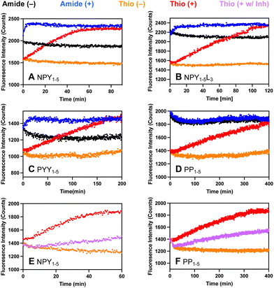
Each probe was incubated with DPP-4 and fluorescence intensity was monitored over time (λex = 325 nm; λem = 390 nm). Significantly, our μ-labeled NPY P2 thioamide sensor, YsPSKPμ, was cleaved by DPP-4 with similar kinetics (Fig. 3A, t1/2 = 26 min) to the full-length NPY P2 thioamide, NPY-PS2 (t1/2 = 30 min). Interestingly, we observed a fluorescence turn-on even without a thioamide, which is presumably due to quenching by the N-terminal Tyr residue. In fact, for both NPY probes (Fig. 3A and B) and the PYY probe (Fig. 3C), we observed the synergic effects of both Tyr and thioamide quenching. In the case of the PP-based probe, since there was no tyrosine, the thiopeptide showed its full utility as the all-amide APLEPμ had no fluorescence quenching, while the thioamide version ASPLEPμ showed good fluorescence turn-on (Fig. 3D). To ensure that the fluorescence assays truly reflect protease cleavage, HPLC and MALDI were used to confirm cleavage product identities (Fig. S14–S17 and Tables S4–S7, ESI†). The P1′ mutated NPY1–5L3 probe, YSPLKPμ, is cleaved 2-fold slower than the native NPY1–5 probe, YSPSKPμ, and PYY1–5, which also differs at the P1′ site (YSPIKPμ), is cleaved 3-fold slower than NPY1–5. These data demonstrate that we can create probes to explore the effects of mutations on the prime sites of a substrate, which could not be achieved with commercially available probes such as GP-AMC.
To ensure that our probes are responsive to the presence of inhibitors, inhibition studies were done with 50 nM sitagliptin, which is a selective inhibitor of DPP-4 with an IC50 between 18–87 nM, depending on the experiment.10,29 In all cases, we observed inhibition by sitagliptin with our probes, as indicated by a decrease in fluorescence signal in the presence of 50 nM sitagliptin consistent with ∼50% inhibition (NPY and PP probe data shown in Fig. 3E and F; NPY-L3 and YY probe data show in Fig. S19, ESI†). At higher concentrations of sitagliptin, we observe complete inhibition (Fig. S20, ESI†). This shows the potential of using our probes in inhibitor screening in biochemical assays, which is an important application for drug development and mechanistic studies.
We also wished to demonstrate that our probes could be used as a tool to quickly detect DPP-4 activity in biological samples. For instance, DPP-4 activity in saliva has been associated with diseases such as periodontitis and the presence of Porphyromonas gingivalis, Sjögren's syndrome, and oral cancers.30–32 Herein, following a very simple protocol, we directly added our thioamide probes to human saliva, then measured fluorescence changes with a plate reader (Fig. 4A). For our NPY- and PYY-based probes, we observed an increase in fluorescence in the presence of saliva in the first two hours of incubation (NPY1–5 probe data shown in Fig. 4B, NPY1–5L3 and PYY1–5 probe data shown in Fig. S22, ESI†). For PP1–5, a slower increase in fluorescence was observed (Fig. 4C), consistent with slower rates in the DPP-4 only assay. The fluorescence increases could be completely blocked by sitagliptin, showing that they are highly specific for DPP-4 (Fig. 4 and Fig. S23, ESI†). To approximate saliva samples from cancer patients, where DPP-4 activity levels are 2–4 fold higher than in controls, we added DPP-4 to the saliva and found that we could easily detect these differences (Fig. 4B and C). These results show the potential for our probes to measure DPP-4 in biological samples.
Here, we have demonstrated that we can create thioamide probes to detect DPP-4 activity using substrate-based probes that not only include the dipeptide recognition motif, but also native sequences on the primed side of the scissile bond – something that is not featured in many of the existing probes. This close mimicking of the native substrate allows our probes to report DPP-4 activity as faithfully as possible. Given the promising results from the human saliva assays, we will further test with actual patient samples. We will take advantage of the thioamide's ability to quench other fluorophores19 to make cellular or in vivo imaging probes with red-shifted dyes, as in many recent cancer imaging probes.33,34
The manuscript was written by H. A. T. P. and E. J. P., with input from all authors. H. A. T. P. designed all experiments and H. A. T. P. and Y. C. performed most experiments. S. A. E. synthesized full-length NPY constructs.
This research was supported by a grant from the National Science Foundation to E. J. P. (NSF CHE-2203909). The MALDI MS was acquired through a National Institutes of Health instrumentation grant (NIH S10-OD030460).
Data availability
The data supporting this article have been included as part of the ESI.†Conflicts of interest
There are no conflicts to declare.Notes and references
- C. F. Deacon, Front. Endocrinol., 2019, 10, 80 CrossRef PubMed.
- W. Jungraithmayr and N. Enz, Cell. Mol. Immunol., 2020, 17, 1208–1209 CrossRef CAS PubMed.
- V. K. Hopsu-Havu and G. G. Glenner, Histochemie, 1966, 7, 197–201 CrossRef CAS PubMed.
- A. M. Lambeir, C. Durinx, S. Scharpe and I. De Meester, Crit. Rev. Clin. Lab. Sci., 2003, 40, 209–294 CrossRef CAS PubMed.
- E. E. Mulvihill and D. J. Drucker, Endocr. Rev., 2014, 35, 992–1019 CrossRef CAS PubMed.
- N. M. Sherwood, S. L. Krueckl and J. E. McRory, Endocr. Rev., 2000, 21, 619–670 CAS.
- J. J. Holst, C. Orskov, O. V. Nielsen and T. W. Schwartz, FEBS Lett., 1987, 211, 169–174 CrossRef CAS PubMed.
- W. Kim and J. M. Egan, Pharmacol. Rev., 2008, 60, 470–512 CrossRef CAS PubMed.
- B. Manandhar and J. M. Ahn, J. Med. Chem., 2015, 58, 1020–1037 CrossRef CAS PubMed.
- D. Kim, L. Wang, M. Beconi, G. J. Eiermann, M. H. Fisher, H. He, G. J. Hickey, J. E. Kowalchick, B. Leiting, K. Lyons, F. Marsilio, M. E. McCann, R. A. Patel, A. Petrov, G. Scapin, S. B. Patel, R. S. Roy, J. K. Wu, M. J. Wyvratt, B. B. Zhang, L. Zhu, N. A. Thornberry and A. E. Weber, J. Med. Chem., 2005, 48, 141–151 CrossRef CAS PubMed.
- A. L. Sun, J. T. Deng, G. J. Guan, S. H. Chen, Y. T. Liu, J. Cheng, Z. W. Li, X. H. Zhuang, F. D. Sun and H. P. Deng, Diab. Vasc. Dis. Res., 2012, 9, 301–308 CrossRef PubMed.
- O. J. Cordero, R. Varela-Calvino, T. Lopez-Gonzalez, C. Calvino-Sampedro, J. E. Vinuela, C. Mourino, I. Hernandez-Rodriguez, M. Rodriguez-Lopez, B. Aspe de la Iglesia and J. M. Pego-Reigosa, PLoS One, 2015, 10, e0131992 CrossRef PubMed.
- G. Firneisz, T. Varga, G. Lengyel, J. Feher, D. Ghyczy, B. Wichmann, L. Selmeci, Z. Tulassay, K. Racz and A. Somogyi, PLoS One, 2010, 5, e12226 CrossRef PubMed.
- X. Guo, S. Mu, J. Li, Y. Zhang, X. Liu, H. Zhang and H. Gao, J. Mater. Chem. B, 2020, 8, 767–775 RSC.
- A. Ogasawara, M. Kamiya, K. Sakamoto, Y. Kuriki, K. Fujita, T. Komatsu, T. Ueno, K. Hanaoka, H. Onoyama, H. Abe, Y. Tsuji, M. Fujishiro, K. Koike, M. Fukayama, Y. Seto and Y. Urano, Bioconj. Chem., 2019, 30, 1055–1060 CrossRef CAS PubMed.
- N. H. Ho, R. Weissleder and C. H. Tung, Bioorg. Med. Chem. Lett., 2006, 16, 2599–2602 CrossRef CAS PubMed.
- L.-W. Zou, P. Wang, X.-K. Qian, L. Feng, Y. Yu, D.-D. Wang, Q. Jin, J. Hou, Z.-H. Liu, G.-B. Ge and L. Yang, Biosens. Bioelectron., 2017, 90, 283–289 CrossRef CAS PubMed.
- K. Kuhn-Wache, J. W. Bar, T. Hoffmann, R. Wolf, J. U. Rahfeld and H. U. Demuth, Biol. Chem., 2011, 392, 223–231 Search PubMed.
- J. M. Goldberg, S. Batjargal, B. S. Chen and E. J. Petersson, J. Am. Chem. Soc., 2013, 135, 18651–18658 CrossRef CAS PubMed.
- J. M. Goldberg, X. Chen, N. Meinhardt, D. C. Greenbaum and E. J. Petersson, J. Am. Chem. Soc., 2014, 136, 2086–2093 CrossRef CAS PubMed.
- T. M. Barrett, X. S. Chen, C. Liu, S. Giannakoulias, H. A. T. Phan, J. Wang, E. K. Keenan, R. J. Karpowicz, Jr. and E. J. Petersson, ACS Chem. Biol., 2020, 15, 774–779 CrossRef CAS PubMed.
- C. Liu, T. M. Barrett, X. Chen, J. J. Ferrie and E. J. Petersson, ChemBioChem, 2019, 20, 2059–2062 CrossRef CAS PubMed.
- H. A. T. Phan, S. G. Giannakoulias, T. M. Barrett, C. Liu and E. J. Petersson, Chem. Sci., 2021, 12, 10825–10835 RSC.
- S. Giannakoulias, S. R. Shringari, C. Liu, H. A. T. Phan, T. M. Barrett, J. J. Ferrie and E. J. Petersson, J. Phys. Chem. B, 2020, 124, 8032–8041 CrossRef CAS PubMed.
- X. Chen, E. G. Mietlicki-Baase, T. M. Barrett, L. E. McGrath, K. Koch-Laskowski, J. J. Ferrie, M. R. Hayes and E. J. Petersson, J. Am. Chem. Soc., 2017, 139, 16688–16695 CrossRef CAS PubMed.
- R. Mentlein, P. Dahms, D. Grandt and R. Krüger, Regul. Pept., 1993, 49, 133–144 CrossRef CAS PubMed.
- S. P. Brothers and C. Wahlestedt, EMBO Mol. Med., 2010, 2, 429–439 CrossRef CAS PubMed.
- K. Aertgeerts, S. Ye, M. G. Tennant, M. L. Kraus, J. Rogers, B.-C. Sang, R. J. Skene, D. R. Webb and G. S. Prasad, Protein Sci., 2004, 13, 412–421 CrossRef CAS PubMed.
- J. Zhang, X. K. Qian, P. F. Song, X. D. Li, A. Q. Wang, H. Huo, J. C. Yao, G. M. Zhang and L. W. Zou, Anal. Methods, 2021, 13, 2671–2678 RSC.
- K. Fukasawa, M. Harada, M. Komatsu, M. Yamaoka, M. Urade, K. Shirasuna and T. Miyazaki, Int. J. Oral Surg., 1982, 11, 246–250 CrossRef CAS PubMed.
- P. Aemaimanan, N. Sattayasai, N. Wara-aswapati, W. Pitiphat, W. Suwannarong, S. Prajaneh and S. Taweechaisupapong, J. Periodontol., 2009, 80, 1809–1814 CrossRef CAS PubMed.
- L. Garreto, S. Charneau, S. C. Mandacaru, O. T. Nobrega, F. N. Motta, C. N. de Araujo, A. C. Tonet, F. M. B. Modesto, L. M. Paula, M. V. de Sousa, J. M. Santana, A. C. Acevedo and I. M. D. Bastos, Front. Immunol., 2021, 12, 686480 CrossRef CAS PubMed.
- R. Sun, W. Ma, M. Ling, C. Tang, M. Zhong, J. Dai, M. Zhu, X. Cai, G. Li, Q. Xu, L. Tang, Z. Yu and Z. Peng, J. Control Rel., 2022, 350, 525–537 CrossRef CAS PubMed.
- M. Tian, R. Wu, C. Xiang, G. Niu and W. Guan, Molecules, 2024, 29, 1420–3049 CrossRef PubMed.
Footnote |
| † Electronic supplementary information (ESI) available: Experimental methods, supplemental figures and tables, and associated references. See DOI: https://doi.org/10.1039/d4cc03309d |
| This journal is © The Royal Society of Chemistry 2024 |


