Nanoprobe-mediated precise imaging and therapy of glioma
Tao
Tang
a,
Baisong
Chang
 a,
Mingxi
Zhang
 *a and
Taolei
Sun
 *ab
*ab
aState Key Laboratory of Advanced Technology for Materials Synthesis and Processing, Wuhan University of Technology, Wuhan 430070, P. R. China. E-mail: mxzhang@whut.edu.cn; suntl@whut.edu.cn
bSchool of Chemistry, Chemical Engineering and Life Science, Wuhan University of Technology, Wuhan 430070, P. R. China
First published on 21st May 2021
Abstract
Gliomas are the most common primary brain tumors in adults, accounting for 80% of primary intracranial tumors. Due to the heterogeneous and infiltrating nature of malignant gliomas and the hindrance of the blood–brain barrier (BBB), it is very difficult to accurately image and differentiate the malignancy grade of gliomas, thus significantly influencing the diagnostic accuracy and subsequent surgery or therapy. In recent years, the rapid development of emerging nanoprobes has provided a promising opportunity for the diagnosis and treatment of gliomas. After rational component regulation and surface modification, functional nanoprobes could efficiently cross the BBB, target gliomas, and realize single-modal or multimodal imaging of gliomas with high clarity. Moreover, these contrast nanoagents could also be conjugated with therapeutic drugs and cure cancerous tissues at the same time. Herein, we focus on the design strategies of nanoprobes for effective crossing of the BBB, and introduce the recent advances in the precise imaging and therapy of gliomas using functional nanoprobes. Finally, we also discuss the challenges and future directions of nanoprobe-based diagnosis and treatment of gliomas.
1. Introduction
Gliomas are the most common primary brain tumors, and their morbidity and mortality rank among the top malignant tumors.1,2 Gliomas are produced by the carcinogenesis of glial cells happening in the brain and spinal cord, which usually grow along nerve fibers and diffuse into surrounding brain regions, with extremely high invasiveness and intracranial heterogeneity.3,4 The World Health Organization (WHO) classifies gliomas on the basis of histologic features into four prognostic grades that have corresponding standard therapies.5 At present, the preferred treatment for gliomas is microsurgical resection, which can resect the tumor more accurately and protect the normal tissue around the tumor. However, it is difficult to define and trace the edge of normal brain tissue and tumor tissue under the microscope due to the invasive crab foot growth, which significantly hinders the rapid and accurate judgment of the tumor boundary and complete resection of cancerous tissues.6–8 Postoperative combined radiotherapy and/or chemotherapy is also an effective treatment for gliomas.9,10 Such methods also require accurate pathological grading of the status of gliomas, so as to achieve precise drug administration and treatment and protect normal brain tissue to a greater extent. Overall, the successful implementation of the above treatment methods depends on the accuracy and reliability of imaging methods.At present, a variety of in vivo imaging techniques, such as ultrasound imaging (USI), magnetic resonance imaging (MRI), computed tomography (CT), positron emission tomography (PET), single photon emission computed tomography (SPECT), fluorescence imaging (FLI), and photoacoustic imaging (PAI), have been widely used to detect structural and functional changes in living bodies.11–14 However, the existing imaging technology is not yet capable of obtaining high-contrast images of gliomas. Conventional contrast agents are unable to effectively cross the blood–brain barrier (BBB) or blood–brain tumor barrier (BBTB) and actively target tumor sites deeply inside the brain, resulting in difficulty in accurately imaging and differentiating the malignancy grade of gliomas.15,16 How to design high-performance probes aiming to obtain the information of gliomas with high fidelity and further improve the treatment effect has become the key to the clinical diagnosis and therapy of gliomas.
In the last decade, the rapid development of emerging advanced nanoprobes provides a promising opportunity for diagnosis and treatment of gliomas.17,18 Compared with conventional contrast agents, the functional nanomaterial-based probes have many unique features: (1) the large specific surface area of nanomaterials facilitates the chemical modification and bioconjugation with specific ligands to improve the BBB-crossing and tumor-targeting efficacies.19 (2) Through the regulation of the components, the nanoprobes can be further tailored with diverse properties to achieve multimodal imaging.20,21 (3) These contrast nanoagents could also realize chemotherapy, chemodynamic therapy, gene therapy, immunotherapy, photothermal therapy (PTT), photodynamic therapy (PDT), etc., and cure cancerous tissues at the same time.22 (4) By controlling the emission wavelength and lifetime of optical nanoprobes, imaging in the near-infrared window or life-time coded imaging could be achieved with high spatial and temporal resolution.23
Herein, we will focus on the very recent advances of precise imaging and therapy of gliomas mediated by functional nanoprobes. We start from the design strategies of nanoprobes for effective crossing of the BBB, and introduce the recent advances in the precise imaging and therapy of gliomas using functional nanoprobes. Finally, a perspective on the challenges and future directions of nanoprobe-based diagnosis and treatment of gliomas is also discussed.
2. Design of nanoprobes for crossing the BBB
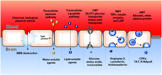
The blood–brain barrier (BBB) is an important defense barrier of the brain to control the microenvironment between the blood and brain tissues. As shown in Fig. 1, the inner layer of the BBB is mainly composed of endothelial cells and tight junctions on the walls of brain capillaries.24 Peripheral cells and matrix are located in the middle layer (i.e., the matrix membrane), while the extracellular matrix and astrocytes compose the outer layer. The unique dense structure of the BBB can prevent about 98% of small molecules and nearly 100% of macromolecules (e.g. peptides, gene drugs and protein drugs) from entering the brain.25 The presence of the BBB also limits the penetration and accumulation of drugs into brain tissue, which greatly reduces the traversing efficacy of contrast agents and medicines.26,27 With the gradual progression of glioma, the integrity of the BBB is impaired to some extent, and then the blood–brain tumor barrier (BBTB) appears between brain tumor tissues and capillaries.28,29 Therefore, the effective crossing of agents through the BBB/BBTB is an essential prerequisite for precise imaging and therapy of gliomas.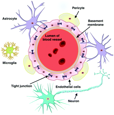 | ||
| Fig. 1 Schematic diagram of the blood–brain barrier structure. Reproduced with permission.24 Copyright 2019, Elsevier. | ||
Nanoprobes entering the brain parenchyma need to cross the BBB and then act on the tumor area. In other words, the design of probes needs upregulation of the receptor targeting specificity and the permeability of the BBB, which requires a dual-targeting probe design strategy. In general, the BBB limits toxic substances, pathogens, and >99% of drugs from entering the brain, yet at the same time selectively controls transport of ions, nutrients, and essential signaling molecules through its highly specialized transport systems. Only some small lipophilic molecules are able to passively permeate the BBB. The molecular weight cut-off is as low as 400–600 Da.30 Therefore, these passive transport routes are inefficient for most of the probes. Currently, the design of trans-BBB probes mainly focuses on active transport into the brain (Fig. 2).
Depending on the transport mechanism, the active penetration through the BBB can be divided into three main pathways (Table 1): carrier-mediated transport (CMT), receptor-mediated transcytosis (RMT) and adsorptive-mediated transport (AMT):
| Approaches of active penetration across the BBB | Ligands for mediating transport | Mechanisms | Existing challenges | Ref. |
|---|---|---|---|---|
| a CMT: carrier-mediated transport. b RMT: receptor-mediated transcytosis. c AMT: adsorptive-mediated transport. | ||||
| CMTa | Glucose | Conformational transition of membrane transporters | (1) Similar structure of the substrate to the transporter | 31, 33 and 125 |
| (2) Different expression of transporters in different cells | ||||
| (3) Competition of endogenous ligands | ||||
| RMTb | (1) Angiopep-2 | Ligand–receptor interaction | (1) Competition of endogenous ligands | 34, 50 and 84 |
| (2) Lactoferrin | (2) Individual and species differences in receptor expression | |||
| (3) Holotransferrin | ||||
| AMTc | Cell penetration peptides (TAT, and K16ApoE) | Electrostatic interaction | (1) The specificity is poor | 38 and 39 |
| (2) An existing risk of immunogenicity | ||||
(1) CMT relies on membrane protein carriers such as the receptors for D-glucose and GLUT1 that are highly expressed in the brain capillaries of the blood–brain barrier to complete the transport of substrate molecules.31–33 The transported substances first selectively bind to one side of the membrane, and thus cause the allosteric effect of carrier proteins to move the bound substrates to the other side. However, the design of probes employing CMT requires a close structural analogy to endogenous carrier substrates. Therefore, such probes may interact with red blood cells and compete with plasma nutrients, thus limiting their brain-active penetration.
(2) RMT is another endogenous transport pathway that depends on the specific binding between specific ligands (angiopep-2, lactoferrin, holotransferrin, etc.) and receptors mediating endocytosis, which are widely expressed on brain capillary endothelial cells.34 Highly expressed receptors in the BBB endothelial cells include three major receptors, including transferrin receptor (TfR), insulin receptor, and low-density lipoprotein receptor (LPR). Fan et al. designed a BBB-crossing probe using human H-ferritin (HFn) as the ligand, which could specifically bind to TfR.35 The results showed that the probe could not only successfully cross the BBB, but also kill glioma cells. Denali Therapeutics developed an antibody transport vehicle (ATV) based on an engineered Fc fragment, which could bind to TfR, efficiently cross the BBB, and generate a direct pharmacodynamic response.36,37 Notably, there are two main aspects that influence the transport effect of RTM-based probes. Several endogenous substances with high concentration in the blood, such as transferrin, would compete with RTM-based probes. Besides, individual and species differences in these receptors expressed on ECs could also influence the transport efficiency.
(3) AMT transport is based on the electrostatic interaction between positively charged ligands (cell penetration peptides, such as TAT) and the negatively charged BBB.38,39 For example, Ahlschwede et al. have shown that the cationic carrier peptide K16ApoE could increase the brain absorption of cisplatin by 34-fold, which could be easily absorbed on the surface of the nanoprobe without covalent binding.40 However, the specificity of AMT is relatively poor because cationic molecules also have strong adsorption capacity on the cell surface of other tissues in vivo. Moreover, cationic molecules are often derived from non-human proteins, which could increase the risk of complement activation and immunogenicity.
The design of probes for the imaging and therapy of gliomas requires not only good permeability across the BBB/BBTB, but also targeting ability to tumors.41,42 In general, nanoprobes can passively accumulate in tumor areas through the enhanced permeability and retention (EPR) effect due to size reasons. Given that the pore size in the BBTB of malignant tumor microvasculatures is around 12 nm, nanoparticles with diameters smaller than 12 nm may have higher permeability in tumor microvasculatures. Several peptides, including c(RGDfK) (a ligand of integrin αvβ3/αvβ5) and Pep-1 (a ligand of IL-13Rα2), have been proved to display active-targeting bioactivity to tumor neovasculature. Moreover, it has been demonstrated that most perivascular cells on BBTB originate from glioma stem cells (GSCs) through transdifferentiation. These GSC-derived pericytes bear tumor-specific genetic alterations that distinguish them from normal pericytes, indicating a possibility to selectively target these neoplastic pericytes.
3. Nanoprobe-mediated precise imaging of gliomas
3.1. MR imaging
MRI is the most widely used non-invasive diagnostic technique based on the interaction of protons with surrounding tissue molecules. The contrast agents (CAs) for MRI are generally divided into T1-positive agents and T2-negative agents. Specifically, T1 CAs, usually including paramagnetic materials containing Gd3+, Mn2+, Fe3+ and some other metal ions, enhance the contrast between tissues.43,44 T2 CAs promote the high feasibility of lesion detection, mainly involving superparamagnetic iron oxide nanoparticles (SPIONs). At present, gadolinium-based CAs have been used for clinical diagnosis of gliomas. However, these T1 CAs suffer from short blood circulation half-life, and may induce nephrogenic systemic fibrosis and neurodegenerative disorder.45 Therefore, T1 CAs with improved relaxivity, while avoiding fast excretion and excessive ion exposure in the brain, are urgently needed.46–48 In order to solve the above problems, research mainly focused on the following aspects: (1) using metal ions with low toxicity (Mn2+, and Fe3+) and a convection-enhanced delivery strategy to avoid nephrotoxicity; (2) forming macrocycles instead of linear chelates to improve the in vivo stability and retention of CAs, while avoiding damage to brain tissue.Xue et al. developed a nano-enabled, iron-based contrast agent (nIBCA) composed of a hydrophilic PEG (polyethylene glycol) tail and a hydrophobic iron-based dendritic oligomer.49 The PEG tail is attached to the dendritic oligomer by iron coordination with the catechol functional groups on nordihydroguaiaretic acid to form a telodendritic structure. This amphiphilic, telodendritic structure can readily self-assemble into a micellar nanostructure, in which the iron coordination endows the nIBCA with enhanced T1 MRI capabilities (Fig. 3a). The core–shell structure by conjugating metal ion chelates onto biocompatible micelles effectively improved the relaxivity of CAs. nIBCA was injected intravenously into a mouse model with a brain tumor and accurately displayed the intracranial tumor with high tumor-to-normal tissue ratio (TNR). The T1 signal of the glioma site was significantly enhanced and lasted at least 24 hours (Fig. 3b-c), exhibiting the potential of nIBCA as a substitute for GBCAs. It also suggests that T1 CAs need to be designed not only to avoid rapid renal clearance to improve brain accumulation, but also to avoid brain exposure due to free release of metal ions.
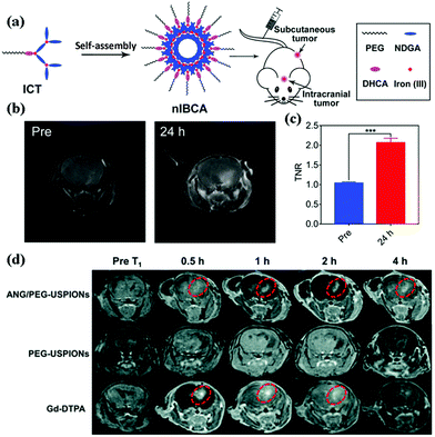 | ||
| Fig. 3 (a) Schematic illustration of the preparation of nlBCAs for MRI. (b) T1-weighted MR images of an orthotopic brain tumor (U251 MG) by using nIBCA as a contrast agent. (c) Quantitative analysis of the T1 MRI signal of an orthotopic brain tumor. Reproduced with permission.49 Copyright 2020, Elsevier. (d) T1-weighted imaging of glioblastoma (U87 MG) bearing mice at different time points (0.5 h, 1 h, 2 h and 4 h) after the injection of ANG/PEG-USPIONs, PEG-USPIONs and Gd-DTPA. Reproduced with permission.50 Copyright 2020, Royal Society of Chemistry. | ||
As the most representative T2 CAs, SPIONs have received extensive attention in the past decades. However, their clinical application is very limited due to the negative contrast effect and the presence of magnetic susceptibility artifacts. A previous report has confirmed that reducing the size of SPIONs could minimize their T2 contrast effect, which could effectively avoid the reduced resolution and spatial specificity caused by the negative contrast effect. Du et al. constructed a T1-weighted dual-targeting MR nanoprobe using the conventional T2 CA ultrasmall Fe3O4 NPs conjugated with Angiopep-2 and PEG for positive MR imaging of intracranial glioblastoma cells.50 Through the mediation of Angiopep-2 peptide, the nanoprobes effectively crossed the BBB, targeted the brain tumor, and achieved high-quality T1-weighted images clearly delineating the brain tumor boundary for at least 4 h (Fig. 3d), which was much longer than commercial Gd-DTPA contrast agents (2 h). It is worth noting that T1 or T2 contrast-enhanced MRI may still have artifacts caused by certain endogenous factors, such as bleeding, calcification, and metal deposition. Therefore, the use of artifact-free MRI for accurate diagnosis and delineation of tumors remains challenging. A new strategy based on dual-mode contrast agents (DMCAs) has been developed to obtain complementary diagnostic information with the same penetration depth and almost simultaneous spatial/temporal resolution at the same time.51,52 In general, DMCAs are prepared by doping T1 components (e.g. Gd3+ and Mn2+ ions) into the T2 matrix (e.g. superparamagnetic iron oxide) or mixing T1 and T2 contrast agents into the same matrix.53–56 Wang et al. reported a one-pot biomimetic method to synthesize KMnF3 nanocrystals mediated by bovine serum protein (KMnF3@BSA NCs) as DMCAs for dual-mode imaging of glial cells in mouse models (Fig. 4a).57 The results show that the relaxation performance of T2 is enhanced by the coupling of albumin and paramagnetic material intermolecular fields, thus greatly eliminating artifacts and improving the MR sensitivity (Fig. 4b). The DMCAs based on a single substance can avoid the interference of multilayer paramagnetic materials, increase the contrast between pathological lesions and normal tissues, and facilitate the accurate detection of invasive gliomas.58
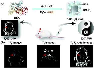 | ||
| Fig. 4 T1–T2 dual-modal MRI of brain gliomas. (a) “One-pot” synthesis of KMnF3@BSA NCs. (b) KMnF3@BSA NCs for T1–T2 dual-modal MRI of brain gliomas with improved sensitivity. Reproduced with permission.57 Copyright 2020, Elsevier. | ||
Notably, cell-based drug delivery systems have exhibited unique advantages for glioma targeting and subsequent treatment. For example, mesenchymal stem cells and neural stem cells have been proven to have intrinsic tumor-homing capacity for the delivery of therapeutic agents.59–61 Most recently, Wu et al. designed biomimetic probes for MRI enhancement using neutrophil-absorbed Fe3O4 NPs.62 In an incompletely resected in situ glioma model, T2-weighted MRI images showed that glioma regions exhibited strong negative signal contrast. Histological analysis of brain tissue sections showed that more nanoprobes were accumulated in the residual glioma area. This excellent targeting ability is attributed to three advantages of neutrophils: (1) the average half-life of neutrophils is 6–7 h, and thus the MRI signal will not be reduced by cell proliferation and exocytosis. (2) Neutrophils can be activated in the blood vessels in the immune response and move along the chemotactic gradient toward the site of inflammation (glioma area). (3) Neutrophils possess the native ability to cross the BBB/BBTB and infiltrate the tumor tissues. In the future, the development of single-substance-based DMCAs, which incorporate various transport-mediated modes across the BBB, will be a promising direction for MRI of gliomas.
3.2. Nuclear medicine imaging
The main problem in the imaging of high-grade gliomas is the arduous interpretation for the radiation necrosis (RN) and glioma recurrence after radiation therapy,63,64 which could cause vasogenic edema, disrupt the BBB, and lead to cavitation. These similar structural alterations are difficult to distinguish by conventional MRI or CT. Thus, nuclear medicine imaging (NMI), including single-photon emission computed tomography (SPCET) and positron emission tomography (PET), are close to being implemented in many centres into clinical routine for planning of biopsy-sampling, differentiation of RN (i.e. pseudoprogression) from tumour progression, and follow-up of the effects of chemotherapy.65 Currently, in the study of Zhao et al, polyethylenimine (PEI) dendrimers were sequentially conjugated with polyethylene glycol (PEG), chlorotoxin (CTX) and 3-(4-hydroxyphenyl)propionic acid-OSu (HPAO) for facile 131I radiolabeling.66 The 131I-labeled CTX-functionalized NPs showed high radiochemical purity and stability, and could be used as SPECT imaging probes to target glioma cells in vivo in a subcutaneous tumor model.Comparing with SPCET, PET enables the quantification of injected tracers, which could achieve more accurate assessment of RN and glioma recurrence.67 Gliomas express various molecular markers for PET imaging. Several new tracers have been introduced for PET imaging of gliomas, such as [124I]mIBG, [18F]mFBG, [18F]FDG, [68Ga]Ga-DOTA peptides, [18F]F-DOPA, and [11C]mHED. [18F]-2-Deoxyfluoro-D-glucose (FDG) is the most widely used positron tracer for cancer diagnosis, which is a glucose analogue labeled with PET radioisotope 18F.68–71 As a metabolic compound, [18F] FDG has high accumulation in tumor tissues with high glucose metabolism. However, the high demand for glucose in the brain can lead to high background signal in normal neurological tissues, which makes FDG-PET imagining unachievable in the diagnosis of brain cancer. To solve the controversy of low sensitivity and specificity of NMI for glioma, high specificity FDG-PET composite imaging NPs were developed by combining radioactive tracer and templated targeted BBB/tumor nanocarrier. Oku et al. applied Ala-Pro-Arg-Pro-Gly (APRPG) (a peptide homing to angiogenic vessels)-PEG modified 100 nm-size liposomes entrapping 18F for the PET imaging of brain tumors.72 A 1 mm diameter brain tumor could be clearly resolved in C6 glioma-bearing rats by PET, which was hardly detected by CT (Fig. 5). Generally, NMI combined with nanotechnology has more advantages in distinguishing between RN and glioma recurrence, which can provide valuable information about metabolism, regional blood flow, and chemical composition.
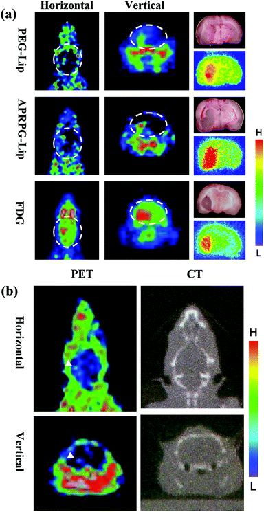 | ||
| Fig. 5 PET imaging of brain tumors. (a) Rats were intravenously injected with 10 MBq of [18F]SteP2-labeled PEG-liposomes (top panel), [18F]SteP2-labeled APRPG-liposomes (middle panel) or [18F]FDG (bottom panel) at day 11 after implantation of C6 glioma cells into the left midbrain. The brains were sliced into 2 mm sections, and the autoradiograms (right lower panels) and the pictures (right upper panels) are shown. (b) PET (left) and CT (right) imaging of 1 mm diameter C6 glioma-bearing model rats. Arrowheads indicate the tumor. Reproduced with permission.72 Copyright 2010, Elsevier. | ||
3.3. Near-infrared fluorescence imaging
Fluorescence imaging can provide direct visualization of biological structures and functions of living organisms. Compared with the conventional visible window (400–700 nm), the near-infrared (NIR) window is more suitable for in vivo imaging with better spatial and temporal resolution, which is also promising for glioma imaging.73–75 Jia et al. prepared biomimetic proteolipid (BLIPO) nanoparticles (NPs) loaded with NIR fluorescent dye indocyanine green (ICG), and embedded membrane proteins of glioma cells into NPs (Fig. 6a).76 Owing to their homotypic targeting and immune escaping characteristics, the obtained BLIPO-ICG NPs could cross the BBB and actively target glioma cells at the early stage. High accumulation in the brain tumor with a signal to background ratio of 8.4 was achieved, and the glioma and its margin were clearly visualized by NIR fluorescence imaging over time (Fig. 6b–d). Wang et al. used the membrane of brain metastasis cells to camouflage ICG-loaded nanoparticles.77 The cell membrane-camouflaged nanoparticles replicated the highly complex functionalities of source cells and exhibited significantly higher ability to traverse both the intact BBB and BBB disrupted to different degrees, allowing NIR fluorescence imaging and phototherapy of brain tumors.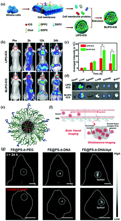 | ||
| Fig. 6 (a) Schematic illustration of the preparation process of the biomimetic proteolipid BLIPO-ICG. (b) In vivo NIR-I fluorescence imaging of BLIPO-ICG in C6 tumor bearing mice before and after intravenous injection of LIPO-ICG and BLIPO-ICG. (c) Semiquantitative fluorescence intensity analysis of the tumor site at different time intervals. (d) Ex vivo fluorescent images of major organs and tumors at 24 h post injection. Reproduced with permission.76 Copyright 2018, American Chemical Society. (e) Schematic illustration of organic NIR-II fluorescence-emitting spherical nucleic acid self-assembled from PS-b-DNA and organic dyes. (f) Illustration of the process of crossing the BBB and targeting glioblastoma. (g) NIR-II fluorescence imaging of mouse heads and ex vivo brain tissues using different NIR-II-emitting materials under 808 nm irradiation. Scale bars: 0.5 cm. Reproduced with permission.81 Copyright 2020, Wiley-VCH. | ||
In the past decade, advances in fluorescence imaging in the NIR range have extended from the traditional NIR window (NIR-I, 700–900 nm) to the second NIR window (NIR-II, 1000–1700 nm).78 Attributed to the suppressed photon scattering and diminished autofluorescence, in vivo fluorescence imaging in the NIR-IIb window can afford high clarity and deep tissue penetration.79,80 Most recently, Xiao et al. reported the use of DNA nanotechnology to transport an NIR-II emitting dye across the BBB for the non-invasive imaging of gliomas. DNA block copolymer was utilized to protect NIR-II emitting dyes from early leakage in an in vivo application, which could also transport the dye across the BBB via the scavenger receptor-mediated transcytosis pathway (Fig. 6e and f).81 Moreover, after being conjugated with glioma cell-specific aptamers, the obtained nanofluorophores could target gliomas with enhanced imaging intensity and resolution (Fig. 6g). The combination of NIR-II imaging and DNA nanotechnology with dual effects will benefit the precise diagnosis and further therapy of gliomas.
3.4. Photoacoustic imaging
Photoacoustic (PA) imaging is a non-ionizing imaging method that relies on a PA effect generated when light is absorbed by exogenous contrast agents, allowing the non-invasive in vivo imaging in deep tissue-regions with high spacial resolution.82,83 Currently, diverse NIR-I CAs have been developed as exogenous CAs for PA imaging of glioma, but there still exist many problems to be solved. It is difficult to distinguish the PA signals of CAs and endogenous hemoglobin. Meanwhile, the relatively low NIR absorbance and narrow absorption spectra of CAs lead to lower imaging sensitivity.84,85 Chen et al. directly prepared ultrathin layered nanostructured molybdenum disulfide (MoS2) nanosheets for PA imaging of brain tumors without further surface modification.86 They found that reducing the number of nanosheet layers could not only significantly improve the absorption of NIR light, but also improve the elastic properties of nanosheets with amplified PA effect, which could serve as an efficient nanoplatform for sensitive PA molecular imaging. On this basis, Liu et al. coupled ICG molecules to the surface of MoS2 nanosheets and improved photothermal/photoacoustic conversion efficiency at the same time (Fig. 7a).87In vivo PA imaging of gliomas clearly revealed a brain tumor 3.5 mm under the scalp of a mouse (Fig. 7b), indicating that the combination of two different NIR-I window CAs significantly improved the optical stability and imaging sensitivity of CAs.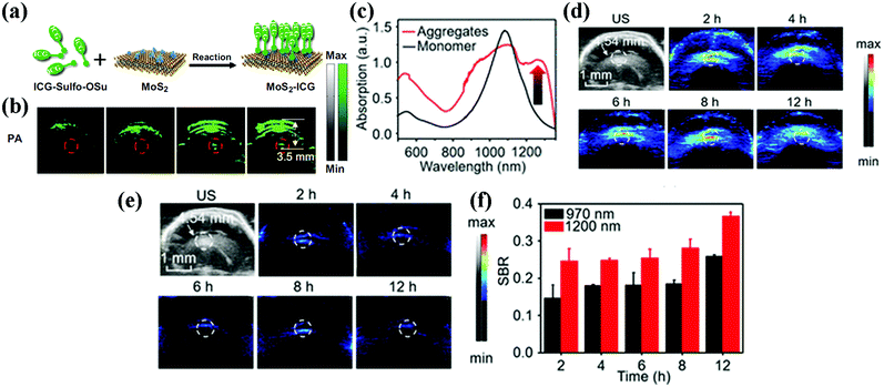 | ||
| Fig. 7 (a) Schematic illustration of the preparation of MoS2-ICG. (b) Photoacoustic images of the brain tumor region before and at 1, 3, and 5 h after intravenous injection of MoS2–ICG. The photoacoustic signals in the red circles show the accumulation and distribution of MoS2–ICG within the brain glioma. Reproduced with permission.87 Copyright 2018, Springer. (c) Absorption spectrum of A1094 monomer and A1094 J-aggregate nanomicelles. (d) PA images of a mouse brain glioma at different time points in vivo at 970 nm and (e) at 1200 nm. (f) SBR of PA signals at 970 and 1200 nm at different time points. Reproduced with permission.92 Copyright 2019, American Chemical Society. | ||
Similar to fluorescence imaging, PA imaging also suffers from strong light scattering and high levels of absorption that interfere in the NIR-I window. Recently, NIR-II PA contrast agents have shown outstanding performance with deeper tissue penetration and better imaging fidelity.88–91 Liu et al. synthesized biocompatible J-aggregate nanoprobes by coating lipophilic small molecule NIR-II dye A1094 (1094 nm) with DSPE-PEG2000 phospholipid membrane.92 Under the close encapsulation of the liposome membrane, the intermolecular distance was significantly shortened and the orbital overlap was further enhanced. The results showed that an unprecedented absorption band of 1.2–1.3 um was obtained in the NIR-II window (Fig. 7c). An ∼4.54 mm deep brain lesion was imaged at 1200 nm with minimized background and increased contrast compared to the NIR-I window (Fig. 7d–f). Conjugated polymer (CP) nanoparticles are another significant class of macromolecular PA contrast agents with NIR-II absorption. Yang et al. reported semiconductor polymer nanoparticles, which encapsulated PDPPTBZ into NPs through nanoprecipitation using 1,2-distearoyl-sn-glycero-3-phosphoethanolamine-N-[methoxy(polyethylene glycol)-2000] (DSPE-PEG2000) with a large mass extinction coefficient (43 mL mg−1 cm−1) at 1064 nm, which realized the PA imaging of the glioma 3.8 mm below the skull.93 The excellent absorbance and imaging depth can avoid the adverse side effects caused by low absorbance requiring high contrast agent concentration and high laser power to produce sufficient signals.
3.5. Multimodal imaging
By rationally controlling the components and structures, the nanoprobes can be further tailored with diverse properties for multimodal imaging, which could realize complementary imaging without the multiple injections of different contrast agents.94 For example, the local surgical failure rate caused by preoperative MRI detection remains high during glioma surgery even with the use of assistive techniques.95–97 However, dual-modal imaging could facilitate the surgeons’ visual detection and tactile feedback to distinguish tumor margins from the surrounding normal tissues. Thawani et al. synthesized a kind of nanocluster encapsulated with ICG (for intraoperative PA imaging) and SPION (for preoperative MR imaging) for dual-modal imaging.98 In an aggressive glioma mouse model, MR/PA dual-modal imaging and surgery were successfully performed, which greatly improved the progression-free survival in mice with malignant gliomas. The surgeons can use PA imaging to depict contrast-enhanced areas in real time on the basis of preoperative MRI, regardless of background signal or rapid clearance in blood circulation, to meet the requirements of overall visualization of gliomas. In addition, several Bi-based nanomaterials, such as Bi2S3 and Bi2Se3 nanostructures, have been developed for CT/PA dual-modal imaging. Lu et al. coated Bi NPs with DSPE-PEG by ultrasonic emulsification, and showed significant enhancement CT and PA signals in C6 glioma model experiments.99 The combination of CT with PA imaging could overcome the low sensitivity of CT to soft tissues and visualize tissue function and disease.Moreover, Kircher et al. proposed novel three-modal MRI-PA-Raman nanoparticles for molecular imaging of glioma (Fig. 8).100 A 60-nm gold core was covered with the Raman molecular tag trans-1,2-bis(4-pyridyl)-ethylene, and further modified with 1,4,7,10-tetraazacyclododecane-1,4,7,10-tetraacetic acid (DOTA)-Gd3+ using a maleimide linkage, resulting in a gold-based surface-enhanced Raman scattering (SERS) nanoparticle coated with Gd3+ ions. The whole brain tumors are localized by MRI before and during operation, and high spatial resolution and three-dimensional imaging by photoacoustic imaging. Meanwhile, Raman imaging further provided the molecular information on the edges of the tumors with high specificity and high resolution, which made the imaging and resection of brain tumors more precise. Duan et al. used cRGD-labeled brain tumor cell membrane as a shell to coated iron oxide nanoparticles encapsulated by conjugated polymers, and achieved MRI-FL-PA multimodal imaging of gliomas.101 The integration of discrete nanoparticles into hetero-nanostructures and conjugation of nanomaterials with functional polymers will open a new avenue for the construction of multimodal contrast agents.
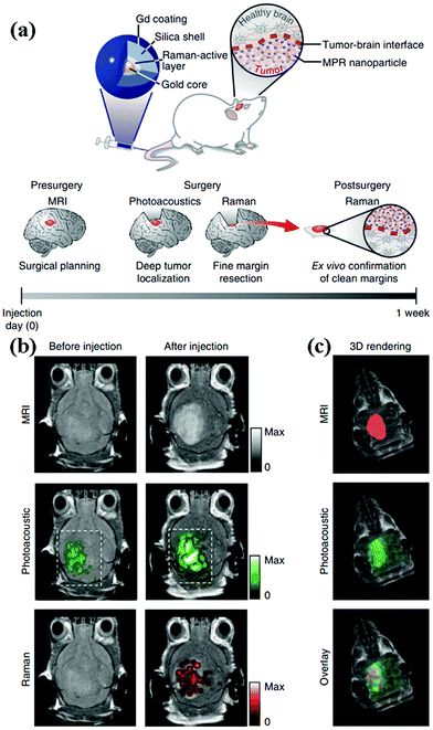 | ||
| Fig. 8 (a) Triple-modality MPR concept. MPRs are injected intravenously into a mouse bearing an orthotopic brain tumor and diffuse through the disrupted BBB and are then sequestered and retained by the tumor (top). The concept of proposed eventual clinical use (bottom). (b) Two-dimensional axial MRI, photoacoustic and Raman images. The post-injection images of all three modalities showed clear tumor visualization (dashed boxes outline the imaged area). (c) A three-dimensional (3D) rendering of magnetic resonance images with the tumor segmented (red; top), an overlay of the three-dimensional photoacoustic images (green) over the MRI (middle) and an overlay of MRI, the segmented tumor and the photoacoustic images (bottom) showing good colocalization of the photoacoustic signal with the tumor. Reproduced with permission.100 Copyright 2012, Nature Publishing Group. | ||
4. Nanoprobe-mediated therapy of gliomas
The difficulty of complete resection of gliomas by surgery often leads to poor prognosis and invariable recurrence, thus requiring adjuvant drug therapy.102,103 Traditional invasive drug delivery strategies rely on opening the BBB by physical methods (osmotic damage, ultrasound damage, magnetic damage, etc.) and injecting therapeutic drugs into the brain parenchyma, which could lead to toxicity and drug expansion beyond the expected location.104–106 Significantly, ultrasound-based techniques for reversible BBB opening have been widely studied. Focused ultrasound (FUS) combined with intravenously administered microbubbles (MB-FUS) can achieve reproducible, focal, transient, and non-invasive BBB/BTB opening to enhance the targeted permeability of therapeutics. Selective accumulation in brain tumors during the optimal time window for transient mediated BBB opening is the most prominent feature of this approach.107–110 Moreover, sonodynamic therapy (SDT) based on ultrasound stimulation and a sonosensitizer, and immunotherapy by converting the immunosuppressive tumor microenvironment (TME) into an immunostimulatory TME via a higher but safe FUS dosage have also been demonstrated, indicating the potential significance of MB-FUS as a novel treatment for SDT and immunotherapy. 111,112 Various non-invasive strategies including ligand-mediated drug delivery and intranasal drug delivery have been developed.113,114 In addition, several nanoprobes based on inorganic mesoporous materials, linear polymer micelles, and supramolecular vesicles have been engineered as the carriers for drug loading and delivery into the brain.115 These composite nanostructures not only facilitate the active penetration of drugs through the BBB, but also prolong the circulation time and retention of drugs. Kang et al. modified docetaxel liposomes with muscone and anti-TfR monoclonal antibody, which could enhance the permeability of the BBB for drug delivery into the cerebrospinal fluid.116 The composite liposomes not only improved the blood circulation time of the DTX, but also increased brain targeting and prolonged the survival time of nude mice bearing gliomas.Intranasal drug delivery could bypass the BBB and intranasally administer into the brain, thus avoiding liver, gastrointestinal tract, serum degradation and renal filtration. Sukumar et al. prepared polyfunctional gold/iron NPs loaded with therapeutic microRNAs, which were injected intranasally into orthotopic glioblastoma-implanted mice.117 The surface modification with chitosan–cyclodextrin hybrid polymers promoted the adhesion of NPs to nasal mucosa upon inhalation, while the coating of T7 peptides improved the targeting ability to glioblastoma cells, thus significantly enhancing the delivery of therapeutic microRNAs to glioblastoma.
Most recently, it has also been demonstrated that PA shock waves generated by the cavitation (growth and collapse) of nanobubbles and microbubbles around nanoparticles heated by laser pulses can cause strong local mechanical damage to the tissues. Liu et al. designed a therapeutic nanoprobe featuring high red absorbance and selective penetration of the BBB at the tumor site (Fig. 9).118 The nanoprobes activated the adenosine receptor on the BBB to allow self-passage and accumulation in the glioblastoma. Upon applying laser pulses with appropriate energy for irradiation, the nanoprobes absorbed the light energy and generated a PA shockwave via PA cavitation, which could achieve localized tumor cell destruction with minimal damage to the adjacent normal tissue, thus realizing precise antitumor treatment for glioblastoma.
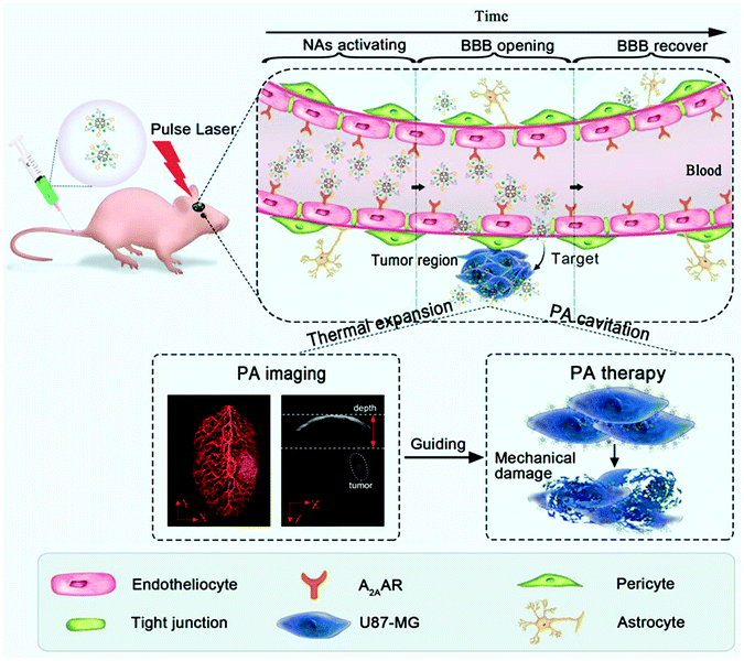 | ||
| Fig. 9 Photoacoustic imaging-guided therapy of gliomas with Den-RGD/CGS/Cy5.5 nanoprobes. Reproduced with permission.118 Copyright 2019, Wiley-VCH. | ||
5. Theranostic nanoprobes for gliomas
The mode of diagnosis first followed by treatment cannot monitor the distribution and metabolic process of therapeutic drugs in patients in real time. Therefore, it is hard to evaluate the efficacy of drugs and their potential toxicity to normal organs.119,120 Furthermore, multiple injections are required when imaging CAs and drugs are used separately, which may lead to additional trauma and increase the metabolic burden of patients. Therefore, the design of theranostic nanoprobes is an important direction to achieve precise diagnosis and treatment of gliomas.121–123 Several imaging nanoprobes have been found to also possess excellent photothermal conversion ability and photodynamic activity, which could be used as efficient photosensitizing agents for photothermal therapy (PTT) and photodynamic therapy (PDT).124–126Guo et al. developed D–A structured biocompatible and photostable conjugated polymer nanoparticles with strong absorption in the NIR-II window for precise PA imaging and PTT of brain tumors through the scalp and skull.127 Taking advantage of a real-time PA imaging system, the NPs assisted clear pinpointing of glioma at a depth of almost 3 mm through the scalp and skull with an ultrahigh signal-to-background ratio of 90 (Fig. 10a and b). After PTT, the glioma progression is effectively inhibited and the survival spans of mice are significantly extended (Fig. 10c). This unique mode of theranostic nanoprobes, which integrates optical imaging and local photothermal ablation therapy is expected to increase the accuracy for the treatment of gliomas. In addition, from the point of view of energy consumption, the dissipated energy from radiation decay can be used for fluorescence imaging, while the part from non-radiation decay provides the feasibility of PTT or PDT. For example, aggregation-induced emission (AIE) materials that can skillfully control the molecular movement of organic molecules through sufficient molecular rotors or vibrators are expected to adjust the balance between radiation attenuation and non-radiation attenuation, and realize an all-around phototherapy diagnosis system for a single substance.128–130
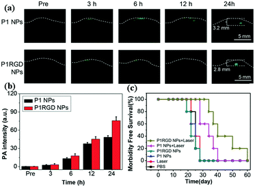 | ||
| Fig. 10 (a) Non-invasive PA imaging of mouse brain tumors through the scalp and skull at different time points upon NP injection. (b) Quantitative results of tumor PA intensity at different time points after administration of P1 NPs, and P1 RGD NPs. (c) Survival rates of orthotopic brain tumor-bearing mice after photothermal treatment. The mice were categorized into six groups composed of PBS, laser, P1 NPs, P1 RGD NPs, P1 NPs + laser, and P1 RGD NPs + laser. Reproduced with permission.127 Copyright 2018, Wiley-VCH. | ||
Moreover, some composite nanoprobes combined with chemodynamic therapy, gene therapy, immunotherapy and other novel alternative therapies have also been developed. Duan et al. designed NIR-responsive polycationic gatekeeper-cloaked hetero-nanoparticles for multimodal imaging (CT/PAI/FLI)-guided triple-combination (PTT/gene therapy/chemotherapy).131 The hierarchical hetero-structure constructed via integration of AuNRs with CdTe QDs through a mesoporous silica intermediate layer enabled the diagnosis of gliomas. The successful loading of p53 (antioncogene) and DOX (chemotherapeutics) was used to inhibit the growth of glioma. Meanwhile, temperature changes caused by NIR not only accelerated the cascade release of chemotherapeutic drugs, but also realized PTT of tumors.
6. Summary and perspective
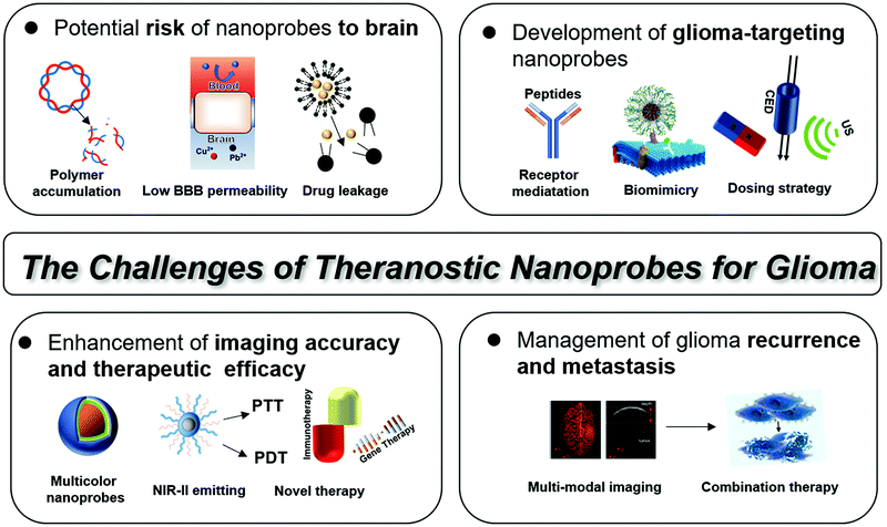
In summary, nanoprobe-mediated imaging and therapy methods have exhibited great potential for the treatment of gliomas (Table 2). The unique physiochemical features of nanoprobes make them a beneficial complement of conventional contrast agents. After conjugation with transmembrane and tumor targeting ligands, the nanoprobes could actively recognize receptors highly expressed in the BBB and glioma cells, thus enhancing the targeting ability and imaging accuracy to gliomas. Moreover, nanoprobes could also act as reliable carriers for the efficient loading and delivery of therapeutic drugs, which could promote the penetration through the BBB and prolong the retention of drugs. Actually, although nanotechnology has made tremendous progress for the preclinical research of glioma, most of the nanoprobes are not officially approved by the Food and Drug Administration (FDA), and no relevant human experimental studies have been carried out yet. To facilitate clinical translation of nanoprobes for glioma, the following issues must be considered carefully (Fig. 11).| Diagnosis/therapy | Nanoprobe | BBB crossing strategy | Glioma targeting method | |
|---|---|---|---|---|
| a EPR: enhanced permeability and retention effect. b RMT: receptor mediated transcytosis. c CDDSs: cell-based drug delivery systems (CDDSs). d CCBA: cancer cell membrane-based bioengineering approach. e BTA: brain-tumor targeting aptamer. | ||||
| MRI | T1 | nIBCA,49 ANG/PEG-USPIONs50 | EPRa![[thin space (1/6-em)]](https://www.rsc.org/images/entities/char_2009.gif) ,49 RMTb ,49 RMTb![[thin space (1/6-em)]](https://www.rsc.org/images/entities/char_2009.gif) 50 50 |
EPR,49 RMT50 |
| T2 | ND-MMSNs62 | CDDSsc | CDDSs | |
| T1/T2 | KMnF3@BSA NCs57 | EPR | EPR | |
| NMI | PET | [18F]FDG-APRPG-Lip72 | RMT | RMT |
| FI | NIR-I | BLIPO-ICG,76 B16-PCL-ICG/4T1-PCL-ICG77 | CCBAd![[thin space (1/6-em)]](https://www.rsc.org/images/entities/char_2009.gif) 76,77 76,77 |
CCBA76,77 |
| NIR-II | NR@PS-b-DNA/Apt81 | DNA nanotechnology | BTAe | |
| PA | NIR-I | Holo-Tf-ICG,84 MoS2-ICG87 | RMT74 | RMT74 |
| EPR77 | EPR77 | |||
| NIR-II | A1094@RGD-HBc,88 PDPPTBZ93 | RMT | RMT | |
| Dual-modal | MRI/FI | RGD-RFP-LBT-Gd94 | EPR | RMT |
| MRI/PA | ISCs98 | EPR | EPR | |
| Tri-modal | MRI/PA/Raman | MPR100 | EPR | EPR |
| MRI/FI/PA | cRGD-CM-CPIO101 | CCBA | CCBA/RMT | |
| Theranostics | PA-PTT | P1RGD127 | RMT | RMT |
| MRI/FI-PDT/PTT | iRGD-ILD126 | RMT | RMT | |
| CT/PAI/FLI-PTT/gene therapy/chemotherapy | ASQ-DOX-PGEA2/p53131 | EPR | EPR | |
6.1 Potential risk of nanoprobes to the brain
Long-term safety issues of these nanoprobes include brain injury, potential systemic toxicity, intracranial residues, and in vivo metabolism, which greatly limit their clinical uses and translation.132,133 Low BBB permeability of nanoprobes often results in multiple injections with large doses, which may lead to additional trauma and increase the metabolic burden. It should be noted that several imaging nanoprobes contain heavy metal elements, which have potential in vivo toxicity and hinder their clinical uses. In addition, nanoprobes coated by self-assembled structures (linear polymer micelles, supramolecular vesicles, etc.) are unstable in the endosome or lysosome.134 The degradation of probes caused by the changes of the environment (pH, enzymatic degradation, etc.) may lead to the release of free metal ions and accumulation of polymers, thus causing damage to nerve cells and brain tissues.134,135 The development of heavy metal-free nanoprobes with in vivo stability is a major challenge in the future.6.2. Development of glioma-targeting nanoprobes
Different from the imaging of other tumors, the contrast agents need to traverse the BBB before targeting gliomas, which raises a higher demand to the design of nanoprobes with high targeting specificity. The advantages and drawbacks of different BBB crossing strategies are summarized in Table 3. Generally, nanoprobes should be conjugated with different ligands for the active crossing of the BBB and targeting of cancer cells, including peptides, proteins, and nucleic acids.81,101 In addition, biomimetic and homotypic modification is also a promising way to promote the delivery across the BBB and into glioma cells.136 Meanwhile, some dual-peptide-modified NPs have a higher BBB penetration or transportation efficiency than that modified with a single peptide. Notably, a long blood circulation time is required for the nanoprobe to improve its targeting efficiency in vivo.137 However, the longer retention of nanoprobes in the body not only leads to the generation of potential toxicity, but also reduces the signal-to-background ratio and the imaging quality. Therefore, how to balance the circulation time and excretion rate is also crucial for improving the targeting efficiency of nanoprobes. Besides optimizing the probe itself, developing an appropriate administration strategy is also important. Intranasal administration bypassing the BBB, MB-FUS, convection-enhanced drug delivery (CED) etc. effectively heighten the intracerebral drug accumulation.107–112 In brief, the BBB-penetrating and tumor-targeting abilities should be considered simultaneously for the design of glioma-targeting nanoprobes.| BBB crossing strategy | Advantages | Existing challenges | Ref. |
|---|---|---|---|
| a RMT: receptor-mediated transcytosis. b CPPs-MT: cell penetrating peptide-mediated transport. c BHM: biomimetic and homotypic modification. d ID: intranasal delivery. e MB-FUS: focused ultrasound combined with intravenous administered microbubbles. f CED: convection-enhanced drug delivery. | |||
| RMTa | Designability and conjugatability of polypeptide ligands | Competition of endogenous ligands and individual differences in targeting receptors | 50, 102, 105 and 113 |
| CPPs-MTb | Direct penetration without energy-dependence or endocytosis | Limited affinity, lack of selectivity, and easy immune clearance | 38 and 39 |
| BHMc | Cancer cell membrane/nucleic acid modification possessing homotypic binding and immune escaping | Few studies on active targeting glioma, needing further evaluation | 62, 76, 77, 81 and 101 |
| IDd | Non-invasive, bypassing BBB | (1) Integrity of nasal mucosa can be broken by repeated administration | 17, 24 and 117 |
| Avoiding non-targeted accumulation and liver metabolism | (2) Significant differences in the administration efficiency of different routes of nasal administration | ||
| MB-FUSe | Transient reversible BBB/BTB opening | (1) Hard to selective accumulation during the optimal time window | 107–112 |
| (2) Allow the delivery of macromolecular substances into CNS, resulting in the neuropathological changes | |||
| CEDf | High intracranial accumulation and limited systemic toxicity | Depends on the external pressure gradient as provided by a syringe pump | 18 and 106 |
6.3. Enhancement of imaging accuracy and therapeutic efficacy
Over the past two decades, several real-time neurosurgical imaging technologies, such as intraoperative neuro-navigation, intraoperative ultrasonography (iUS), intraoperative MRI (iMRI) and fluorescence imaging have been applied for the diagnosis and treatment of gliomas. As shown in Table 4, different imaging modalities have their own characteristics. How to improve the imaging accuracy for the diagnosis of gliomas is the most arduous issue. For example, the clinical applications of nanoprobes for MRI are quite limited because of the negative contrast effect and magnetic susceptibility artifacts.52 With the development of optical imaging nanomaterials, imaging in the NIR-II window becomes promising for the precise diagnosis of gliomas. Several NIR-II emitting nanoprobes have been developed for the in vivo imaging of deep tissues with high clarity.138,139 Combined with high-resolution imaging techniques, including light-sheet microscopy and time-resolved fluorescence microscopy, in vivo imaging at the cellular or molecular level could also be realized. In addition, the use of an NIR-II laser source can generate the lowest PA background noise due to the low optical absorption of deoxygenated hemoglobin, which greatly improves the resolution of PA imaging.140 Besides the improvement of luminescence or absorption performance of imaging nanoprobes, multicolour nanoprobes with longer wavelength are also needed for multiplexed imaging to ensure the in vivo qualitative and quantitative accuracy.| Precise imaging modes | Advantages | Existing challenges | Ref. |
|---|---|---|---|
| a MRI: magnetic resonance imaging. b NMI: nuclear medicine imaging. c FLI: fluorescence imaging. d PAI: photoacoustic imaging. | |||
| MRIa | Complementary advantages of T1/T2 | Negative contrast effect and magnetic susceptibility artifacts | 46–48 and 50–52 |
| NMIb | Differentiating between RN and recurrence | (1) Short circulation time of radiotracer | 65, 66 and 72 |
| (2) Radiation brain injury | |||
| FLIc | (1) High clarity and deep tissue penetration of NIR II window | Influence of tissue scattering and absorption | 76, 77 and 79–81 |
| (2) Multicolour nanoprobes with longer wavelength for multiplexed imaging to ensure the in vivo qualitative and quantitative accuracy. | |||
| PAId | Multiscale and multidimensional high resolution and the lowest PA background noise in NIR II imaging | (1) Difficult to distinguish the PA signals of CAs and endogenous haemoglobin | 86 and 88–92 |
| (2) The relatively low NIR absorbance and narrow absorption spectra of CAs lead to lower imaging sensitivity | |||
Furthermore, several therapeutic strategies have emerged based on the unique and prominent physicochemical properties of the NPs. For example, the excellent photoconversion effect of the NPs produces a series of phototherapy methods such as PTT and PDT; nanocarrier-based transport of gene drugs and temperature-responsive controlled release realizes gene therapy; NP-based ultrasound mediation initiates immunogenicity of the glioma microenvironment, which is promising for immunotherapies. It can be assumed that an all-in-one nanoprobe incorporated with imaging and therapeutic functions may provide a prospective platform for the whole diagnosis, therapy and prognosis process of gliomas.
6.4 Management of glioma recurrence and metastasis
Currently, surgical resection remains the mainstay of treatment for gliomas. The relationship between the extent of glioma resection and actual clinical benefit for the patient is predicated on the balance between cytoreduction and neurological morbidity.8 Therefore, glioma recurrence and metastasis are often inevitable.141,142 The development of theranostic nanoprobes integrating accurate preoperative diagnosis and therapeutic effects of non-invasive methods (including PTT, PDT, chemodynamic therapy,143,144 gene therapy,131,145,146 and immunotherapy112,147) could be a promising direction for the treatment of glioma recurrence and metastasis.In summary, the adoption of functional nanoprobes for precise diagnosis and personalized therapy of glioma is moving forward steadily. The interdisciplinary fusion of nanotechnology, in vivo imaging techniques, and tumor therapeutics will offer a great opportunity for the preclinical research of gliomas.
Conflicts of interest
The authors declare no conflicts of interest.Acknowledgements
This work was supported by the National Natural Science Foundation of China (21974104, 51873168, and 51803161), and the Fundamental Research Funds for the Central Universities (WUT: 2020III035GX).Notes and references
- M. Weller, W. Wick, K. Aldape, M. Brada, M. Berger, S. M. Pfister, R. Nishikawa, M. Rosenthal, P. Y. Wen, R. Stupp and G. Reifenberger, Nat. Rev. Dis. Primers, 2015, 1, 15017 Search PubMed.
- J. N. Rich and D. D. Bigner, Development of novel targeted therapies in the treatment of malignant glioma, Nat. Rev. Drug Discovery, 2004, 3, 430–446 Search PubMed.
- A. S. Achrol, R. C. Rennert, C. Anders, R. Soffietti, M. S. Ahluwalia, L. Nayak, S. Peters, N. D. Arvold, G. R. Harsh, P. S. Steeg and S. D. Chang, Nat. Rev. Dis. Primers, 2019, 5, 5 Search PubMed.
- H. A. Fine, Malignant gliomas: Simplifying the complexity, Cancer Discovery, 2019, 9, 1650–1652 Search PubMed.
- D. Sturm, S. M. Pfister and D. T. W. Jones, J. Clin. Oncol., 2017, 35, 2370–2377 Search PubMed.
- G. Nie, H. J. Hah, G. Kim, Y. E. K. Lee, M. Qin, T. S. Ratani, P. Fotiadis, A. Miller, A. Kochi and D. Gao, Small, 2012, 8, 884–891 Search PubMed.
- Y. Liu, J. Tan, Y. Zhang, J. Zhuang, M. Ge, B. Shi, J. Li, G. Xu, S. Xu, C. Fan and C. Zhao, Biomaterials, 2018, 173, 1–10 Search PubMed.
- N. Sanai and M. S. Berger, Nat. Rev. Clin. Oncol., 2018, 15, 112–125 Search PubMed.
- M. J. van den Bent, M. Smits, J. M. Kros and S. M. Chang, J. Clin. Oncol., 2017, 35, 2394–2401 Search PubMed.
- A. D. Norden, J. Drappatz and P. Y. Wen, Lancet Neurol., 2008, 7, 1152–1160 Search PubMed.
- F. G. Dhermain, P. Hau, H. Lanfermann, A. H. Jacobs and M. J. van den Bent, Lancet Neurol., 2010, 9, 906–920 Search PubMed.
- Z. H. Houston, J. Bunt, K. S. Chen, S. Puttick, C. B. Howard, N. L. Fletcher, A. V. Fuchs, J. Cui, Y. Ju, G. Cowin, X. Song, A. W. Boyd, S. M. Mahler, L. J. Richards, F. Caruso and K. J. Thurecht, ACS Central Sci., 2020, 6, 727–738 Search PubMed.
- B. Ling, J. Lee, D. Maresca, A. Lee-Gosselin, D. Malounda, M. B. Swift and M. G. Shapiro, ACS Nano, 2020, 14, 12210–12221 Search PubMed.
- X. Gao, Q. Yue, Y. Liu, D. Fan, K. Fan, S. Li, J. Qian, L. Han, F. Fang, F. Xu, D. Geng, L. Chen, X. Zhou, Y. Mao and C. Li, Theranostics, 2018, 8, 3126–3137 Search PubMed.
- G. Sharma, A. R. Sharma, S. S. Lee, M. Bhattacharya, J. S. Nam and C. Chakraborty, Int. J. Pharm., 2019, 559, 60–372 Search PubMed.
- C. Chen, Z. Duan, Y. Yuan, R. Li, L. Pang, J. Liang, X. Xu and J. Wang, ACS Appl. Mater. Interfaces, 2017, 9, 5864–5873 Search PubMed.
- X. Gao and C. Li, Small, 2014, 10, 426–440 Search PubMed.
- X. Wu, H. Yang, W. Yang, X. Chen, J. Gao, X. Gong, H. Wang, Y. Duan, D. Wei and J. Chang, J. Mater. Chem. B, 2019, 7, 4734–4750 Search PubMed.
- H. Yan, L. Wang, J. Wang, X. Weng, H. Lei, X. Wang, L. Jiang, J. Zhu, W. Lu, X. Wei and C. Li, ACS Nano, 2012, 6, 410–420 Search PubMed.
- S. He, S. Chen, D. Li, Y. Wu, X. Zhang, J. Liu, J. Song, L. Liu, J. Qu and Z. Cheng, Nano Lett., 2019, 19, 2985–2992 Search PubMed.
- Q. Miao and K. Pu, Adv. Mater., 2018, 30, 1801778 Search PubMed.
- Y. C. Tsai, P. Vijayaraghavan, W. H. Chiang, H. H. Chen, T. I. Liu, M. Y. Shen, A. Omoto, M. Kamimura, K. Soga and H. C. Chiu, Theranostics, 2018, 8, 1435–1448 Search PubMed.
- S. Duan, Y. Yang, C. Zhang, N. Zhao and F. J. Xu, Small, 2017, 13, 1603133 Search PubMed.
- J. Xie, Z. Shen, Y. Anraku, K. Kataoka and X. Chen, Biomaterials, 2019, 224, 119491 Search PubMed.
- W. Zhou, C. Chen, Y. Shi, Q. Wu, R. C. Gimple, X. Fang, Z. Huang, K. Zhai, S. Q. Ke, Y. F. Ping, H. Feng, J. N. Rich, J. S. Yu, S. Bao and X. W. Bian, Cell Stem Cell, 2017, 21, 591–604 Search PubMed.
- J. K. Rad, A. R. Mahdavian, S. Khoei and S. Shirvalilou, ACS Appl. Mater. Interfaces, 2018, 10, 19483–19493 Search PubMed.
- J. Li, A. Xi, H. Qiao and Z. Liu, Cancer Lett., 2020, 475, 92–98 Search PubMed.
- F. Ma, L. Yang, Z. Sun, J. Chen, X. Rui, Z. Glass and Q. Xu, Sci. Adv., 2020, 6, eabb4429 Search PubMed.
- S. I. Ahn, Y. J. Sei, H. J. Park, J. Kim, Y. Ryu, J. J. Choi, H. J. Sung, T. J. MacDonald, A. I. Levey and Y. T. Kim, Nat. Commun., 2020, 11, 75 Search PubMed.
- Z. Zhao and B. V. Zlokovic, Cell, 2020, 182, 267–269 Search PubMed.
- A. Beduneau, P. Saulnier and J. P. Benoit, Biomaterials, 2007, 28, 4947–4967 Search PubMed.
- S. Bhunia, V. Vangala, D. Bhattacharya, H. G. Ravuri, M. Kuncha, S. Chakravarty, R. Sistla and A. Chaudhuri, Mol. Pharmaceutics, 2017, 14, 3834–3847 Search PubMed.
- X. Jiang, H. Xin, Q. Ren, J. Gu, L. Zhu, F. Du, C. Feng, Y. Xie, X. Sha and X. Fang, Biomaterials, 2014, 35, 518–529 Search PubMed.
- S. Ruan, L. Qin, W. Xiao, C. Hu, Y. Zhou, R. Wang, X. Sun, W. Yu, Q. He and H. Gao, Adv. Funct. Mater., 2018, 28, 802227 Search PubMed.
- K. Fan, X. Jia, M. Zhou, K. Wang, J. Conde, J. He, J. Tian and X. Yan, ACS Nano, 2018, 12, 4105–4115 Search PubMed.
- J. C. Ullman, A. Arguello, J. A. Getz, A. Bhalla, C. S. Mahon, J. Wang, T. Giese, C. Bedard, D. J. Kim, J. R. Blumenfeld, N. Liang, R. Ravi, A. A. Nugent, S. S. Davis, C. Ha, J. Duque, H. L. Tran, R. C. Wells, S. Lianoglou, V. M. Daryani, W. Kwan, H. Solanoy, H. Nguyen, T. Earr, J. C. Dugas, M. D. Tuck, J. L. Harvey, M. L. Reyzer, R. M. Caprioli, S. Hall, S. Poda, P. E. Sanchez, M. S. Dennis, K. Gunasekaran, A. Srivastava, T. Sandmann, K. R. Henne, R. G. Thorne, G. Di Paolo, G. Astarita, D. Diaz, A. P. Silverman, R. J. Watts, Z. K. Sweeney, M. S. Kariolis and A. G. Henry, Sci. Transl. Med., 2020, 12, eaay1163 Search PubMed.
- M. S. Kariolis, R. C. Wells, J. A. Getz, W. Kwan, C. S. Mahon, R. Tong, D. J. Kim, A. Srivastava, C. Bedard, K. R. Henne, T. Giese, V. A. Assimon, X. Chen, Y. Zhang, H. Solanoy, K. Jenkins, P. E. Sanchez, L. Kane, T. Miyamoto, K. S. Chew, M. E. Pizzo, N. Liang, M. E. K. Calvert, S. L. DeVos, S. Baskaran, S. Hall, Z. K. Sweeney, R. G. Thorne, R. J. Watts, M. S. Dennis, A. P. Silverman and Y. J. Y. Zuchero, Sci. Transl. Med., 2020, 12, eaay1359 Search PubMed.
- Y. Cheng, Q. Dai, R. A. Morshed, X. Fan, M. L. Wegscheid, D. A. Wainwright, Y. Han, L. Zhang, B. Auffinger, A. L. Tobias, E. Rincon, B. Thaci, A. U. Ahmed, P. C. Warnke, C. He and M. S. Lesniak, Small, 2014, 10, 5137–5150 Search PubMed.
- H. Zhang, Y. Wu, J. Wang, Z. Tang, Y. Ren, D. Ni, H. Gao, R. Song, T. Jin, Q. Li, W. Bu and Z. Yao, Small, 2018, 14, 1702951 Search PubMed.
- K. M. Ahlschwede, G. L. Curran, J. T. Rosenberg, S. C. Grant, G. Sarkar, R. B. Jenkins, S. Ramakrishnan, J. F. Poduslo and K. K. Kandimalla, Nanomed. Nanotechnol., 2019, 16, 258–266 Search PubMed.
- H. Gao, C. Chu, Y. Cheng, Y. Zhang, X. Pang, D. Li, X. Wang, E. Ren, F. Xie, Y. Bai, L. Chen, G. Liu and M. Wang, ACS Appl. Mater. Interfaces, 2020, 12, 26880–26892 Search PubMed.
- S. Watkins, S. Robel, I. F. Kimbrough, S. M. Robert, G. Ellis-Davies and H. Sontheimer, Nat. Commun., 2014, 5, 4196 Search PubMed.
- C. Fu, X. Duan, M. Cao, S. Jiang, X. Ban, N. Guo, F. Zhang, J. Mao, T. Huyan, J. Shen and L. M. Zhang, Adv. Healthcare Mater., 2019, 8, 1900047 Search PubMed.
- M. Tan, Z. Ye, E. K. Jeong, X. Wu, D. L. Parker and Z. R. Lu, Bioconjugate Chem., 2011, 22, 931–937 Search PubMed.
- Y. Luo, J. Yang, Y. Yan, J. Li, M. Shen, G. Zhang, S. Mignani and X. Shi, Nanoscale, 2015, 7, 14538–14546 Search PubMed.
- Y. Du, M. Qian, C. Li, H. Jiang, Y. Yang and R. Huang, Int. J. Pharm., 2018, 552, 84–90 Search PubMed.
- J. Zhang, Y. Yuan, M. Gao, Z. Han, C. Chu, Y. Li, P. C. M. van Zijl, M. Ying, J. W. M. Bulte and G. Liu, Angew. Chem., Int. Ed., 2019, 58, 9871–9875 Search PubMed.
- H. L. Chen, F. T. Hsu, Y. C. J. Kao, H. S. Liu, W. Z. Huang, C. F. Lu, P. H. Tsai, A. A. A. Ali, G. A. Lee, R. J. Chen and C. Y. Chen, J. Nanobiotechnol., 2017, 15, 86 Search PubMed.
- X. Xue, R. Bo, H. Qu, B. Jia, W. Xiao, Y. Yuan, N. Vapniarsky, A. Lindstrom, H. Wu, D. Zhang, L. Li, M. Racci, Z. Ma, Z. Zhu, T. Lin, A. Louie and Y. Li, Biomaterials, 2020, 257, 120234 Search PubMed.
- C. Du, X. Liu, H. Hu, H. Li, L. Yu, D. Geng, Y. Chen and J. Zhang, J. Mater. Chem. B, 2020, 8, 2296–2306 Search PubMed.
- H. Yang, Y. Zhuang, Y. Sun, A. Dai, X. Shi, D. Wu, F. Li, H. Hu and S. Yang, Biomaterials, 2011, 32, 4584–4593 Search PubMed.
- X. Sun, G. Zhang, R. Du, R. Xu, D. Zhu, J. Qian, G. Bai, C. Yang, Z. Zhang, X. Zhang, D. Zou and Z. Wu, Biomaterials, 2019, 194, 151–160 Search PubMed.
- L. Xu, S. H. Hong, Y. Sun, Z. Sun, K. Shou, K. Cheng, H. Chen, D. Huang, H. Xu and Z. Cheng, Nanomed. Nanotechnol., 2018, 14, 1743–1752 Search PubMed.
- X. Sun, R. Du, L. Zhang, G. Zhang, X. Zheng, J. Qian, X. Tian, J. Zhou, J. He, Y. Wang, Y. Wu, K. Zhong, D. Cai, D. Zou and Z. Wu, ACS Nano, 2017, 11, 7049–7059 Search PubMed.
- Y. Si, G. Zhang, D. Wang, C. Zhang, C. Yang, G. Bai, J. Qian, Q. Chen, Z. Zhang, Z. Wu, Y. Xu and D. Zou, Chem. Eng. J., 2019, 360, 289–298 Search PubMed.
- L. Gao, J. Yu, Y. Liu, J. Zhou, L. Sun, J. Wang, J. Zhu, H. Peng, W. Lu, L. Yu, Z. Yan and Y. Wang, Theranostics, 2018, 8, 92–108 Search PubMed.
- X. Wang, Y. Hu, R. Wang, P. Zhao, W. Gu and L. Ye, Chem. Eng. J., 2020, 395, 125066 Search PubMed.
- D. Niu, X. Luo, Y. Li, X. Liu, X. Wang and J. Shi, ACS Appl. Mater. Interfaces, 2013, 5, 9942–9948 Search PubMed.
- L. Y. Chien, J. K. Hsiao, S. C. Hsu, M. Yao, C. W. Lu, H. M. Liu, Y. C. Chen, C. S. Yang and D. M. Huang, Biomaterials, 2011, 32, 3275–3284 Search PubMed.
- S. Q. Wu, C. X. Yang and X. P. Yan, Adv. Funct. Mater., 2017, 27, 1604992 Search PubMed.
- Y. Cheng, R. Morshed, S. H. Cheng, A. Tobias, B. Auffinger, D. A. Wainwright, L. Zhang, C. Yunis, Y. Han, C. T. Chen, L. W. Lo, K. S. Aboody, A. U. Ahmed and M. S. Lesniak, Small, 2013, 9, 4123–4129 Search PubMed.
- M. Wu, H. Zhang, C. Tie, C. Yan, Z. Deng, Q. Wan, X. Liu, F. Yan and H. Zheng, Nat. Commun., 2018, 9, 4777 Search PubMed.
- J. Ge, Q. Zhang, J. Zeng, Z. Gu and M. Gao, Biomaterials, 2020, 228, 119553 Search PubMed.
- S. B. Lee, J. Lee, S. J. Cho, J. Chin, S. K. Kim, I. Lee, S. Lee, J. Lee and Y. H. Jeon, Nano–Micro Lett., 2019, 11, 36 Search PubMed.
- Z. Bar-Sever, L. Biassoni, B. Shulkin, G. Kong, M. S. Hofman, E. Lopci, I. Manea, J. Koziorowski, R. Castellani, A. Boubaker, B. Lambert, T. Pfluger, H. Nadel, S. Sharp and F. Giammarile, Eur. J. Nucl. Med. Mol. Imaging, 2018, 45, 2009–2024 Search PubMed.
- L. Zhao, Y. Li, J. Zhu, N. Sun, N. Song, Y. Xing, H. Huang and J. Zhao, J. Nanobiotechnol., 2019, 19, 30 Search PubMed.
- A. Samim, G. A. M. Tytgat, G. Bleeker, S. T. M. Wenker, K. L. S. Chatalic, A. J. Poot, N. Tolboom, M. M. van Noesel, M. G. E. H. Lam and B. de Keizer, J. Pers. Med., 2021, 11, 270–292 Search PubMed.
- S. Venneti, M. P. Dunphy, H. Zhang, K. L. Pitter, P. Zanzonico, C. Campos, S. D. Carlin, G. La Rocca, S. Lyashchenko, K. Ploessl, D. Rohle, A. M. Omuro, J. R. Cross, C. W. Brennan, W. A. Weber, E. C. Holland, I. K. Mellinghoff, H. F. Kung, J. S. Lewis and C. B. Thompson, Sci. Transl. Med., 2015, 7, 274ra17 Search PubMed.
- I. Yang and M. K. Aghi, Nat. Rev. Clin. Oncol., 2009, 6, 648–657 Search PubMed.
- R. R. Kudchadkar, M. C. Lowe, M. K. Khan and S. M. McBrien, Ca-Cancer J. Clin., 2020, 70, 78–85 Search PubMed.
- N. L. Albert, M. Weller, B. Suchorska, N. Galldiks, R. Soffietti, M. M. Kim, C. la Fougere, W. Pope, I. Law, J. Arbizu, M. C. Chamberlain, M. Vogelbaum, B. M. Ellingson and J. C. Tonn, Neuro-Oncology, 2016, 18, 1199–1208 Search PubMed.
- N. Oku, M. Yamashita, Y. Katayama, T. Urakami, K. Hatanaka, K. Shimizu, T. Asai, H. Tsukad, S. Akai and H. Kanazawa, Int. J. Pharm., 2011, 403, 170–177 Search PubMed.
- G. Li, J. Wang, M. Xu, H. Zhang, C. Tu, J. Yang, X. Chen, Q. Yao, P. Lan and M. Xie, App. Mater. Today, 2020, 20, 100723 Search PubMed.
- J. Lai, G. Deng, Z. Sun, X. Peng, J. Li, P. Gong, P. Zhang and L. Cai, Biomaterials, 2019, 211, 48–56 Search PubMed.
- S. Dixit, T. Novak, K. Miller, Y. Zhu, M. E. Kenney and A. M. Broome, Nanoscale, 2015, 7, 1782–1790 Search PubMed.
- Y. Jia, X. Wang, D. Hu, P. Wang, Q. Liu, X. Zhang, J. Jiang, X. Liu, Z. Sheng, B. Liu and H. Zheng, ACS Nano, 2019, 13, 386–398 Search PubMed.
- C. Wang, B. Wu, Y. Wu, X. Song, S. Zhang and Z. Liu, Adv. Funct. Mater., 2020, 30, 1909369 Search PubMed.
- F. Ren, H. Liu, H. Zhang, Z. Jiang, B. Xia, C. Genevois, T. He, M. Allix, Q. Sun, Z. Li and M. Gao, Nano Today, 2020, 34, 100905 Search PubMed.
- Z. Liu, F. Ren, H. Zhang, Q. Yuan, Z. Jiang, H. Liu, Q. Sun and Z. Li, Biomaterials, 2019, 219, 119364 Search PubMed.
- X. Zhang, S. He, B. Ding, C. Qu, Q. Zhang, H. Chen, Y. Sun, H. Fang, Y. Long, R. Zhang, X. Lan and Z. Cheng, Chem. Eng. J., 2020, 385, 123959 Search PubMed.
- F. Xiao, L. Lin, Z. Chao, C. Shao, Z. Chen, Z. Wei, J. Lu, Y. Huang, L. Li, Q. Liu, Y. Liang and L. Tian, Angew. Chem., Int. Ed., 2020, 59, 9702–9710 Search PubMed.
- J. Jo, C. H. Lee, J. Folz, J. W. Y. Tan, X. Wang and R. Kopelman, ACS Nano, 2019, 13, 14024–14032 Search PubMed.
- D. Gao, D. Hu, X. Liu, Z. Sheng and H. Zheng, Nanoscale Horiz., 2019, 4, 1037–1045 Search PubMed.
- M. Zhu, Z. Sheng, Y. Jia, D. Hu, X. Liu, X. Xia, C. Liu, P. Wang, X. Wang and H. Zheng, ACS Appl. Mater. Interfaces, 2017, 9, 39249–39258 Search PubMed.
- S. Li, W. Su, H. Wu, T. Yuan, C. Yuan, J. Liu, G. Deng, X. Gao, Z. Chen, Y. Bao, F. Yuan, S. Zhou, H. Tan, Y. Li, X. Li, L. Fan, J. Zhu, A. T. Chen, F. Liu, Y. Zhou, M. Li, X. Zhai and J. Zhou, Nat. Biomed. Eng., 2020, 4, 704–716 Search PubMed.
- J. Chen, C. Liu, D. Hu, F. Wang, H. Wu, X. Gong, X. Liu, L. Song, Z. Sheng and H. Zheng, Adv. Funct. Mater., 2016, 26, 8715–8725 Search PubMed.
- C. Liu, J. Chen, Y. Zhu, X. Gong, R. Zheng, N. Chen, D. Chen, H. Yan, P. Zhang, H. Zheng, Z. Sheng and L. Song, Nano–Micro Lett., 2018, 10, 48 Search PubMed.
- Y. Liu, H. Liu, H. Yan, Y. Liu, J. Zhang, W. Shan, P. Lai, H. Li, L. Ren, Z. Li and L. Nie, Adv. Sci., 2019, 6, 1801615 Search PubMed.
- G. Wen, X. Li, Y. Zhang, X. Han, X. Xu, C. Liu, K. W. Y. Chan, C. S. Lee, C. Yin, L. Bian and L. Wang, ACS Appl. Mater. Interfaces, 2020, 12, 33492 Search PubMed.
- K. Shou, Y. Tang, H. Chen, S. Chen, L. Zhang, A. Zhang, Q. Fan, A. Yu and Z. Cheng, Chem. Sci., 2018, 9, 3105–3110 Search PubMed.
- B. Guo, J. Chen, N. Chen, E. Middha, S. Xu, Y. Pan, M. Wu, K. Li, C. Liu and B. Liu, Adv. Mater., 2019, 31, 1808355 Search PubMed.
- H. Liu, X. Wang, Y. Huang, H. Li, C. Peng, H. Yang, J. Li, H. Hong, Z. Lei, X. Zhang and Z. Li, ACS Appl. Mater. Interfaces, 2019, 11, 30511–30517 Search PubMed.
- Y. Yang, J. Chen, Y. Yang, Z. Xie, L. Song, P. Zhang, C. Liu and J. Liu, Nanoscale, 2019, 11, 7754–7760 Search PubMed.
- H. Zhao, H. Zhao, Y. Jiao, Y. Zhu, C. Liu, F. Li, Y. Wang, Z. Gu and D. Yang, Biomaterials, 2020, 256, 120220 Search PubMed.
- A. Tomitaka, S. Ota, K. Nishimoto, H. Arami, Y. Takemura and M. Nair, Nanoscale, 2020, 12, 20546 Search PubMed.
- K. Karimian-Jazi, P. Munch, A. Alexander, M. Fischer, K. Pfleiderer, M. Piechutta, M. A. Karreman, G. M. Solecki, A. S. Berghoff, M. Friedrich, K. Deumelandt, F. T. Kurz, W. Wick, S. Heiland, M. Bendszus, F. Winkler, M. Platten and M. O. Breckwoldt, Theranostics, 2020, 10, 1873–1883 Search PubMed.
- L. Zhang, X. Q. Yang, J. An, S. D. Zhao, T. Y. Zhao, F. Tan, Y. C. Cao and Y. D. Zhao, J. Mater. Chem. B, 2018, 6, 2574–2583 Search PubMed.
- J. P. Thawani, A. Amirshaghaghi, L. Yan, J. M. Stein, J. Liu and A. Tsourkas, Small, 2017, 13, 1701300 Search PubMed.
- S. T. Lu, D. Xu, R. F. Liao, J. Z. Luo, Y. H. Liu, Z. H. Qi, C. J. Zhang, N. L. Ye, B. Wu and H. B. Xu, Comput. Struct. Biotechnol. J., 2019, 17, 619–627 Search PubMed.
- M. F. Kircher, A. de la Zerda, J. V. Jokerst, C. L. Zavaleta, P. J. Kempen, E. Mittra, K. Pitter, R. Huang, C. Campos, F. Habte, R. Sinclair, C. W. Brennan, I. K. Mellinghoff, E. C. Holland and S. S. Gambhir, Nat. Med., 2012, 18, 829–834 Search PubMed.
- Y. Duan, M. Wu, D. Hu, Y. Pan, F. Hu, X. Liu, N. Thakor, W. H. Ng, X. Liu, Z. Sheng, H. Zheng and B. Liu, Adv. Funct. Mater., 2020, 30, 2004346 Search PubMed.
- Y. Zou, X. Sun, Y. Wang, C. Yan, Y. Liu, J. Li, D. Zhang, M. Zheng, R. S. Chung and B. Shi, Adv. Mater., 2020, 32, 2000416 Search PubMed.
- H. Zhu, X. Cao, X. Cai, Y. Tian, D. Wang, J. Qi, Z. Teng, G. Lu, Q. Ni, S. Wang and L. Zhang, Biomaterials, 2020, 232, 119677 Search PubMed.
- D. Coluccia, C. A. Figueiredo, M. Y. J. Wu, A. N. Riemenschneider, R. Diaz, A. Luck, C. Smith, S. Das, C. Ackerley, M. O’Reilly, K. Hynynen and J. T. Rutka, Nanomedicine, 2018, 14, 1137–1148 Search PubMed.
- W. Qian, M. Qian, Y. Wang, J. Huang, J. Chen, L. Ni, Q. Huang, Q. Liu, P. Gong, S. Hou, H. Zhu, Z. Jia, D. Shen, C. Zhu, R. Jiang, J. Sun, J. Yao, Z. Tang, X. Ji, J. Shi, R. Huang and W. Shi, Small, 2018, 14, 1801905 Search PubMed.
- A. Gaudin, E. Song, A. R. King, J. K. Saucier-Sawyer, R. Bindra, D. Desmaele, P. Couvreur and W. M. Saltzman, Biomaterials, 2016, 105, 136–144 Search PubMed.
- C. Fan, C. Ting, H. Lin, C. Wang, H. Liu, T. Yen and C. Yeh, Biomaterials, 2013, 34, 3706–3715 Search PubMed.
- N. McDannold, Y. Zhang, J. G. Supko, C. Power, T. Sun, C. Peng, N. Vykhodtseva, A. J. Golby and D. A. Reardon, Theranostics, 2019, 9, 6284–6299 Search PubMed.
- C. Fan, T. Wang, Y. Hsieh, C. Wang, Z. Gao, A. Kim, Y. Nagasaki and C. Yeh, ACS Appl. Mater. Interfaces, 2019, 11, 11144–11156 Search PubMed.
- N. D. Sheybani, A. J. Batts, A. S. Mathew, E. A. Thim and R. J. Price, Theranostics, 2020, 10, 7436–7447 Search PubMed.
- F. Qu, P. Wang, K. Zhang, Y. Shia, Y. Li, C. Li, J. Lu, Q. Liu and X. Wang, Autophagy, 2020, 16, 1413–1435 Search PubMed.
- K. Chen, W. Chai, Y. Lin, C. Lin, P. Chen, H. Tsai, C. Huang, J. S. Kuo, H. Liu and K. Wei, Sci. Adv., 2021, 7, eabd0772 Search PubMed.
- O. A. Marcos-Contreras, C. F. Greineder, R. Y. Kiseleva, H. Parhiz, L. R. Walsh, V. Zuluaga-Ramirez, J. W. Myerson, E. D. Hood, C. H. Villa, I. Tombacz, N. Pardi, A. Seliga, B. L. Mui, Y. K. Tam, P. M. Glassman, V. V. Shuvaev, J. Nong, J. S. Brenner, M. Khoshnejad, T. Madden, D. Weissmann, Y. Persidsky and V. R. Muzykantov, Proc. Natl. Acad. Sci. U. S. A., 2020, 117, 3405–3414 Search PubMed.
- H. Lajous, R. Riva, B. Lelievre, C. Tetaud, S. Avril, F. Hindre, F. Boury, C. Jerome, P. Lecomte and E. Garcion, Biomater. Sci., 2018, 6, 2386–2409 Search PubMed.
- L. Bai, Y. Liu, K. Guo, K. Zhang, Q. Liu, P. Wang and X. Wang, ACS Appl. Mater. Interfaces, 2019, 11, 14576–14587 Search PubMed.
- S. Kang, W. Duan, S. Zhang, D. Chen, J. Feng and N. Qi, Theranostics, 2020, 10, 4308–4322 Search PubMed.
- U. K. Sukumar, R. J. C. Bose, M. Malhotra, H. A. Babikir, R. Afjei, E. Robinson, Y. Zeng, E. Chang, F. Habte, R. Sinclair, S. S. Gambhir, T. F. Massoud and R. Paulmurugan, Biomaterials, 2019, 218, 119342 Search PubMed.
- L. Liu, Q. Chen, L. Wen, C. Li, H. Qin and D. Xing, Adv. Funct. Mater., 2019, 29, 1808601 Search PubMed.
- Y. B. Pan, S. Wang, X. He, W. Tang, J. Wang, A. Shao and J. Zhang, J. Mater. Chem. B, 2019, 7, 7683–7689 Search PubMed.
- M. Zhao, J. Ding, Q. Mao, Y. Zhang, Y. Gao, S. Ye, H. Qin and H. Shi, Nanoscale, 2020, 12, 6953–6958 Search PubMed.
- J. Tan, X. Duan, F. Zhang, X. Ban, J. Mao, M. Cao, S. Han, X. Shuai and J. Shen, Adv. Sci., 2020, 7, 2003036 Search PubMed.
- H. Wang, Q. Mu, K. Wang, R. A. Revia, C. Yen, X. Gu, B. Tian, J. Liu and M. Zhang, Appl. Mater. Today, 2019, 14, 108–117 Search PubMed.
- Y. Hao, B. Zhang, C. Zheng, R. Ji, X. Ren, F. Guo, S. Sun, J. Shi, H. Zhang, Z. Zhang, L. Wang and Y. Zhang, J. Controlled Release, 2015, 220, 545–555 Search PubMed.
- Q. Jia, J. Ge, W. Liu, X. Zheng, S. Chen, Y. Wen, H. Zhang and P. Wang, Adv. Mater., 2018, 30, 1706090 Search PubMed.
- X. Li, J. F. Lovell, J. Yoon and X. Chen, Nat. Rev. Clin. Oncol., 2020, 17, 657–674 Search PubMed.
- B. Li, H. Xiao, M. Cai, X. Li, X. Xu, S. Wang, S. Huang, Y. Wang, D. Cheng, P. Pang, H. Shan and X. Shuai, Adv. Funct. Mater., 2020, 30, 1909117 Search PubMed.
- B. Guo, Z. Sheng, D. Hu, C. Liu, H. Zheng and B. Liu, Adv. Mater., 2018, 30, 1802591 Search PubMed.
- Z. Zhang, W. Xu, M. Kang, H. Wen, H. Guo, P. Zhang, L. Xi, K. Li, L. Wang, D. Wang and B. Z. Tang, Adv. Mater., 2020, 32, 2003210 Search PubMed.
- W. Qin, N. Alifu, J. W. Y. Lam, Y. Cui, H. Su, G. Liang, J. Qian and B. Z. Tang, Adv. Mater., 2020, 32, 2000364 Search PubMed.
- M. Liu, B. Gu, W. Wu, Y. Duan, H. Liu, X. Deng, M. Fan, X. Wang, X. Wei, K. T. Yong, K. Wang, G. Xu and B. Liu, Chem. Mater., 2020, 32, 6437–6443 Search PubMed.
- S. Duan, Y. Yang, C. Zhang, N. Zhao and F. Xu, Small, 2017, 13, 131603133 Search PubMed.
- Y. Zou, S. Ito, F. Yoshino, Y. Suzuki, L. Zhao and N. Komatsu, ACS Nano, 2020, 14, 7216–7226 Search PubMed.
- Z. Xie, T. Fan, J. An, W. Choi, Y. Duo, Y. Ge, B. Zhang, G. Nie, N. Xie, T. Zheng, Y. Chen, H. Zhang and J. S. Kim, Chem. Soc. Rev., 2020, 49, 8065–8087 Search PubMed.
- X. Wang, G. Liu, N. Chen, J. Wu, J. Zhang, Y. Qian, L. Zhang, D. Zhou and Y. Yu, ACS Appl. Mater. Interfaces, 2020, 12, 12143–12154 Search PubMed.
- R. H. Kang, J. E. Jang, E. Huh, S. J. Kang, D. R. Ahn, J. S. Kang, M. J. Sailor, S. G. Yeo, M. S. Oh, D. Kim and H. Y. Kim, Nanoscale Horiz., 2020, 5, 1213–1225 Search PubMed.
- H. S. Min, H. J. Kim, M. Naito, S. Ogura, K. Toh, K. Hayashi, B. S. Kim, S. Fukushima, Y. Anraku, K. Miyata and K. Kataoka, Angew. Chem., Int. Ed., 2020, 59, 8173–8180 Search PubMed.
- S. R. Kumar, T. F. Miles, X. Chen, D. Brown, T. Dobreva, Q. Huang, X. Ding, Y. Luo, P. H. Einarsson, A. Greenbaum, M. J. Jang, B. E. Deverman and V. Gradinaru, Nat. Methods, 2020, 17, 541–557 Search PubMed.
- C. Li, L. Cao, Y. Zhang, P. Yi, M. Wang, B. Tan, Z. Deng, D. Wu and Q. Wang, Small, 2015, 11, 4517–4525 Search PubMed.
- J. Li, R. Jiang, Q. Wang, X. Li, X. Hu, Y. Yuan, X. Lu, W. Wang, W. Huang and Q. Fan, Biomaterials, 2019, 217, 119304 Search PubMed.
- M. Wu, W. Chen, Y. Chen, H. Zhang, C. Liu, Z. Deng, Z. Sheng, J. Chen, X. Liu, F. Yan and H. Zheng, Adv. Sci., 2018, 5, 1700474 Search PubMed.
- C. Verry, S. Dufort, B. Lemasson, S. Grand, J. Pietras, I. Tropres, Y. Cremillieux, F. Lux, S. Meriaux, B. Larrat, J. Balosso, G. L. Duc, E. L. Barbier and O. Tillement, Sci. Adv., 2020, 6, eaay5279 Search PubMed.
- J. Xue, Z. Zhao, L. Zhang, L. Xue, S. Shen, Y. Wen, Z. Wei, L. Wang, L. Kong, H. Sun, Q. Ping, R. Mo and C. Zhang, Nat. Nanotechnol., 2017, 12, 692–703 Search PubMed.
- J. Tan, X. Duan, F. Zhang, X. Ban, J. Mao, M. Cao, S. Han, X. Shuai and J. Shen, Adv. Sci., 2020, 7, 2003036 Search PubMed.
- H. Wang, L. An, C. Tao, Z. Ling, J. Lin, Q. Tian and S. Yang, Nanoscale, 2020, 12, 5139–5150 Search PubMed.
- F. M. Mendez, P. Kadiyala, F. J. Nunez, S. Carney, F. Nunez, J. C. Gauss, R. Ravindran, S. Pawar, M. Edwards, M. B. Garcia-Fabiani, S. Haase, P. R. Lowenstein and M. G. Castro, Clin. Cancer Res., 2020, 26, 4080–4092 Search PubMed.
- L. A. Carvalho, J. Teng, R. L. Fleming, E. I. Tabet, M. Zinter, R. A. de Melo Reis and B. A. Tannous, J. Natl. Cancer Inst., 2019, 111, 283–291 Search PubMed.
- H. Wang, T. Xu, Q. Huang, W. Jin and J. Chen, Trends Pharmacol. Sci., 2020, 41, 123–138 Search PubMed.
| This journal is © The Royal Society of Chemistry 2021 |

