
 Open Access Article
 This Open Access Article is licensed under a
 Creative Commons Attribution 3.0 Unported Licence
Multifunctional azo-BODIPY-functionalised upconversion nanoparticles as sensors of hypoxia in biological environments†
Jingke
Yao
hi,
Silvia
Simón-Fuente
 b,
Gabriel
Lopez-Peña
c,
Silvia
Gómez-Pastor
d,
Santiago
Guisan-Ceinos
b,
Riccardo
Marin
ae,
Emma
Martín Rodríguez
cefg,
Daniel
Jaque
aeg,
Francisco
Sanz-Rodríguez
*dg,
Maria
Ribagorda
*be and
Dirk H.
Ortgies
*aefg
b,
Gabriel
Lopez-Peña
c,
Silvia
Gómez-Pastor
d,
Santiago
Guisan-Ceinos
b,
Riccardo
Marin
ae,
Emma
Martín Rodríguez
cefg,
Daniel
Jaque
aeg,
Francisco
Sanz-Rodríguez
*dg,
Maria
Ribagorda
*be and
Dirk H.
Ortgies
*aefg
aNanomaterials for BioImaging Group, Departamento de Física de Materiales, Universidad Autónoma de Madrid, C/Francisco Tomás y Valiente 7, 28049 Madrid, Spain. E-mail: dirk.ortgies@uam.es
bFacultad de Ciencias, Departamento de Química Orgánica, Universidad Autónoma de Madrid, 28049 Madrid, Spain. E-mail: maria.ribagorda@uam.es
cNanomaterials for BioImaging Group, Departamento de Física Aplicada, Universidad Autónoma de Madrid, C/Francisco Tomás y Valiente 7, 28049 Madrid, Spain
dDepartamento de Biología, Universidad Autónoma de Madrid, C/Darwin 2, 28049 Madrid, Spain. E-mail: francisco.sanz@uam.es
eInstitute for Advanced Research in Chemical Sciences (IAdChem), Universidad Autónoma de Madrid, 28049 Madrid, Spain
fInstituto de Ciencia de Materiales Nicolás Cabrera, Universidad Autónoma de Madrid, 28049 Madrid, Spain
gNanomaterials for BioImaging Group, Instituto Ramón y Cajal de Investigación Sanitaria IRYCIS, Ctra de Colmenar km 9300, 28034 Madrid, Spain
hNingbo Eye Institute, Ningbo Eye Hospital, Wenzhou Medical University, Ningbo 315040, China
iNational Engineering Research Center of Ophthalmology and Optometry, Eye Hospital, Wenzhou Medical University, Wenzhou 325027, China
First published on 27th November 2024
Abstract
In this work, a hypoxia-sensitive nanoprobe is developed by coating the surface of upconverting core/shell nanoparticles (NaGdF4: 2%Yb3+, 3%Nd3+, 0.2%Tm3+/NaYF4) with a non-fluorescent azo-dye based on a boron-dipyrromethene functionalized azo compound. Azo-dyes are able to quench fluorescence emissions due to their N![[double bond, length as m-dash]](https://www.rsc.org/images/entities/char_e001.gif) N azo bond, which results in the absorption of most visible emissions of the nanoparticles. However, in a biological environment suffering hypoxia, the azo bond is reduced, which allows the recovery of the nanoparticles’ upconversion emissions. Thereby a near-infrared excitable sensor with an azo-dye is created and for the first time not only enables excitation via NIR at biocompatible 808 nm but also continuous imaging and tracking of the probe in the infrared due to NIR-emissions enabled by the dopant combination since quenching only occurs in the visible. These multifunctional (imaging and sensing) nanoparticles are characterized, their behaviour in reductive and hypoxic environments is determined and the detection of reducing conditions in a hypoxic environment is demonstrated in cells.
N azo bond, which results in the absorption of most visible emissions of the nanoparticles. However, in a biological environment suffering hypoxia, the azo bond is reduced, which allows the recovery of the nanoparticles’ upconversion emissions. Thereby a near-infrared excitable sensor with an azo-dye is created and for the first time not only enables excitation via NIR at biocompatible 808 nm but also continuous imaging and tracking of the probe in the infrared due to NIR-emissions enabled by the dopant combination since quenching only occurs in the visible. These multifunctional (imaging and sensing) nanoparticles are characterized, their behaviour in reductive and hypoxic environments is determined and the detection of reducing conditions in a hypoxic environment is demonstrated in cells.
Introduction
Hypoxia, or low oxygen levels, is a common characteristic of solid tumours due to their rapid growth and inadequate blood supply.1 This oxygen deficiency (<3% O2) leads to a hostile tumour microenvironment, promoting cancer cell survival, resistance to therapy, and aggressive behavior.2 As a consequence, common therapeutic strategies that rely on reactive oxygen species generation from oxygen are hampered.3 Therefore, diagnosing hypoxia in tumour biology is crucial for developing targeted therapies. Early localization of hypoxic tumours can improve staging cancer patients and help enhance treatment outcomes.The most accurate, yet invasive, method for measuring the local O2 concentration in a tumour makes use of a needle electrode that provides a readout of partial oxygen pressure (pO2) in the tissue.4 Up to now, many imaging strategies have been developed to visualise hypoxic tumours such as positron emission tomography (PET),5 and single-photon emission computed tomography (SPECT).6 However, PET and SPECT imaging are based on ionising radiation, are expensive, and rely on radiotracers with a stringent supply chain. Therefore, fluorescence-based hypoxia-responsive molecular probes have attracted considerable attention in recent years.7–11 Under hypoxic conditions, several reductases, such as azoreductases, are overexpressed due to an excess of reducing cofactors, including flavin adenine dinucleotide (FADH2) and nicotinamide adenine dinucleotide (NADH).12–14 Based on this knowledge, various probes and pro-drugs whose fluorescence is sensitive to hypoxic conditions in tumours have been developed, relying on these reductases. This includes hypoxia-responsive nitro-functionalized probes,15–22 as well as hypoxia-responsive azo-functionalized probes.23–32
The azo moiety can quench the emission of fluorophores in close proximity through non-radiative processes, either via Förster resonance energy transfer (FRET) or non-FRET processes typically associated with the ultrafast trans/cis isomerization of the –NN– double bond.25,33–35 The overexpression of azoreductases in the cellular environment under hypoxia enables the reductive cleavage of the azo bond (–NN–) of these dyes thereby restoring the emission of the released fluorophore.
The combination of an azocompound with near-infrared (NIR) emitters enable the use of excitation pathways compatible with biological tissues, in contrast to classical dye-based sensors. Rare-earth-doped nanoparticles (RENPs) are particularly suited for this application because depending on the dopants they present the ability to convert two or more NIR photons into one photon of higher energy (typically in the visible range) by upconversion. This makes it possible to excite the sensor in the NIR transparency windows, i.e., wavelength ranges where the photon-tissue interaction is minimal and hence photons can penetrate biological tissues up to a few centimetres. This capability has become a pivotal aspect of Förster resonance energy transfer (FRET) to dyes and photosensitizers from RENPs in close proximity, enabling their indirect excitation with NIR radiation. Thereby the structure of visible-light-absorbing chromophores can be maintained, which usually results in improved photostability and less reactivity in biological media. Furthermore, RENPs generally show good biocompatibility and allow a wide range of surface functionalization, which can help with the biodistribution and transport of organic chromophores that act as biosensors.36,37
Although the use of azocompounds as fluorescent quenchers is well-known, to the best of our knowledge, there have been just a few reports employing upconversion-based hypoxia sensors, excited at 980 nm.38–40 The simple combination of an azo quenching dye and nanoparticles excitable at the biocompatible wavelength of 800 nm would be useful for the development of novel NIR sensors for hypoxia sensing, because reductive cleavage of the azo bond would reestablish the emission properties of the nanoparticles and excitation at 800 nm would impart the nanosensor with improved biocompatibility. We recently described a new family of non-toxic 3-azo-conjugated BODIPY dyes as fluorescent biosensors of hypoxia-like conditions.41 Herein, we report the preparation and combination of a modified 3-arylazo-BODIPY with a pendant carboxylic acid with upconverting RENPs, discuss their photophysical properties, and demonstrate their employment as NIR probes for hypoxia conditions (Fig. 1). The initial non-emissive azo-conjugated BODIPY is turned on under bioreductive conditions and the recuperation of the sensor's emission is also visualized in HeLa cells by fluorescence imaging techniques.
 | ||
| Fig. 1 Schematic depiction of the preparation of UCNP-BDP-azo as NIR fluorescent nanoprobe for hypoxia. (A) Nonfluorescent azo-BODIPY (BDP-azo) is bound to PEG-functionalized upconverting nanoparticles (UCNP) resulting in nanoparticles with quenched visible emissions (UCNP-BDP-azo). Under hypoxia conditions azo cleavage occurs and the resulting nanoparticles (UCNP-BDP-NH2) have their visible emission restored [drawn with ChemDraw 22]. (B) Extinction and emission spectra demonstrating the overlap of the dye's absorbance (BDP-azo, turquoise line) with the upconversion emission of the UCNPs (magenta line). The absorbance of the product of azo reductive cleavage, BDP-NH2 (yellow line), in contrast does only partially affect the UCNP's emissions. | ||
Experimental
Synthetic procedures
All materials were used as received without further purification. Y2O3 (99.99%), Gd2O3 (99.99%), Yb2O3 (99.9%), Nd2O3 (99.9%), Tm2O3 (99.99%), hexane (97%), 1-octadecene (ODE, >90%), nitrosonium tetrafluoroborate (NOBF4, 97%), hydrochloric acid (37%), 1-ethyl-3-(3′-dimethylaminopropyl)carbodiimide HCl (EDC·HCl, ≥98%), and N,N-dimethylformamide (DMF) were purchased from Sigma-Aldrich. Phosphate-buffered saline (PBS, pH 7.4), sodium formate (98%), zinc powder (>97%), oleic acid (OA, 90%), and 1-octadecene (ODE, 90%) were purchased from Alfa Aesar. NH2-PEG12-COOH (≥95%, Thermo Fisher Scientific).Characterizations
Cellular experiments
![[thin space (1/6-em)]](https://www.rsc.org/images/entities/char_2009.gif) 000 U mL−1] and streptomycin sulfate [10000 mg mL−1] (Gibco)). DMEM supplemented with FCS and antibiotics will be referred to as complete medium. The cells were grown in a Thermo FORMA Direct Heat cell incubator (Thermo Scientific) with a 5% CO2 atmosphere, 95% relative humidity, and a constant temperature of 37 °C.
000 U mL−1] and streptomycin sulfate [10000 mg mL−1] (Gibco)). DMEM supplemented with FCS and antibiotics will be referred to as complete medium. The cells were grown in a Thermo FORMA Direct Heat cell incubator (Thermo Scientific) with a 5% CO2 atmosphere, 95% relative humidity, and a constant temperature of 37 °C.
Results and discussion
Core/shell nanoparticles were prepared by a modified co-precipitation method (see the Experimental section for details).43,44 We synthesised NaGdF4: 2%Yb3+, 3%Nd3+, 0.2%Tm3+ (core) and NaGdF4: 2%Yb3+, 3%Nd3+, 0.2%Tm3+@NaYF4 (core/shell) nanoparticles. These small nanoparticles with intense upconversion emissions, excitable in the first NIR transparency window, were also endowed with dispersibility in biological media allowing them to reach a hypoxic environment. Additionally, their stable NIR emissions enabled the visualisation and tracking of the nanoparticles in biological tissues. Their emission mechanism featuring both upconversion and NIR emissions with long photoluminescence lifetime, was described for the first time in 2014.45 An energy level diagram depicting the different emissions is included in the ESI† (Fig. S1).The as-prepared core and core/shell nanoparticles were spherical and highly monodispersed, with average sizes of (12 ± 2) nm and (17 ± 2) nm, respectively (Fig. 2A and B). The optical properties of the synthesised core and core/shell structures were investigated. As expected, the emission intensities of both upconversion and downshifting (i.e., NIR) luminescence of core/shell nanoparticles were stronger than those of core-only, due to the reduction in surface defects by the inert NaYF4 shell (Fig. 2C and D).46,47 Additionally, DLS experiments in hexane were performed (Fig. S2, ESI†), and the results are in concordance with the TEM sizes, providing a mean diameter of 11 nm for the core nanoparticles and 14 nm for the core–shell nanoparticles, which are very close to the actual sizes of the nanoparticles. Moreover, the polydispersity indices (PdI) of the core and core/shell nanoparticles are both 0.2, meaning that both nanoparticles present a monodisperse character (PdI < 0.7).48
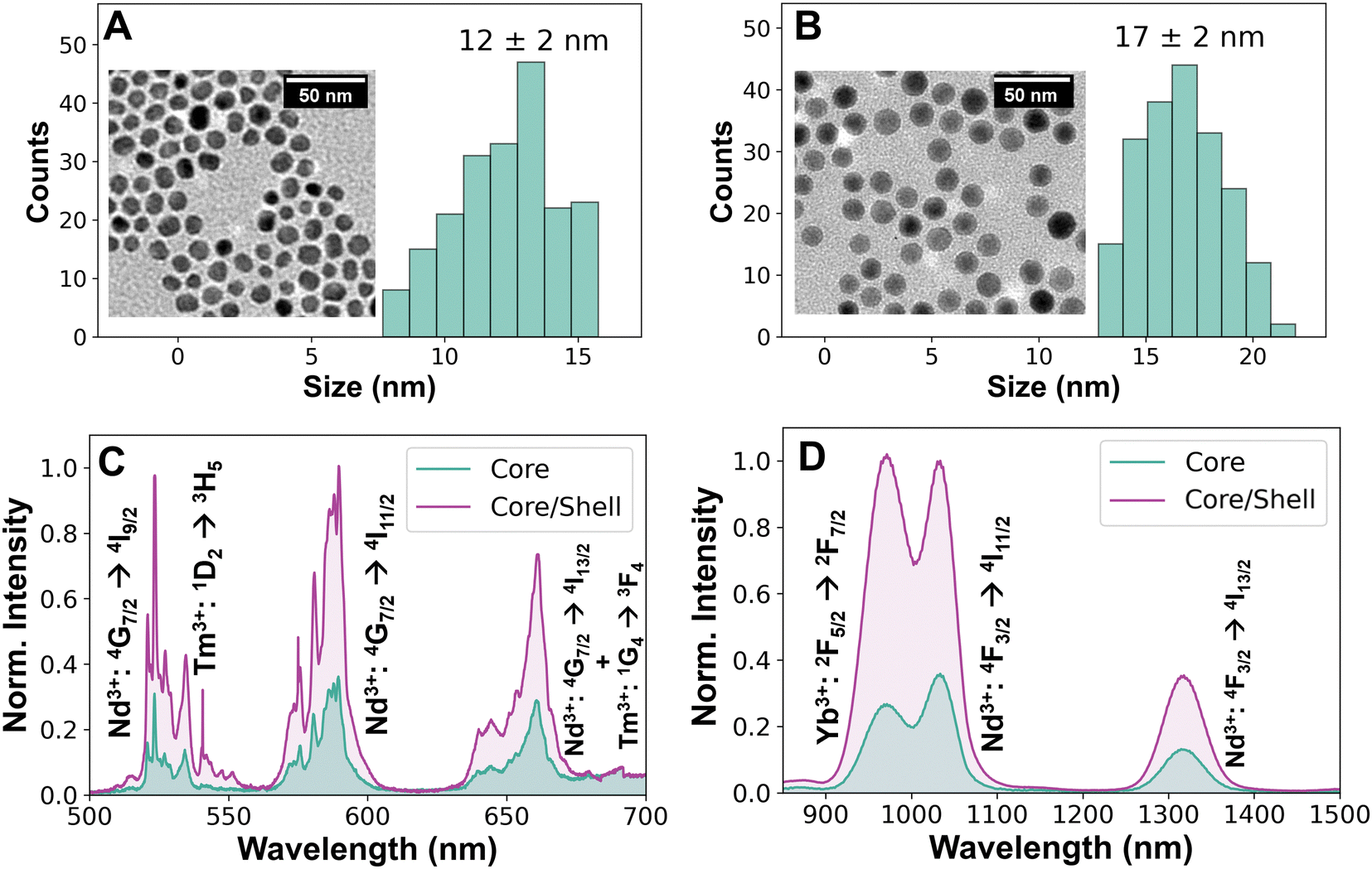 | ||
| Fig. 2 Characterization of core/shell nanoparticles. (A) TEM image and size distribution of core nanoparticles. (B) TEM and size distribution of core/shell nanoparticles. (C) Emission spectrum of a 1.0 mg mL−1 hexane dispersion of upconversion emissions in the visible of core and core/shell nanoparticles excited at 790 nm. (D) Comparison of infrared emissions of a 1.0 mg mL−1 hexane dispersion of core and core/shell nanoparticles. | ||
The BDP-azo fluorescence quencher was prepared following our previous work, from 3,5-dichloro-BODIPY by a two-step protocol followed by nucleophilic addition of 3-mercaptopropionic acid enabling the conjugation to the NPs (see detailed synthesis in Fig. S3, ESI†). This dye was chosen due to the overlap of its absorbance with the NPs’ upconversion emission as demonstrated in Fig. 1B. It is noteworthy to mention that the BDP-NH2, obtained after reductive cleavage of BDP-azo using Zn/HCO2NH4 (Fig. S4, ESI†), does not exhibit strong absorbance overlapping with the upconversion emissions. Therefore, the absorption spectra of BDP-azo and free BDP-NH2 included in Fig. 1B, suggested that under hypoxic conditions, leading to cleavage of the azo-bond, the upconversion emissions should be recovered at least partially. Hence, to convert our NPs into the desired hypoxia sensors, ligand transfer was first performed by exchanging oleic acid on the surface by treating the NPs with NOBF4 to generate BF4−-stabilised core/shell NPs.49,50 Second, the short linker HOOC-PEG12-NH2 was added to obtain UCNP-PEG (from now on referred to as UNCP for simplicity), binding through the interaction of the negatively charged carboxylate with the surface metal ion on the surface of the NPs, and bearing the amino group for the next step.51 The polyethylene glycol groups in the linker also improved their dispersibility in phosphate-buffered saline (PBS). Finally, UCNP-BDP-azo was obtained by EDC/NHS (N-ethyl-N′-(3-(dimethylamino)propyl)carbodiimide/N-hydroxysuccinimide) mediated coupling reaction in dimethyl sulfoxide (DMSO).52
Characterisation of the final UCNP-BDP-azo revealed spherical and highly monodisperse NPs with an average size of (20 ± 1) nm (Fig. 3A). Their dispersion in DMSO showed a distinct blue colour owing to the strong absorption of BDP-azo (Fig. 3B). This property was also demonstrated in the absorption spectrum of the UCNP (Fig. 3C). After binding BDP-azo to the surface of the UCNP, there were two absorption peaks in the UCNP-BDP-azo, consistent with the absorption bands of free BDP-azo (compare Fig. 1B), while the UCNP-BDP-NH2 show a broad shoulder around 490 nm associated with BDP-NH2. Thereby it was demonstrated that the absorption properties of the azo dye were maintained during conjugation to the UCNP.
 | ||
| Fig. 3 Characterization of UCNP-BDP-azo. (A) TEM and size distribution of dye-functionalised UCNP-BDP-azo. (B) Cuvette images of UCNPs (left) and UCNP-BDP-azo (right) illustrating the blue colour of UCNP-BDP-azo. (C) Extinction spectra of UCNP (magenta), showing mostly scattering from the nanoparticles, functionalized UCNP-BDP-azo (turquoise), where the absorption of the organic dye is dominating, and the UCNP-BDP-NH2 (yellow) presenting less extinction, and one broad band associated with BDP-NH2. (D) FTIR spectra of the nanoparticles during the functionalisation steps towards the UCNP-BDP-azo. | ||
The surface chemistry of the UCNP was further investigated using FTIR spectroscopy, as shown in Fig. 3D. The double peaks at 2920 and 2854 cm−1 indicate the presence of CH2 in the spectrum of the oleate-capped UCNP (UCNP@OA), and the interaction between the carboxyl group and the positively charged metal atoms on the surface of the nanoparticles resulted in the presence of peaks at 1455 and 1560 cm−1 due to the asymmetric and symmetric vibration modes of the –COO− groups, respectively. After replacing oleic acid with NOBF4, the –CH2 and CO peaks disappeared and a characteristic peak of BF4− appeared at 1084 cm−1. In the FTIR spectrum of UCNP, there was a broad peak at 3400 cm−1, and the vibration of C–O at 1085 cm−1 confirmed that the peak at 3400 cm−1 was mostly –OH overshadowing the N–H vibration, which indicated that HOOC-PEG12-NH2 had successfully replaced OA on the surface of the NPs. The decrease in the O–H vibration in UCNP and UCNP-BDP-azo indicated a reduction in the presence of hydrogen bonding to the N–H and a decreased accessibility of water to the carboxylates on the NPs surface once the azo dye was attached. These results indicated the successful preparation of UCNP-BDP-azo.
To evaluate the optical properties of the multifunctional UCNP-BDP-azo as hypoxia sensors, the azo bond was reduced to the amino groups employing ammonium formate and zinc powder in DMSO as a mild source of hydrogen (analogous to Fig. S4, ESI†). After chemical reduction, the colour of UCNP-BDP-azo changed from blue to nearly colourless. Additionally, an optical characterisation of the BDP-azo and BDP-NH2 molecules was performed, demonstrating that BDP-azo shows two main absorption bands around 370 and 600 nm and BDP-NH2 shows one broad absorption band around 500 nm (Fig. S5A, ESI†). Additionally, only BDP-NH2 was able to show a broad emission band around 560 nm when excited at 500 nm (Fig. S5B, ESI†).
The fluorescence emission of UCNP, UCNP-BDP-azo, and UCNP-BDP-NH2 at a concentration of 1 mg mL−1 was investigated. The visible emission of UCNP-BDP-azo was strongly quenched, especially the bands at 523 nm (Nd3+: 4G7/2 → 4I9/2), 542 nm (Tm3+: 1D2 → 3H5), 588 nm (Nd3+: 4G7/2 → 4I11/2), and 662 nm (Nd3+: 4G7/2 → 4I13/2 and Tm3+: 1G4 → 3F4), because they overlap with the main broad absorption band of BDP-azo, as shown in Fig. 1A.53 Due to the weaker quenching ability of the BDP-NH2 dye after reduction, compared to the BDP-azo dye, the emission of UCNP-BDP-NH2 was not equal to that of bare UCNP but partially recovered (Fig. 4A). These experiments demonstrated that a reductive environment, such as the one found in hypoxia, stopped the quenching of the visible emission of the nanoparticles and led to partial recovery of the upconversion emission of the UCNP. Therefore, the developed UCNP clearly demonstrated their potential as hypoxia-sensitive nanosensors.
 | ||
| Fig. 4 Change in fluorescence emission of UCNP due to reductive environment. (A) Upconversion emission of dispersions of free UCNP and of UCNP-BDP-azo prior and after reduction to UCNP-BDP-NH2. While the UCNP-BDP-azo signal is nearly completely quenched respective the free UCNP the signal is practically recovered for UCNP-BDP-NH2. (B) Emission spectra in the near infrared. The UCN-BDP-azo demonstrate higher NIR emission which is not affected by reduction to UCNP-BDP-NH2. (C) Fluorescence lifetimes of the three samples measured at 980 nm under 800 nm excitation. | ||
The NIR emissions of UCNP, UCNP-BDP-azo, and UCNP-BDP-NH2 (1 mg mL−1) were also investigated under 800 nm laser irradiation. The NIR emission of UCNP-BDP-azo decreased compared to the UCNP with only PEG on the surface but after the azo reduction, the NIR emission of UCNP-BDP-NH2 remained stable compared to that of UCNP-BDP-azo (Fig. 4B). These results demonstrate that even though the visible emission of the nanoprobe depend on the reductive conditions in the environment, its NIR emissions are independent of the oxygen levels or presence of reducing enzymes in the tissues. Indeed, this confers our nanosensors with a capability to act as multifunctional (sensing and imaging) contrast agent for deep-tissue imaging and thereby allows their tracking prior and during hypoxia conditions in tissue. Fig. S6 (ESI†) demonstrates the capability for deep-tissue imaging showing a dispersion of UCNP-BDP-NH2 under 5 mm of lipid-rich phantom tissue. Even though the imaging experiment only employed a low, biocompatible power density (50 mW cm−2) and a short exposure time (0.1 s), it still allows to identify the emission from the cuvette. This highlights thereby the potential for in vivo imaging in the NIR of our probe, which is a consequence of the particular dopant combination as previously demonstrated.36,54
Subsequently, the NIR fluorescence lifetimes of UCNP, UCNP-BDP-azo, and UCNP-BDP-NH2 (1 mg mL−1) were measured at 980 nm under 800 nm pulsed-laser excitation (Fig. 4C). The fluorescence lifetime obtained from the decay curves of the UCNP was 1.8 ms. For the UCNP-BDP-azo, the fluorescence lifetime decreased to 0.6 ms. This decrease in fluorescence lifetime stems from the FRET process between UCNP and BDP-azo molecules on their surface. After azo reduction, the fluorescence lifetime recovered nearly to its initial value of 1.8 ms. The results showed that FRET, which resulted in partial non-radiative deexcitation by the dye, ceased after the azo reduction had occurred. From these lifetimes, the FRET efficiency can be calculated using eqn (1):
 | (1) |
Subsequently, the biocompatibility of the nanosensors (Fig. S7, ESI†) was evaluated in cultured cells. The cytotoxicity of UCNP and UCNP-BDP-azo was tested by incubating HeLa cells with different concentrations (2, 10, 20 and 50 μg mL−1 in complete medium) of the nanosensors for 2 h. To exclude DMSO toxicity, a control group was incubated with vehicle only (5% DMSO in complete medium). Fig. S7 (ESI†) shows the results of this toxicity experiment 24 hours after treatments, demonstrating no appreciable toxicity, with a maximum decrease in cell viability of 18% after incubation with UCNP-BDP-azo at a concentration of 50 μg mL−1. Therefore, UCNP-BDP-azo showed good biocompatibility, which served as a basis for the following experiments.
Fig. 5 illustrates the study of the hypoxic response of the nanosensors in cell cultures. HeLa cells were incubated with UCNP and UCNP-BDP-azo for 2 h and prepared for analysis by multiphoton confocal microscopy (see Experimental section). To generate the hypoxia environment, before starting the analysis, the coverslips on which the cells were seeded were sealed, depriving them of oxygen and generating an anoxic environment as a model of the hypoxic conditions that the probes might encounter in a tumour environment.57 This was visualized throughout in different excitation and emission channels in the multiphoton confocal microscope to detect the visible emissions of the UCNP-BDP-NH2 that should form in that process due to azo group cleavage. Fig. 5A follows the cleavage of the azo bond in UCNP-BDP-azo when exciting in the NIR at 808 nm and via direct excitation at 561 nm. Both channels demonstrate an increase in signal intensity in their respective emission range (false green: 508–615 nm, false red: 570–641 nm) over time as illustrated by the intensity diagram in Fig. 5C. The third line of images in Fig. 5A shows the bright-field vision of the cells so that the signal from the top can be localized as stemming from inside the cells.
 | ||
| Fig. 5 Confocal microscope images showing the hypoxic response in sealed HeLa cell cultures of (A) UCNP-BDP-azo, where the first two rows present two different excitation channels and the third the bright-field view and (B) UCNP (3 middle rows). (C) Representation of the increase in intensity over time obtained from the images in (A) and (B) showing the increase in intensity for the cells incubated with UCNP-BDP-azo under anoxia over time. | ||
In the control experiment UCNP without any dye were employed, shown in Fig. 5B. Here the signal fluctuates lightly between images but no increase over time due to the anoxic conditions can be observed, as visualized in Fig. 5C. Interestingly, not only the upconversion excitation (λex = 808 nm, first row) results in emissions but also a direct excitation of the UCNP at 561 nm (second row) leading to weak emissions from the Nd3+ 4G7/2 level is observed. This signal stemming from the endocytosed nanoparticles was possible to observe due to aggregation inside endosomes/vesicles improving their absorbance.36 Comparison of the intensities under these conditions with the broken UCNP-BDP-azo shows a clear difference in the signal, illustrating the emission recovery over time due to removal of the quenching azo group. A series of control images of cells without nanoparticles is included as Fig. S8 (ESI†) showing no relevant emissions. These results demonstrated that the UCNP-BDP-azo can be employed as a hypoxia sensitive probe in cells because its visible emission are recovered in an anoxic cellular environment thereby switching the signal on in case of hypoxia.
An additional fluorescence microscopy experiment was designed, where the emission was observed in the visible region, but excitation was performed in the NIR by a simple diode laser in an inverted fluorescence microscope (Fig. S9, ESI†). This experiment demonstrated that luminescence recovery was possible even without the highly focused irradiation source of a confocal microscope. Further confirming UCNP-BDP-azo is a promising tool for the detection of hypoxia.
Conclusions
In this study, a hypoxia-responsive upconversion nanosensor was developed. The probe was obtained via an EDC/NHS coupling reaction between the UCNP and the hypoxia-sensitive azo dye. The optimised probe exhibited a negligible fluorescence signal in the visible region under normal conditions. However, chemical reduction experiments, as well as cellular anoxia experiments for the creation of a reductive environment, demonstrated that the reduction of the azo bond was possible, and that quenching could be stopped. Confocal fluorescence microscopy confirmed that UCNP-BDP-azo were localised inside the cells and recuperated emissions in the visible region stemming from upconversion when exposed to a hypoxic environment. Additionally, the UCNP-BDP-azo probe maintained their NIR-emission independently of the azo-bond, conferring the particles with the capability to act as contrast agent in deep-tissue imaging. Therefore, the developed nanosensor was revealed to be a promising probe for detecting hypoxic environments that includes tracking capability owing to its maintained NIR emissions. Future work will focus on more hypoxia-sensitive organic dyes that could show improved solubility and liberation of the BDP-NH2 dye after reduction of the azo bond to improve recovery from quenching.Author contributions
J. Yao: investigation, data curation; S. Simon: investigation, data curation; G. Lopez-Peña: data curation, visualization, writing; S. Gómez-Pastor: investigation; S. Guisan-Ceinos: investigation; R. Marin: supervision, visualization, writing, resources, funding; E. Martín Rodríguez: methodology, formal analysis, writing, funding; D. Jaque: conceptualization, methodology, funding, resources; F. Sanz-Rodríguez: investigation, data curation, visualization, resources, funding, writing; M. Ribagorda: supervision, methodology, resources, funding, writing D. H. Ortgies: supervision, methodology, resources, funding, writing.Data availability
The authors declare that the data supporting this article will be made available at https://zenodo.org/.Conflicts of interest
There are no conflicts to declare.Acknowledgements
This work was financed by the Spanish Ministerio de Ciencia e Innovación MCIN/AEI/10.13039/501100011033 under projects PID2019-106211RB-I00 (nanonerv), PID2020-113059GB-C22, PID2020-118878RB-I00 (RETINanoTHERMIA) and PID2023-146801NB-C32, by the Instituto de Salud Carlos III (PI19/00565), by the Comunidad Autónoma de Madrid (CAM) S2022/BMD-7403 RENIM-CM grant and co-financed by the European structural and investment fund. Additional funding was provided by the European Union Horizon 2020 FETOpen project NanoTBTech (801305), the Comunidad Autónoma de Madrid and Universidad Autónoma de Madrid young investigator project SI3/PJI/2021-00211, the Fundación para la Investigación Biomédica del Hospital Universitario Ramón y Cajal projects IMP21_A4 (2021/0427) and IMP18_38 (2018/0265), and also by COST action CA17140. R. M. acknowledges the support of the European Union's Horizon 2020 research and innovation program under the Marie Skłodowska-Curie Grant Agreement no. 797945 (LANTERNS), and is grateful to the Spanish Ministerio de Ciencia, Innovación y Universidades for support to research through a Ramón y Cajal Fellowship (RYC2021-032913-I) and Project PID2022-14210NA-I00 (NAMASTEPS) funded by MCIN/AEI/10.13039/501100011033 and by FEDER, EU. J. Y. acknowledges the support from the China Scholarship Council (CSC File no. 201704910867). D. H. O. is grateful for Ramón y Cajal Fellowship RYC2022-036732-I funded by MCIN/AEI/10.13039/501100011033 and by “ESF investing in your future”. E. M. R. and D. H. O. acknowledge Grant CPP2021-008902 funded by MCIN/AEI/10.13039/501100011033 and by the “European Union NextGenerationEU/PRTR”. Silvia Gómez-Pastor is a predoctoral fellow of the Autonomous Community of Madrid (Grant PIPF-2022/SAL-GL-25806). We thank the “Unidad de Microscopía e Imagen Dinámica del Centro Nacional de Investigaciones Cardiovasculares Carlos III” facility for their assistance and are grateful to Paloma Rodríguez Sevilla for help with the group's microscopes.Notes and references
- M. C. Brahimi-Horn, J. Chiche and J. Pouysségur, J. Mol. Med., 2007, 85, 1301–1307 CrossRef PubMed.
- M. V. Blagosklonny, Cancer Cell, 2004, 5, 13–17 CrossRef CAS PubMed.
- O. Trédan, C. M. Galmarini, K. Patel and I. F. Tannock, J. Natl. Cancer Inst., 2007, 99, 1441–1454 CrossRef.
- P. Vaupel, K. Schlenger, C. Knoop and M. Höckel, Cancer Res., 1991, 51, 3316–3322 CAS.
- E. E. Verwer, R. Boellaard and A. A. van der Veldt, World J. Clin. Oncol., 2014, 5, 824–844 CrossRef.
- X. Lin, Q. Ruan, L. Lin, X. Zhang, X. Duan, Y. Teng and J. Zhang, J. Radioanal. Nucl. Chem., 2018, 317, 1463–1468 CrossRef CAS.
- L. Ge, Y. Tang, C. Wang, J. Chen, H. Mao and X. Jiang, Nat. Commun., 2024, 15, 153 CrossRef CAS PubMed.
- X.-B. Zhao, J.-Y. Kang and Y.-P. Shi, Anal. Chem., 2022, 94, 6574–6581 CrossRef CAS.
- S. Acharya and R. Misra, Nanomedicine, 2022, 42, 102549 CrossRef CAS.
- A. L. D. Wallabregue, H. Bolland, S. Faulkner, E. M. Hammond and S. J. Conway, J. Am. Chem. Soc., 2023, 145, 2572–2583 CrossRef CAS.
- L. Gai, Y. Liu, Z. Zhou, H. Lu and Z. Guo, Coord. Chem. Rev., 2023, 481, 215041 CrossRef CAS.
- J. Liu, W. Bu and J. Shi, Chem. Rev., 2017, 117, 6160–6224 CrossRef CAS PubMed.
- G. G. Dias, A. King, F. de Moliner, M. Vendrell and E. N. da S. Júnior, Chem. Soc. Rev., 2018, 47, 12–27 RSC.
- H. Komatsu, Y. Shindo, K. Oka, J. P. Hill and K. Ariga, Angew. Chem., Int. Ed., 2014, 53, 3993–3995 CrossRef CAS PubMed.
- S. Karan, M. Y. Cho, H. Lee, H. Lee, H. S. Park, M. Sundararajan, J. L. Sessler and K. S. Hong, J. Med. Chem., 2021, 64, 2971–2981 CrossRef CAS PubMed.
- F. Xu, H. Li, Q. Yao, H. Ge, J. Fan, W. Sun, J. Wang and X. Peng, Chem. Sci., 2019, 10, 10586–10594 RSC.
- Y. Li, Y. Sun, J. Li, Q. Su, W. Yuan, Y. Dai, C. Han, Q. Wang, W. Feng and F. Li, J. Am. Chem. Soc., 2015, 137, 6407–6416 CrossRef CAS.
- S. Karan, M. Y. Cho, H. Lee, H. M. Kim, H. S. Park, E. H. Han, J. L. Sessler and K. S. Hong, J. Med. Chem., 2023, 66, 14175–14187 CrossRef CAS.
- X. Meng, J. Zhang, Z. Sun, L. Zhou, G. Deng, S. Li, W. Li, P. Gong and L. Cai, Theranostics, 2018, 8, 6025–6034 CrossRef CAS.
- Y. Liu, W. Liu, H. Li, W. Yan, X. Yang, D. Liu, S. Wang and J. Zhang, Anal. Chim. Acta, 2018, 1024, 177–186 CrossRef CAS PubMed.
- K. Morihiro, T. Ishinabe, M. Takatsu, H. Osumi, T. Osawa and A. Okamoto, J. Am. Chem. Soc., 2021, 143, 3340–3347 CrossRef CAS.
- R. Zou, Q. Gong, Z. Shi, J. Zheng, J. Xing, C. Liu, Z. Jiang and A. Wu, Nanoscale, 2020, 12, 14870–14881 RSC.
- X. Tian, Z. Li, Y. Sun, P. Wang and H. Ma, Anal. Chem., 2018, 90, 13759–13766 CrossRef CAS PubMed.
- A. Chevalier, P. Y. Renard and A. Romieu, Chem. – Asian J., 2017, 12, 2008–2028 CrossRef CAS PubMed.
- W. Piao, S. Tsuda, Y. Tanaka, S. Maeda, F. Liu, S. Takahashi, Y. Kushida, T. Komatsu, T. Ueno, T. Terai, T. Nakazawa, M. Uchiyama, K. Morokuma, T. Nagano and K. Hanaoka, Angew. Chem., Int. Ed., 2013, 52, 13028–13032 CrossRef CAS.
- W. Liu, X. Yao, W. Zhu, J. Wang, F. Zhou, X. Qian, A. Tiemuer, S. Yang, H.-Y. Wang and Y. Liu, ACS Appl. Bio Mater., 2021, 4, 2752–2758 CrossRef CAS PubMed.
- N. Ding, Z. Li, X. Tian, J. Zhang, K. Guo and P. Wang, Chem. Commun., 2019, 55, 13172–13175 RSC.
- S. Yao, Y. Chen, H. Xu, F. Qi, Y. Zhang, T. Yang, Y. Wu, H. Fang, W. He and Z. Guo, Dyes Pigm., 2022, 206, 110583 CrossRef CAS.
- J. Xiong, P. Wang, S. Son, C. Zhong, F. Zhang, Z. Mao, Z. Liu and J. S. Kim, Matter, 2022, 5, 1502–1519 CrossRef CAS.
- C. Wang, S. Wang, Y. Wang, H. Wu, K. Bao, R. Sheng and X. Li, Sci. Rep., 2020, 10, 12127 CrossRef CAS.
- D. Xiao, L. Liu, F. Xie, J. Dong, Y. Wang, X. Xu, W. Zhong, H. Deng, X. Zhou and S. Li, Angew. Chem., Int. Ed., 2024, 63, e202310318 CrossRef CAS.
- H. Liu, X. Zeng, H. Yin, J. Chen, W. Xie, G. Zhou, G. Mao, Y. Zhou and S. Yang, Sens. Actuators, B, 2023, 381, 133431 CrossRef CAS.
- A. Chevalier, C. Massif, P.-Y. Renard and A. Romieu, Chem. – Eur. J., 2013, 19, 1686–1699 CrossRef CAS PubMed.
- K. Kiyose, K. Hanaoka, D. Oushiki, T. Nakamura, M. Kajimura, M. Suematsu, H. Nishimatsu, T. Yamane, T. Terai, Y. Hirata and T. Nagano, J. Am. Chem. Soc., 2010, 132, 15846–15848 CrossRef CAS.
- T. Myochin, K. Hanaoka, T. Komatsu, T. Terai and T. Nagano, J. Am. Chem. Soc., 2012, 134, 13730–13737 CrossRef CAS.
- G. López-Peña, S. Simón-Fuente, D. H. Ortgies, M. Á. Moliné, E. Martín Rodríguez, F. Sanz-Rodríguez and M. Ribagorda, Cancers, 2023, 15, 102 CrossRef.
- J. Wu, J. Wu, W. Wei, Y. Zhang and Q. Chen, Small, 2024, 20, 2311729 CrossRef CAS.
- X. Zhang, W. Shu, M. Cheng, L. Wang and X. Ran, Nanotechnology, 2023, 34, 415502 CrossRef CAS.
- S. Xu, X. Zhang, H. Xu, B. Dong, X. Qu, B. Chen, S. Zhang, T. Zhang, Y. Cheng, S. Xu and H. Song, Sci. Rep., 2016, 6, 22350 CrossRef CAS.
- J. Liu, Y. Liu, W. Bu, J. Bu, Y. Sun, J. Du and J. Shi, J. Am. Chem. Soc., 2014, 136, 9701–9709 CrossRef CAS PubMed.
- S. Guisán-Ceinos, A. R. Rivero, F. Romeo-Gella, S. Simón-Fuente, S. Gómez-Pastor, N. Calvo, A. H. Orrego, J. M. Guisán, I. Corral, F. Sanz-Rodriguez and M. Ribagorda, J. Am. Chem. Soc., 2022, 144, 8185–8193 CrossRef.
- N. Shin, K. Hanaoka, W. Piao, T. Miyakawa, T. Fujisawa, S. Takeuchi, S. Takahashi, T. Komatsu, T. Ueno, T. Terai, T. Tahara, M. Tanokura, T. Nagano and Y. Urano, ACS Chem. Biol., 2017, 12, 558–563 CrossRef CAS.
- F. Wang, R. Deng and X. Liu, Nat. Protoc., 2014, 9, 1634–1644 CrossRef CAS.
- F. Wang, R. Deng, J. Wang, Q. Wang, Y. Han, H. Zhu, X. Chen and X. Liu, Nat. Mater., 2011, 10, 968–973 CrossRef CAS.
- X. Zhang, Z. Zhao, X. Zhang, D. B. Cordes, B. Weeks, B. Qiu, K. Madanan, D. Sardar and J. Chaudhuri, Nano Res., 2015, 8, 636–648 CrossRef CAS.
- F. Wang, J. Wang and X. Liu, Angew. Chem., Int. Ed., 2010, 49, 7456–7460 CrossRef CAS PubMed.
- A. Skripka, A. Benayas, C. D. S. Brites, I. R. Martín, L. D. Carlos and F. Vetrone, Nano Lett., 2020, 20, 7648–7654 CrossRef CAS PubMed.
- M. Danaei, M. Dehghankhold, S. Ataei, F. Hasanzadeh Davarani, R. Javanmard, A. Dokhani, S. Khorasani and M. R. Mozafari, Pharmaceutics, 2018, 10, 57 CrossRef PubMed.
- V. Muhr, S. Wilhelm, T. Hirsch and O. S. Wolfbeis, Acc. Chem. Res., 2014, 47, 3481–3493 CrossRef CAS.
- V. Muhr, C. Würth, M. Kraft, M. Buchner, A. J. Baeumner, U. Resch-Genger and T. Hirsch, Anal. Chem., 2017, 89, 4868–4874 CrossRef CAS.
- G. B. Kauffman, J. Chem. Educ., 1988, 65, 28–31 CrossRef CAS.
- J. Bart, R. Tiggelaar, M. Yang, S. Schlautmann, H. Zuilhof and H. Gardeniers, Lab Chip, 2009, 9, 3481 RSC.
- L. Wu, C. Huang, B. P. Emery, A. C. Sedgwick, S. D. Bull, X.-P. He, H. Tian, J. Yoon, J. L. Sessler and T. D. James, Chem. Soc. Rev., 2020, 49, 5110–5139 RSC.
- M. Tan, B. Del Rosal, Y. Zhang, E. Martín Rodríguez, J. Hu, Z. Zhou, R. Fan, D. H. Ortgies, N. Fernández, I. Chaves-Coira, Á. Núñez, D. Jaque and G. Chen, Nanoscale, 2018, 10, 17771–17780 RSC.
- Y. Wang, K. Liu, X. Liu, K. Dohnalová, T. Gregorkiewicz, X. Kong, M. C. G. Aalders, W. J. Buma and H. Zhang, J. Phys. Chem. Lett., 2011, 2, 2083–2088 CrossRef CAS.
- S. Bhuckory, E. Hemmer, Y. Wu, A. Yahia-Ammar, F. Vetrone and N. Hildebrandt, Eur. J. Inorg. Chem., 2017, 5186–5195 CrossRef CAS.
- E. Takahashi and M. Sato, Am. J. Physiol., 2010, 299, C1318–C1323 CrossRef CAS.
Footnote |
| † Electronic supplementary information (ESI) available: Energy scheme of the nanoparticles, DLS, synthesis of the azo-dye and its reduction, NIR imaging, and dark toxicity data. See DOI: https://doi.org/10.1039/d4tc03302g |
| This journal is © The Royal Society of Chemistry 2025 |