
 Open Access Article
 This Open Access Article is licensed under a
 Creative Commons Attribution 3.0 Unported Licence
A review on the role of deep eutectic solvents in mango (Mangifera indica) extraction
Ahmad Mukhlis Abdul Rahman a,
Amirul Ridzuan Abu Bakar*a,
Ang Qian Yeea,
Mohd Asraf Mohd Zainudina,
Nik Muhammad Azhar Nik Dauda,
Ahmad Anas Nagoor Gunnya,
Mohd Sharizan Md Saripa,
Ryan Vitthaya Perona and
Nurul Husna Khairuddinb
a,
Amirul Ridzuan Abu Bakar*a,
Ang Qian Yeea,
Mohd Asraf Mohd Zainudina,
Nik Muhammad Azhar Nik Dauda,
Ahmad Anas Nagoor Gunnya,
Mohd Sharizan Md Saripa,
Ryan Vitthaya Perona and
Nurul Husna Khairuddinb
aFaculty of Chemical Engineering & Technology, Universiti Malaysia Perlis, Jejawi, Perlis 02600, Malaysia. E-mail: amirulridzuan@unimap.edu.my
bM. Kandiah Faculty of Medicine and Health Sciences, University Tunku Abdul Rahman, Bandar Sungai Long, Kajang, Selangor 43000, Malaysia
First published on 10th February 2025
Abstract
The present review attempts to evaluate the applicability of deep eutectic solvents (DES) as a green technique for the extraction of phytochemicals from Mangifera indica L. and their therapeutic potential. Mango has been reported to show numerous therapeutic activities, which are attributed to its abundant source of bioactive compounds. Thus, the therapeutic potential of phytochemicals in mangoes is reviewed based on different reported bioactivity tests. The use of DESs is considered a green approach for the extraction of bioactive compounds from natural sources utilizing two or more components and a safe alternative for application in the nutritional, pharmaceutical and other sectors. The trends in the extraction of phytochemicals from mango using different DES components and different extraction parameters of the optimum protocol are reviewed. Hence, DESs are considered potential solvents with selective and efficient properties for extracting bioactive ingredients from mango. However, there are several knowledge gaps that need to be assessed for DES-based bioactive compound extraction from mango such as information on the local and specific varieties of mangoes, standardization of the extraction protocols and use of other parts of the mango plant as alternatives to its peel as bioactive sources. Accordingly, the extraction of bioactive compounds from mango using DESs will provide useful information for subsequent agricultural, pharmaceutical and nutraceutical applications in the future.
1. Introduction
Phytochemicals are naturally occurring compounds in plants that can be divided into primary and secondary metabolites. Primary metabolites such as nucleic acids, carbohydrates, fatty acids and proteins are required for the normal growth and development of plants.1 Alternatively, secondary metabolites are compounds produced by plants for their protection and to enhance their ability to survive in harsh environments. Phytochemicals including secondary metabolites can be derived from various sources such as medicinal plants,2,3 vegetables,4,5 fruits,6–8 and agricultural by-products.9–11 Secondary metabolites are important plant constituents with effective therapeutic potential, especially exhibiting antioxidant and anthelmintic activities.12–14 Phytochemicals can be found in different parts of a target plant, such as stems, roots, seeds, fruits, leaves and flowers. The extraction of phytochemicals using different plant parts usually produces different extraction yields and types of phytochemical compounds.15,16To date, the recovery of bioactive compound extracts from several natural sources is highly associated with the conventional extraction process using conventional extraction techniques such as percolation, maceration, digestion, decoction and Soxhlet extraction in combination with traditional organic solvents.1,17–19 Examples of traditional organic solvents are acetone, ethyl acetate, methanol and ethanol.17,20–23 However, conventional extraction techniques are usually based on highly pollutant, non-renewable organic solvents and often related to increased energy consumption, toxicity and volatility.24–26 Therefore, there is an urgent need to apply green extraction approaches, which are considered safer and more environmentally friendly as well as reduce water, solvent and energy consumption.26,27
Ionic liquids (ILs) are the first multimolecular generation of solvents being used as green solvents for the extraction of bioactive compounds. The advantages of IL solvents include their wide range of miscibility and solubility, very low vapor pressure, non-flammability and good thermal properties and recyclability.20,27,28 However, ILs do possess several disadvantages such as high production cost, high toxicity, poor water stability, difficult purification and low biodegradability.27–30 Thus, over the past few years, the scientific community has been committed to applying deep eutectic solvents (DESs) for the recovery of bioactive compounds. DESs have been developed as analogous to ILs with similar physicochemical properties. The greener and environmentally friendly DESs have been reported to have additional advantages compared to ILs including biodegradable properties, chemical inertness with water, lower toxicity and lower cost.20,31
However, they differ from ILs in several aspects such as the source of their starting materials and chemical formation process. The formation of DESs is a result from the chemical reaction between a halide salt or another hydrogen bond acceptor (HBA) and a hydrogen bond donor (HBD). Natural deep eutectic solvents (NADESs), new derivatives of DESs, have been developed from the combination of primary metabolites and bio-renewable materials such as sugar, alcohols, organic acids and amino acids.31,32 Accordingly, NADESs may be very useful for solubilizing, storing or transporting non-water-soluble metabolites in living cells and organisms. Several factors that affect the extraction yield of bioactive compounds from different parts of mango by DESs such as the combination of materials for the preparation of DESs and NADES, application of innovative extraction techniques and selected critical extraction parameters are reviewed in this article.
Mango (Mangifera indica) is widely consumed worldwide and is rich in bioactive compounds such as polyphenols, carotenoids, vitamins and minerals. It has been used for medicinal purposes for centuries and has potential in various therapeutic effects such as antioxidant,33 anti-inflammatory,34 anti-cancer35 and anti-diabetic36 activities. Several different parts of mango have been used for the extraction of bioactive compounds. The main source of mango that has been used for the extraction of bioactive compounds by DES is the mango peel,37–39 while other parts of the mango plant have not been widely exploited such as its leaves, kernels, and seeds, which will be useful targets for DES extraction to gain new knowledge on the additional and complementary data of bioactive compounds. This review article also describes the therapeutic potential of bioactive compounds extracted from different sources of mango samples.
2. Background of DESs
2.1 Historical development of DES
DESs are environmentally friendly solvents similar to ionic liquids (ILs) and are often compared to ILs because they share some common characteristics, such as high thermal stability, low volatility and adjustable polarity. This unique class of solvents gained attention following the discovery by Abbott and colleagues.40 These researchers found that when certain substances capable of forming hydrogen bonds, known as hydrogen bond donors (HBDs), are mixed with others that can accept these bonds, referred to as hydrogen bond acceptors (HBAs), in specific proportions, the melting point of the mixture decreases significantly.This surprising behaviour was first noticed when choline chloride powder (typically melts at a high temperature of about 302 °C) was combined with crystalline urea (with a melting point of around 133 °C) in a 1![[thin space (1/6-em)]](https://www.rsc.org/images/entities/char_2009.gif) :2 ratio. Remarkably, this mixture turned into a liquid at room temperature, with a low melting point of its eutectic composition of about 12 °C.40 Abbott and team named this unique liquid a “deep” eutectic mixture (DEM). DEMs, due to their ability to melt at much lower temperatures, have opened up new possibilities for creating innovative solutes and electrolytes for various chemical applications.40–48 Scientists often refer to these liquids as DES when discussing their roles as solvents in specific applications.
:2 ratio. Remarkably, this mixture turned into a liquid at room temperature, with a low melting point of its eutectic composition of about 12 °C.40 Abbott and team named this unique liquid a “deep” eutectic mixture (DEM). DEMs, due to their ability to melt at much lower temperatures, have opened up new possibilities for creating innovative solutes and electrolytes for various chemical applications.40–48 Scientists often refer to these liquids as DES when discussing their roles as solvents in specific applications.
Ionic liquids (ILs), which are composed of anions and cations and have a melting point below 100 °C, have captured the attention of organic chemists looking for environmentally friendly and sustainable methods since their discovery in 1914. At that time, no one could have predicted that ILs would become a prominent topic in chemistry a century later. Today, some key perspectives differ from the original concepts as our understanding of ILs has deepened. For example, ILs were initially thought to be non-volatile, non-flammable and stable in air and water, making them appear as green solvents. However, it is now widely recognized that many ILs are indeed volatile, flammable, unstable, and sometimes toxic.49
This complexity arises from the vast combinations of cations and anions that meet the definition of ILs, leading to a wide range of behaviours.49 In contrast, DESs have several advantages over ILs. Although ILs can be expensive, harmful to the environment and potentially toxic, DESs are typically affordable,50,51 environmentally friendly,52,53 and safe to use.53–55 For instance, choline, a common component in DESs, is found in vitamin B and widely produced for livestock nutrition, making it readily available and harmless.56,57 In contrast, urea, a typical component in DESs, is often used in fertilizers but does not share the same eco-friendly characteristics.58
2.2 Principles and classification of DESs
Deep eutectic solvents (DESs) consist of multiple compounds, with a hydrogen bond donor (HBD) and a hydrogen bond acceptor (HBA), exhibit a considerably lower eutectic point than an ideal liquid mixture and remain in a liquid state at ambient temperature.59–61 The physiochemical properties of DESs, encompassing attributes such as their melting point, density, conductivity and viscosity, undergo variations depending on the specific structures of these solvents. The melting point of DESs is primarily determined by the proportion of hydrogen bond donor present in them, resulting in a significantly lower melting point compared to their individual constituents.29,62An increased concentration of hydrogen bond interaction with anionic groups leads to diminished interactions with cationic groups. This shift in interaction, characterized by a reduced lattice energy, contributes to the reduction of the melting point.63 The general formula for deep eutectic solvents can be expressed as Cat+X−zY. The cation, denoted as Cat+, can be an ammonium, phosphonium or sulfonium ion, while X− represents a Lewis base, often a halide anion. The variable Y signifies a Lewis or Brønsted acid, and the value of z corresponds to the number of Y molecules interacting with the anion X−.26,29 Most DESs can be grouped into four main types, as discussed in the next subtopic.
Type I DESs include well-studied combinations such as chloroaluminate/imidazolium. They are also comprised of less common ionic liquids, such as those made by mixing imidazolium salts with different metal halides, including FeCl2.66 Additionally, the research by Scheffler and Thomson involved type I eutectics, including EMIC combined with various metal halides, including AgCl.67 However, the availability of nonhydrated metal halides suitable for type I DESs is limited due to their higher melting points. Thus, improving deep eutectic solvents involves mixing choline chloride with hydrated metal-halides, resulting in type II DESs. These hydrated metal salts are cost-effective and exhibit resistance to air and moisture, making them suitable for large-scale industrial use.68 Type III DESs refer to a category of eutectic mixture where the hydrogen bond donor (HBD) typically consists of an organic molecular component such as amides, carboxylic acids, amino acids, sugars and polyols.
Type III DESs, resulting from the combination of choline chloride with hydrogen bond donors such as amides and carboxylic acids, have attracted increasing attention owing to their versatility and favourable properties. Their capability to effectively dissolve a wide range of transition metal compounds, including chlorides and oxides, contributes to their widespread use across various applications.29,69–71 Furthermore, they are easy to prepare, have limited reactivity with water, are often environmentally friendly and are cost-effective. The wide range of available hydrogen bond donors makes this category of deep eutectic solvents versatile and adaptable. Regardless of the specific type, DESs are believed to be formed through interactions involving factors such as the entropy of mixing, van der Waals forces, hydrogen bonding and/or ionic bonding.29,69–71 However, further investigation is required to gain a comprehensive understanding of how these factors interact and contribute to the properties of DESs.
Some researchers have proposed expanding the classification of deep eutectic solvents (DESs) by introducing two additional categories, namely, therapeutic DESs (THEDES, type VI) and amino acid-based DESs (AADES, type VII).72 Additional classification based on function or dominant components has distinct advantages, such as aiding readers and the scientific community in understanding the relevant types of DESs. Nevertheless, some groups of DESs, which are organized based on important functions or dominant components, such as NADES and VODES, have not been explicitly included in this classification. This matter can be resolved through two approaches.
The first approach involves including NADES and VODES as additional categories within the existing classification. However, this approach may lead to substantial overlap, with some DESs potentially falling into multiple distinct categories, making the situation more complex. Accordingly, the second approach can be considered to avoid this confusion. It involves introducing a new set of classifications based on specific functions, applications and the dominant components of DESs. This new classification is meticulously organized according to the primary functions and dominant components of the relevant DESs. Through this approach, NADES, THEDES, VODES and AADES can be systematically categorized. However, given that this article is not focused on the in-depth examination of new classifications, a thorough analysis of this matter can be systematically discussed by authoritative scientific communities in the future. This may give rise to specialized review papers dedicated to this topic.
The examples of binary DESs include choline chloride/triethylene glycol,73 choline chloride/glycerol,74 zinc nitrate hexahydrate/glycerol,75 tetraethylammonium chloride/ethylene glycol,76 and choline chloride/levulinic acid.77 Alternatively, ternary DESs are composed of three main components, i.e., an HBD, an HBA and a third component, often a metal salt or another substance. This additional component adds complexity to the DES composition and can result in distinctive properties or improved performance in specific applications. The examples of ternary DESs include dichloroacetic acid/L-menthol/n-butanol,78 choline chloride/anhydrous oxalic acid/ethylene glycol, choline chloride/anhydrous oxalic acid/glycerol and choline chloride/anhydrous oxalic acid/lactic acid.79
Deep eutectic solvents (DESs), which are created by combining hydrogen bond donors and acceptors, represent a promising and innovative category of solvents. Both hydrophilic and hydrophobic binary DESs have a tendency to absorb water, resulting in the formation of ternary mixtures. Consequently, the presence of a minor water content is practically unavoidable under normal ambient conditions.80 Hydrophobic deep eutectic solvents (DESs) are a type of custom-designed solvent known for their limited or even absent water miscibility and low vapor pressure. These DESs are typically created by combining hydrophobic hydrogen bond acceptors (HBAs) and hydrogen bond donors (HBDs).
As a result, hydrophobic DESs tend to separate from water, forming distinct layers or phases due to their limited solubility in aqueous environments. Hydrophobic deep eutectic solvents (DESs), such as the combination of tetrabutyl ammonium chloride and decanoic acid, are characterized by their low water miscibility. Even a minimal water content in these DESs can trigger dynamic nanoscale phase segregation, resulting in reduced solvent viscosity and increased fragility. This phenomenon also leads to higher self-diffusion coefficients and conductivity within the solvent, ultimately enhancing the local dynamics and making hydrophobic DESs valuable in various applications.80
Alternatively, hydrophilic deep eutectic solvents are made up of components that have a strong affinity for water. These DESs are formulated by combining hydrophilic HBAs and HBDs, resulting in stable solutions that are highly soluble in water. Choline chloride (ChCl) and lactic acid are typically considered hydrophilic components when used in deep eutectic solvents (DESs).81 These components have an affinity for water and can readily form hydrophilic DESs. Hydrophilic DESs are valuable in situations where maintaining a water-compatible environment is essential. They can be employed as environmentally friendly solvents for extracting hydrophilic compounds or as suitable media for hydrophilic chemical reactions that require an aqueous environment.
2.3 Properties of density and viscosity in DESs
Changes in the density of DESs also result from the dynamics of molecular interactions and the availability of free volume in the DES. As the temperature of the DES increases, more free space becomes available, allowing molecules to move more rapidly. This increased molecular motion leads to a reduction in the density of the DES. The density of deep eutectic solvents (DESs) is clearly affected by the characteristics of the HBD such as the presence of different halide ions in the HBA.84–86 For example, DESs containing the bromide salt of tetraalkylammoniums tend to exhibit higher densities compared to that with chloride salts.87–89 Additionally, when considering the alkyl chain length of HBD, ranging from ethyl to butyl, in tetraalkylammonium salts, the density of DESs is the highest when using TEABr/Cl. This is primarily due to the compact structure of the DESs.89,90
Another example involves ChCl-based DESs, where a range of deep eutectic solvents (DESs) was created by combining choline chloride (ChCl) with various carboxylic acids having different alkyl chain lengths, including oxalic acid, glycolic acid, malonic acid, glutaric acid and levulinic acid.91 Particularly, DESs formulated with glutaric acid and levulinic acid exhibited lower densities compared to those composed of other acids. This decline in density was primarily attributed to the longer C5 alkyl chain found in these compounds, and an increase in alkyl chain length resulted in a higher molar volume, subsequently leading to a decrease in the DES density.91 Also, the density of DESs is significantly influenced by the molar ratio of HBA to HBD.86,92–94 For example, in TBABr:PEG-based DESs, an increase in the molar ratio of PEG resulted in a slight elevation in density.88 DESs with a 1:3 HBA to HBD ratio displayed higher densities compared to that with a 1:2 ratio. Specifically, the densities of TBABr:PEG 200 at 1:2 and 1:3 ratios were recorded to be 1.11077 g cm−3 and 1.11360 g cm−3, respectively.
Temperature affects the density of deep eutectic solvents (DESs), and this relationship is described by the isobaric thermal expansion coefficient, which indicates the available free volume in DESs. This coefficient helps explain how DESs compress.94 The isobaric thermal expansion coefficient (αP) is calculated based on experimental data. DESs exhibit a linear decrease in density with an increase in temperature. This drop is due to the increased free space between the hydrogen bond acceptor (HBA) and hydrogen bond donor (HBD) components. Although the αP values of DESs are lower than that of common solvents, they are similar to that of imidazolium-based ionic liquids (ILs). DESs show less expansion or compression with temperature compared to ILs and organic solvents.94 Numerous researchers have extensively investigated the density of DESs at different temperature levels.95–97
For example, the density of ChCl-based deep eutectic solvents (DESs) was measured at a constant pressure of 101.3 kPa in the temperature range of 293.15 K to 333.15 K.98 These DESs were formulated using ChCl as the hydrogen bond acceptor (HBA) and various hydrogen bond donors (HBDs), including ethylene glycol, levulinic acid and phenol, maintaining a consistent 1:2 HBA-to-HBD mole ratio. The results consistently revealed that with an increase in temperature, the density of these DESs decreased due to thermal expansion. Among the examined DESs, the ChCl:levulinic acid-based DES demonstrated remarkable thermal stability, which is primarily attributed to the robust intermolecular interactions between ChCl and levulinic acid. These interactions resulted in higher density compared to the other DESs.98
Shah and Mjalli conducted a study to explore the impact of water content on the density of a ChCl:urea deep eutectic solvent (DES) at two different temperatures.99 To gain insights into the interactions within pure DESs and their aqueous solutions, they used molecular dynamics simulations. The findings revealed significant insights into the behaviour of the DESs. Notably, the presence of urea and Cl anion interactions affected the melting point of the DES. Interestingly, the Cl anions exhibited higher levels of hydration compared to urea and choline cations. These interactions had a direct influence on the melting point, given that they interfered with the interactions between the urea molecules and choline cations.
The impact of water on density was divided into three distinct phases. As the content of water increased, the number of hydrogen bonds within the DES decreased. At higher concentrations of water, the anion of the hydrogen bond donor exhibited increased hydration compared to its cation.70,94 These findings were consistent with the observations for ChCl:urea under high-pressure and temperature conditions, ranging from 298.15 to 323.15 K. Yadav and Pandey conducted a study on aqueous solutions of this deep eutectic solvent in the temperature range of 298.15 K to 363.15 K. They formulated a quadratic equation to demonstrate the decrease in density as the temperature increased.100
The viscosity of DESs is primarily determined by the chemical properties of the substances involved, which are known as hydrogen bond acceptors (HBAs) and hydrogen bond donors (HBDs). For example, when ChCl is mixed with ethylene glycol in a 1:4 ratio, the resulting DES has a low viscosity of 19 cP at 293.15 K. However, if the HBD is replaced with ZnCl2, the viscosity of the DES significantly increases to 8500 cP at 298.15 K.102 Similarly, DESs that include carbohydrates such as xylitol and sorbitol or carboxylic acids such as malonic acid tend to have much higher viscosities. For example, a ChCl/sorbitol DES can have a viscosity of 12730 cP at 293.15 K and a ChCl/malonic acid DES can have a viscosity of 1124 cP at 298.15 K.103,104 This increase in viscosity is due to the formation of a network of intermolecular hydrogen bonds. It is also important to note that the molecular structure of the HBD, including its size and weight, can significantly affect how the DES flows. In some cases, such as ChCl:ethylene glycol, ChCl:glycerol and ChCl:urea DESs, the “choline cation” moves more slowly than the HBD. This can be observed when comparing the movement of ChCl with malonic acid, where malonic acid forms longer molecular chains, resulting in slower motion.
The molar ratio of hydrogen bond acceptor (HBA) to hydrogen bond donor (HBD) is a significant factor affecting the viscosity of DESs. In general, when the molar ratio of HBA to HBD is higher, DESs tend to exhibit increased viscosity. This phenomenon can be attributed to the reduction in free volume within DESs, given that they adopt a more compact structure. This decrease in available free volume results in slower molecular motion, and consequently higher DES viscosity.105 The introduction of choline chloride (ChCl) in glycerol disrupts the hydrogen bonding, leading to a reduction in the viscosity of the DES.106 This trend is similarly observed in DESs such as ChCl:1,4-butanediol and TBABr:PEG.102,107 The water content in deep eutectic solvents (DESs) is crucial for understanding their viscosity. DESs have a strong affinity for water, and thus it is essential to account for their water content. For instance, in a choline chloride (ChCl):urea (1:2) DES, the viscosity decreased from 527.3 cP to 200.6 cP with just 0.1 mol fraction of water.100 The presence of water can significantly reduce the viscosity of DESs by factors ranging from 10 to 30, depending on the specific hydrogen bond donors (HBDs) used. In the case of the ChCl:oxalic acid (1:1) DES, its viscosity dropped by approximately 44 cP when it absorbed 19.40% moisture. Thus, it is important to note that the viscosity of a DES can be controlled by adding a known amount of water.91
Temperature also impacts the viscosity of DESs. Generally, as the temperature increases, the viscosity of DESs decreases. This means that DESs with a high viscosity at room temperature can be used at higher temperatures. For example, the viscosity of ChCl:urea (1:2), ChCl:glycerol (1:2), ChCl:malonic acid and ChCl:glucose decreased from 750 to 95 mPa s, 259 to 52 mPa s, 1124 to 161 mPa s, and 7992 mPa s to 262 mPa s in the temperature range of 298 K to 328 K.102,104 The temperature-dependent viscosity of DESs can be explained using the hole theory, a concept explored by researchers including Abbott and colleagues.48 According to this theory, the viscosity and electrical conductivity of DESs depend on the presence of “holes” within the liquid, which facilitate the movement of ionic compounds within the network. Instead of strong interactions between hydrogen bond acceptors (HBA) and hydrogen bond donors (HBD), this theory emphasizes that the viscosity is mainly influenced by volume-related factors. The hole theory also helps in understanding the impact of steric effects and interactions on the viscosity of DESs.108
These “holes” or empty spaces have been studied in choline and tetrabutylammonium bromide-based DESs containing substances such as phenol, glycerol, ethylene glycol and malonic acid.61 The distribution of these holes varies depending on the types of HBA and HBD. The hole theory suggests that when ionic materials melt, they create tiny empty spaces due to the change in liquid density with temperature. These small holes are in constant motion. At lower temperatures, they are very small compared to the components of DESs, making it difficult for the components to fit. This restricts their mobility and results in higher viscosity, typically in the range of 100 to 1000 Pa at lower temperatures. As the temperature increased, the average hole size became more compatible with the size of the DES components. This allowed smaller components to move more freely into these holes, enhancing the overall mobility of the DES. The hole theory assumes that these cavities within the DES move in the opposite direction to the solvent molecules. Thus, at a given temperature, a component of the DES can only move if there is a hole available that matches its size.
Researchers often use two models to study how temperature affects the viscosity of deep eutectic solvents (DESs), i.e., the Arrhenius model and the Vogel–Fulcher–Tammann (VFT) model.50,100,109 The Arrhenius model connects the viscosity of DESs with temperature using parameters such as pre-exponential constants and activation energy (Ea). It reveals that less viscous DESs, such as ChCl:ethylene glycol, have lower Ea values, while highly viscous DESs such as ChCl:glucose have higher Ea values. For instance, ChCl DES combined with oxalic acid, malonic acid and glutaric acid show Ea values of −65.2, −46.7, and −47.6 kJ mol−1, respectively. Larger Ea values indicate strong hydrogen bonding, which significantly influences the viscosity of DESs. The VFT model offers an alternative way to describe the connection between the viscosity and temperature of DESs. It uses parameters such as A, B, and T0, where T0 represents the Vogel temperature.100,110 Researchers have also developed empirical models, such as the Eyring-based viscosity model, to establish links between temperature and composition-dependent viscosity.111 The choice among these models depends on the specific DES composition.
2.4 Applications of DESs
 | ||
| Fig. 1 Application of DESs in various fields, including natural product extraction, macromolecule extraction, electrochemistry, nanomaterials, biomass processing, and trace metal removal. | ||
The hydrogen bonding between DESs and phenolic compounds prevents the degradation of the phenolic molecules by reducing their movement, thus decreasing their contact time with air, and simultaneously avoiding oxidative degradation.114 DESs can be designed for the highly efficient extraction of specific compounds, improving their bioactivity and stability compared to traditional solvents.115 Importantly, DESs/NADESs have demonstrated high efficiency in extracting phenolic compounds and offer an eco-friendly alternative to toxic organic solvents. The success of the extraction process is influenced by factors such as solvent composition, component structure, molar ratio, extraction temperature, solid–liquid ratio and water content in DESs/NADESs.
The uses of DESs as extraction solvents for proteins falls into two main categories, i.e., solid–liquid extraction and liquid–liquid extraction. In solid–liquid extraction, analytes are dissolved in a DES, which serves as the liquid solvent, originating from solid samples. Alternatively, liquid–liquid extraction involves the partitioning of compounds of interest into one of two immiscible phases, with one of these phases being the DES.119,120 In a separate study, Saravana and team applied a combination of subcritical water and DES (ChCl:glycerol) to extract polysaccharides from brown seaweed, specifically Saccharina japonica.121 Researchers developed three hydrophobic deep eutectic solvents by combining oleic acid with terpenes such as menthol, geraniol and thymol. These novel solvents were designed for extracting astaxanthin, a carotenoid pigment, from the microalgae Haematococcus pluvialis.122
DESs have gained significant attention for their versatile properties, including impressive thermal stability, effective dispersion capabilities, substantial ionic conductivity and wide electrochemical window. These attributes have led to their utilization in various applications, such as serving as dispersants, exfoliants and templates for nanomaterials. Their role in facilitating the synthesis of nanoparticles, both through chemical and electrochemical methods, is similar to the use of ionic liquids in these contexts.112 The ongoing progress in nanotechnology with the help of DESs is likely to be highly valuable for advancing various fields such as medicine, energy storage, catalysis and carbon capture. This is achieved through the creation of new and innovative nanomaterials.125,126
The existence of trace metals in soil poses substantial risks to both food safety and the environment. These trace metals can have natural origins or result from the excessive application of metal-based fertilizers, pesticides and other human activities. Consequently, the evaluation of trace metal concentrations in the soil plays a vital role in ensuring environmental safety and advancing agricultural practices. DESs have demonstrated their effectiveness in extracting heavy metals such as Cu, Pb, Cd, As and Mn, from various sources, including food water, and soil, achieving removal rates exceeding 90%.136–138 Traditional analysis techniques, including dry/wet digestion, ultrasonic-assisted extraction, microwave-assisted acid digestion and similar methods, often involve the use of dangerous chemicals such as sulfuric acid, hydrochloric acid, nitric acid and oxidants with halogen ions.136–138 Thus, to address the environmental and health risks associated with these hazardous substances, there is an urgent requirement to develop eco-friendly reagent preparation methods.
2.5 Critical perspectives on DES development and applications
Deep eutectic solvents (DESs) have emerged as a groundbreaking innovation in green chemistry, offering a versatile and environmentally friendly alternative to conventional solvents. Their unique characteristics, such as low toxicity, affordability, biodegradability, and ease of preparation, have positioned them at the forefront of sustainable extraction technologies. However, despite these advantages, the full potential of DESs has yet to be realized. The broad range of compositions and classifications of DESs, while enabling tailored solutions for specific applications, presents challenges in standardization and reproducibility. For example, the overlapping properties of various DES categories, such as natural DESs (NADES) and therapeutic DESs (THEDES), often blur the boundaries of their classification, complicating their systematic study. Moreover, the physicochemical properties of DESs, including high viscosity and sensitivity to water content, may limit their performance in certain industrial processes, necessitating further optimization.From a practical perspective, the hydrogen-bond-based formation mechanism of DESs underscores their adaptability, but the reliance on these interactions also makes them susceptible to variations in external conditions, such as temperature and humidity. These characteristics highlight both the strength and limitations of DESs, particularly compared to ionic liquids, which share similar attributes but often lack the biodegradability and cost-efficiency of DESs. Moving forward, research efforts should focus on addressing these challenges by exploring novel DES compositions, improving their scalability for industrial use, and enhancing their compatibility with diverse chemical and biological systems. By bridging these gaps, DESs can unlock new opportunities in sustainable extraction processes, particularly for bioactive compounds from natural sources, and set a new standard for green chemistry solutions.
3. DESs for extraction of bioactive compounds from plant
Fig. 2 shows the important elements of DESs for the extraction of bioactive compounds from plants. The growing focus on healthy nutrition has driven the rapid expansion of the global market for functional foods and dietary supplements. Simultaneously, there is an increasing interest in substituting synthetic additives and colorants with natural alternatives. This shift extends beyond the food industry, with similar demands in the pharmaceutical and cosmetic sectors. Bioactive compounds in plants, such as terpenoids, alkaloids, sulfur-containing compounds, nitrogen-containing compounds and phenolic compounds, have significant effects on humans and animals.139,140 Phenolic compounds, a specific group of bioactive compounds, are gaining recognition for their value in enhancing functional food production and replacing synthetic additives due to their numerous health benefits. As a result, numerous studies have been conducted to refine the extraction, isolation, separation and analysis of these compounds from natural sources. However, due to the diverse and complex structures of phenolic compounds, there is no universally applicable method for extracting all the subclasses of plant polyphenols.141 Table 1 provides insights into the application of DESs for the extraction of bioactive compounds from plants in selected research. | ||
| Fig. 2 Important elements of DESs for the extraction of bioactive compounds from plants. | ||
| Plant/biological sample | Type of DES | Target compound | Comments | Citation | Reference number |
|---|---|---|---|---|---|
| Pyrola incarnata Fisch. | DES-MAE | Phenolic compounds | Choline chloride:1,4-butanediol (1:4), 30% water |
Yao et al., 2015 | 142 |
| Acanthopanax senticosus | DES-UAE | Phytochemicals | Choline chloride:lactic acid (1:2), 20% water (v/v) |
Shi et al., 2020 | 143 |
| Herba Epimedii | DES-UAE | Flavonoids | L-Proline–ethylene glycol (1:4) |
Guo et al., 2020 | 144 |
| Oroxylum indicum Vent. | DES-TSE | Flavonoids | Choline chloride:1,4-butanediol (1:3), 40% water |
Yin et al., 2020 | 145 |
| Prunella vulgaris L. | DES | Phenolic acids | Choline chloride:ethylene glycol (1:4), 30–36% vol water |
Xia et al., 2015 | 146 |
| Chamaecyparis obtusa | HS-SDME | Bioactive terpenoids | Choline chloride:ethylene glycol (1:4) |
Tang et al., 2014 | 147 |
| Traditional Chinese medicines | DES-HF-LPME | Cinnamic acid derivatives | Chloride:hexanoic acid (1:3) |
Zhang etal., 2020 | 148 |
| Olive leaves | DES-HSE | Phenolic compound | Choline chloride based:acetic acid (1:2), 50% water addition |
de Almeida Pontes et al., 2021 | 149 |
| Ampelopsis grossedentata | DES-UAE | Flavanoids | Choline chloride:glucose (4:1), 20% water |
Zhen etal., 2022 | 150 |
| Mate tea | DES-UAE | Phenolic compound | Lactic acid:glycine:water (3:1:3) |
Rebocho et al., 2022 | 151 |
| Lactic acid:glucose:water (5:1:3) |
|||||
| Moringa oleifera L. | DES-UAE | Phenolic compound | Proline:glycerol (2:5), 37% water content |
Wu et al., 2020 | 152 |
| Eucommia ulmoides | DES-MAE | Phenolic acids (chlorogenic acid) | ChCl–1,4-butanediol–ascorbic acid (1:1:0.2) |
Yu etal., 2021 | 153 |
The extraction and separation of bioactive compounds from plants using deep eutectic solvents (DES) primarily involve direct solid–liquid extraction supported by techniques such as mixing, heating, ultrasound and microwaving. Following this extraction, further separation of the compounds is typically carried out through chromatography, with a focus on high-performance liquid chromatography (HPLC) equipped with either ultraviolet (UV) or mass spectrometric (MS) detectors. Several studies have explored DES-based extraction followed by the quantification of bioactive compounds in herbal samples. In a study in 2015, the polyol-based DES of choline chloride:1,4-butanediol combined with microwave-assisted extraction was employed to extract five phenolic compounds from Pyrola incarnata Fisch., identifying the optimal DES with specific conditions that enhanced the extraction efficiency compared to heat-stirring and ultrasonic extraction.142 Another study conducted in 2020 employed a DES of choline chloride:lactic acid for the ultrasonic-assisted extraction (UAE) of compounds from Acanthopanax senticosus roots, which enabled the extraction of a wide range of phytochemicals, making DESs potential replacements for strong organic solvents in certain situations.143 An efficient DES-UAE procedure using L-proline–ethylene glycol (1:4) for evaluating the quality of Herba Epimedii, a herbal mixture, was reported in 2020, selecting a particular DES with reduced solvent use and extraction time, showcasing its advantages over traditional methods.146
A tissue-smashing extraction (TSE) procedure was introduced for extracting flavonoids from seeds, demonstrating an effective DES that yielded higher extraction efficiency and shorter extraction time compared to methanol.145 DES has found application in various microextraction techniques. A DES-based procedure for extracting rosmarinic acid and salviaflaside from solid Prunella vulgaris L. samples was developed utilizing a specific alcohol-based DES (choline chloride–ethylene glycol at a 1:4 M ratio).146 This method, employing ultra-performance liquid chromatography (UPLC) for quantification, outperformed traditional techniques such as ultrasonic and maceration. An effective headspace single-drop microextraction (HS-SME) method was developed for extracting bioactive compounds, showing a superior performance compared to other extraction techniques.147 Another study investigated the application of deep eutectic solvent-based hollow fibre liquid-phase microextraction (DES-based HF-LPME) for analyzing compounds in Chinese medicines, providing valuable insights into the extraction and determination mechanisms.148 The extraction of phenolic compounds from olive leaves was assessed, comparing the efficiency of a specific deep eutectic solvent (DES) composed of choline chloride and acetic acid to ethanol. The results emphasized the superior performance of the ChCl:acetic acid DES when exposed to specific temperature and water addition conditions, resulting in a 15% higher extraction of phenolic compounds from olive leaves compared to ethanol.149
Ultrasound-assisted extraction with a natural deep eutectic solvent (NADES) also has been employed for efficient flavonoid extraction from Ampelopsis grossedentata leaves. The most suitable NADES consisted of choline chloride and glucose (4:1 molar ratio, 20% water), resulting in an 83.93% flavonoid yield and significant bioactivities. This approach makes it a promising option for plant-derived functional foods.151 In a different study, they used a new way to extract antioxidants from mate tea leaves, even the decaffeinated ones, using natural deep eutectic systems (NADES). This method produced two separate extracts, i.e., one with pigments and the other with polyphenols, and it was more effective than traditional methods, extracting 30% more polyphenols. Also, the antioxidants remained stable for at least three months when placed in NADES, and the absence of caffeine did not affect this.151 A study improved the extraction of antioxidant-rich compounds from Moringa oleifera L. leaves using DES-based ultrasonic extraction under specific conditions. These conditions included a water content of 37%, power of 144 W and temperature of 40 °C, which outperformed other methods. HPLC analysis found 14 phenolic compounds, including high levels of vicenin-2 and orientin.152
4. Therapeutic potential of bioactive compounds extracted from mango
Mango (Mangifera indica L.) belongs to the Anacardiaceae family and has a history of cultivation in Asia for over 4000 years.154 The key mango varieties dominating the global export market include Tommy Atkins, Haden, Ataulfo, Kent, Keitt, and Alphonso.155–158 In Southeast Asia, various mango varieties are cultivated, such as ‘Chokanan’, ‘Golden Phoenix’, ‘Water Lily’, ‘Harumanis’, ‘Sala’, ‘Masmuda’, and others.159–163 Mangoes are popular for their bioactive compounds such as polyphenols, carotenoids, and vitamins. Mango bark has been traditionally used to treat diarrhea, cancer, diabetes, and skin infections, and as a diuretic and antiseptic.164 With a history of centuries of medicinal uses, mangoes exhibit significant promise in offering a range of therapeutic benefits, such as antioxidative, anti-inflammatory, anti-cancer and anti-diabetic effects. Globally, a significant challenge arises from the generation of substantial agricultural and food waste due to industrial processes and consumer consumption, leading to environmental contamination. Mango by-products stand out for their abundant bioactive compounds, including mangiferin, phenolic acids, flavonoids and tannins. These compounds provide antioxidant properties that can combat diseases such as cancer and cardiovascular issues. Moreover, these by-products exhibit antimicrobial effectiveness against a range of harmful microorganisms. Table 2 provides insights into the potential health benefits, usage of diverse mango components, and mango varieties that have been examined in selected research. Mangoes are a great source of antioxidants such as carotenoids, vitamin C (ascorbic acid) and different types of phenolic compounds.165,166 These phenolic compounds include flavonoids, phenolic acids, xanthones, and gallotannins.167,168 It is important to note that the composition of these compounds can differ depending on the type of mango. Two specific compounds of interest are ascorbic acid (vitamin C) and mangiferin. Ascorbic acid plays a key role in protecting against oxidative damage caused by reactive oxygen species (ROS) and has neuroprotective properties. Mangiferin, found in mangoes, is most concentrated in their fruit peel and stem bark.168,169 It helps prevent oxidative reactions and can neutralize various types of ROS, similar to vitamin C.| Therapeutic potential | Example of variety/cultivar and sample parts used | Citation | Reference number |
|---|---|---|---|
| Antioxidant properties | Tommy Atkins (peel, seed, and leaves), Kent (peel, seed, and leaves), Keitt (peel, seed, and leaves), and Haden (peel, seed, and leaves), and Ataulfo mango (seed and peel) | Manthey et al., 2009 | 165, 170 and 171 |
| Anti-inflammatory | Mahajanaka mango, Keitt mango, Willard mango (peel, pulp and seed kernel), Vellai Colomban mango (peel, pulp and seed kernel), and Karutha Colomban mango (peel, pulp and seed kernel) | Kim et al., 2018; Kim et al., 2017, Kuganesan et al., 2017 | 172–175 |
| Anti-cancer properties | Hagar mango (peel and seed kernel), Francis mango, Kent mango, Ataulfo mango, Tommy Atkins mango, Haden mango and Okrong mango (leaves) | Shaban et al., 2023; Noratto et al., 2010; Ganogpichayagrai et al., 2017 | 176–178 |
| Anti-diabetic properties | Variety information is not clear (stem bark and flowers), Anwar Ratol mango (leaves) and Okrong mango (leaves) | Akram et al., 2014; Mistry et al., 2023; Saleem et al., 2019; Ganogpichayagrai et al., 2017 | 164 and 178–180 |
Inflammation is a natural and important immune system response to injuries, infections, or stress, serving as a fundamental part of the healing and defence mechanisms in the body. There are instances where inflammation can persist for extended periods and lead to various health problems. “Anti-inflammatory” involves methods or substances aimed at mitigating or preventing excessive inflammation within the body. Mango extracts and compounds such as gallic acid and mangiferin were studied for their anti-inflammatory effects in mice with colitis. This study showed that mango polyphenols improved intestinal health, reduced pro-inflammatory substances, and eased colitis symptoms by influencing specific pathways.172,173 Gallic acid blocks inflammation by binding to the IGF-1R receptor.181 Mangiferin reduces inflammation in mouse colitis models through various mechanisms.182 In another study, mango leaf extracts from three different varieties (Morakot, Nam Doc Mai, and Mahajanaka) were examined to explore their potential in reducing inflammation and acting as antioxidants.174 To assess inflammation, nitric oxide release was measured in a lab setting, and various antioxidant tests were performed. The MeOH extract from the Mahajanaka variety (M-MeOH) demonstrated the most significant effect and was further evaluated for its ability to reduce inflammation in rats, where it showed noteworthy results. Additionally, the presence of mangiferin, a beneficial compound found in mango leaves, was confirmed in both the MeOH and aqueous extract.
The formation of tumors disrupts normal cell growth and reduces cell death. Recent research highlights a significant link between tumor development and the inhibition of a crucial process called apoptosis.183 Apoptosis is typically identified by specific morphological features and energy-dependent biochemical processes. Apoptosis is a regulated cellular mechanism that typically occurs in various natural situations, such as growth, development, reproduction and the maintenance of healthy organs and tissues.176 Mango extracts combated DMBA-induced breast cancer through distinct methods. These extracts served as potent antioxidants, reducing oxidative stress and restoring the antioxidant balance. They also displayed anti-proliferative and pro-apoptotic effects by increasing caspase-3 activity, promoting DNA fragmentation and elevating caspase-3 expression.176 Each extract offered therapeutic benefits against DMBA-induced mammary tumors, supporting apoptosis and countering oxidative stress, proliferation and estrogenic effects. Their role in pharmacology is significant.177 Research has been conducted to explore the potential of polyphenolic extracts from different mango varieties, including Francis, Kent, Ataulfo, Tommy Atkins, and Haden, in inhibiting cancer cell growth.176,177 Extracts from Ataulfo and Haden, selected for their strong antioxidant properties, delivered the most promising results. Specifically, Ataulfo, at a concentration of 5 mg GAE per L, significantly suppressed the growth of SW-480 colon carcinoma cells by 72%, without affecting noncancer colonic myofibroblast CCD-18Co cells. This growth inhibition in SW-480 cells was associated with increased pro-apoptotic markers, cell cycle regulation, cell cycle arrest and a reduction in the production of reactive oxygen species.177
Diabetic animals show significant elevations in blood sugar, cholesterol and triglycerides, accompanied by decreased body weight and glycogen reserves. The quest for effective diabetes treatments is a worldwide concern. Researchers are investigating non-pharmaceutical methods to complement or substitute traditional treatments. Furthermore, in traditional medicine, different parts of M. indica are employed for managing diabetes. Mango peel extract and mangiferin improved the condition of rats with alloxan-induced diabetes.179 These rats initially lost weight but began to gain it back after the second week, which the control group did not experience. After 21 days of treatment, their high blood sugar levels significantly decreased. The treatments also increased the glycogen levels in muscles and the liver, aiding in diabetes management. Additionally, the extracts had useful effects on the cholesterol and triglyceride levels and helped restore pancreatic function in diabetic rats.179 In a different study, it was found that an extract from M. indica cv. Anwar Ratol leaves contained helpful compounds such as mangiferin.180 This extract showed promise for managing diabetes by lowering blood sugar after meals, improving glucose tolerance, increasing body weight, improving lipid profiles and reducing harm to beta cells. Thus, the use of this leaf extract from the plant can be beneficial for diabetes management and related issues such as weight loss and lipid profile changes. Research has been conducted to explore the potential of M. indica cv. Okrong, specifically its leaf extract and mangiferin, in contributing to the management of diabetes and potentially combating cancer.178 They were tested for their ability to inhibit enzymes related to diabetes, and both showed dose-dependent inhibition. Additionally, the mango leaf extract displayed potential in killing cancer cells at higher concentrations. This suggests that M. indica cv. Okrong can be beneficial for diabetes management and potentially for fighting cancer.178
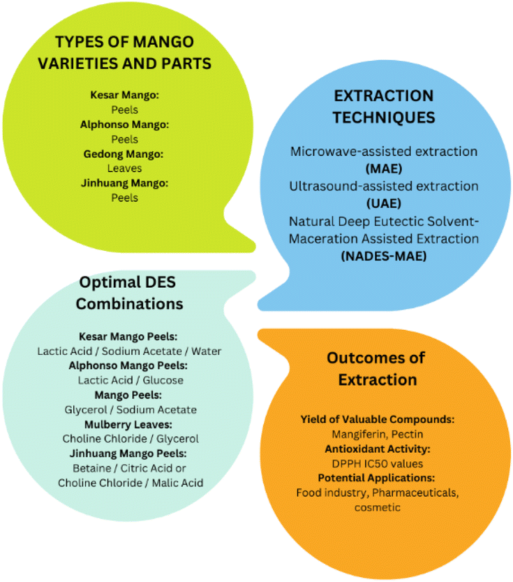
5. Phytochemical extraction from different varieties and parts of mango by DES
The information regarding the selected optimum DESs used for the extraction of phytochemicals from different mango varieties and the specific parts employed for extraction is presented in Table 3. The study conducted by Pal and Jadeja in 2020 focused on the extraction of valuable compounds from ripe Kesar mango peels.37 The researchers used a unique mixture of lactic acid, sodium acetate and water as the extracting solution, enhancing the process with the use of a microwave. Through a systematic approach, this team determined the most effective extraction conditions, which included using a microwave with a power of 436.45 W, an extraction time of 19.66 min, and a specific amount of liquid for every gram of peel. Under these optimal conditions, a significant quantity of valuable compounds was successfully extracted, with mangiferin identified as the predominant compound.37 The combination of microwave assistance and the specialized liquid demonstrated remarkable efficiency in the extraction process. Additionally, when these extracts were added to sunflower oil, they significantly extended its shelf life. This discovery underscores the potential of mango peels, which are often discarded, as a valuable source of antioxidants when utilizing this specific solution and the microwave technique.| Sample | DES | Extraction procedure | Compound extract | Operating conditions | Extraction yield/conversion efficiency | Findings | Citation & reference number |
|---|---|---|---|---|---|---|---|
| Mango peel (Kesar mango) | Lactic acid:sodium acetate:water (3:1:4) |
HSE and MAE | Phenolic compounds | HSE, magnetic stirrer: 500 rpm, temperature: 60 °C, time: 90 min, liquid-to-solid ratio: 50:1 (mL g−1) |
HSE > 80 mg GAE per g DW | - HPLC analysis discovered mangiferin as the prominent phenolic compound in the mango peel extracts | Pal and Jadeja, 2020 (ref. 37) |
| MAE 56.17 mg GAE per g DW | - Lactic acid:sodium acetate DES system in combination with MAE showed remarkable effects on the extraction efficiency of phenolic compounds |
||||||
| MAE, power of 436.45 W, time of 19.66 min, liquid-to-solid ratio: 59.82 mL g−1 | |||||||
| Mango peel (Jinhuang mango) | Betaine:citric acid (3:1), 30% water (before optimization) |
HSE and UAE | Pectin | Bet:CA pH: 0.85, liquid–solid ratio of 27:1, temperature: 80 °C, water content of Bet–CA: 70% |
Bet–CA 27.63% | - Two eco-friendly solvents extracted higher pectin yields than conventional hydrochloric acid | Chen et al., 2022 (ref. 38) |
| Choline chloride:malic acid (1:2), 30% water (before optimization) |
|||||||
| ChCl:MaA pH: 0.40, temperature: 85 °C, time: 120 min, water content of ChCl–MaA: 70%, liquid–solid ratio: 27:1 |
ChCl:MaA 30.01% |
- High intensity ultrasound treatment remarkably increased the extraction yield of low-ester pectins, but decreased their molecular weight and particles size | |||||
| Mango peel (Alphonso mango) | Lactic acid:glucose (5:1), 20% water |
UAE | Phenolic compounds | Water content: 20%, duty cycle: 50%, acoustic density: 2 W cm−3, liquid:solid ratio 30:1 (v/w), particle size: 0.3 mm, time: 30 min |
69.85 mg GAE per g | - UAE in combination with NADES technique offered 1.4 times TPC, 1.7 times TFC, 1.9 times antioxidant activity | Lanjekar et al., 2022 (ref. 39) |
| - UA-NADES also offered 50% shorter time, and 25% reduced solvent consumption compared to the batch method with 80% ethanol | |||||||
| Mango leaves (Gedong mango) | Lactic acid:sodium acetate (3:1) in distilled water |
MAE | Alkaloids, flavonoids, saponins and tannins | Temperature: 70 °C, time: 1 h | Qualitative test of the secondary metabolites | - Screening of phytochemical compounds found that the extract of Gedong mango leaves contains polyphenol | Rizikiyan et al., 2022 (ref. 184) |
| - The use of Gedong mango and mulberry extract in spray gel preparation revealed the sunscreen activity | |||||||
| Mango peel | Glycerol:sodium acetate (3:1), with 20% water |
MAE | Phenolic compounds | MAE, power: 440.18 W, time: 12.10 min, liquid to solid ratio: 59.99 mL g−1 | MAE 155.28 mg GAE per g DW | -The extraction rate was very slow for Gly:SA DES system (without the combination of MAE methods) due to their higher viscosity |
Pal and Jadeja, 2022 (ref. 185) |
| -Higher than other organic solvents like distilled water or 70% (v/v) methanol |
Fig. 3 shows the extraction of phytochemicals from different varieties and parts of mango using deep eutectic solvents (DES). In 2022, the same research team explored the use of microwave-assisted extraction to convert mango peels into valuable polyphenolic compounds.185 This innovative process not only utilized clean energy but also aimed to repurpose discarded peels for bioenergy production, including biofuels, heat and electricity. Mango peels, rich in potent antioxidant mangiferin, are often overlooked despite the environmental concerns related to their disposal. The researchers used a DES and created a technique called microwave-assisted deep eutectic solvent extraction (MADESE). Among the different DES options, glycerol:sodium acetate proved to be the most effective for extracting the polyphenolic content from mango peels. They employed advanced techniques to optimize the process, achieving the best conditions with 155.28 mg of polyphenolic content per gram of dry weight, outperforming other solvents.185 Although the glycerol:sodium acetate DES had a slower extraction rate due to its higher viscosity, MADESE holds significant potential for the large-scale extraction of bioactive compounds, particularly in the food and medical industries.
 | ||
| Fig. 3 Phytochemical extraction from different varieties and parts of mango using DESs. | ||
Lanjekar and colleagues highlighted the potential of Alphonso mango peels as a valuable source of compounds, with a specific focus on polyphenol extraction.39 Their research effectively addressed the limitations associated with traditional organic solvents by introducing natural deep eutectic solvents (NADESs) as an eco-friendlier and more non-toxic alternative. The extraction process was optimized by combining NADES with ultrasound, leading to impressive results. Using an NADES mixture containing lactic acid and glucose in a 5:1 ratio, this study achieved the highest total phenolic content (TPC) at 69.85 mg GAE per g of mango peel, a total flavonoid content (TFC) of 16.5 mg QE per g of mango peel, and a DPPH IC50 value of 35.37 μg mL−1, indicating its potent antioxidant activity.39 The ultrasound-assisted NADES method significantly improved the efficiency by providing higher TPC and TFC levels, increased antioxidant activity, shorter extraction time, and reduced solvent consumption compared to traditional ethanol-based extraction. The characterization and quantification of the polyphenolic profile revealed that gallic acid was the predominant antioxidant, followed by chlorogenic acid, with quercetin and kaempferol as the major flavonoids.
Rizikiyan et al. (2022) initiated a research project with the primary goal of formulating an effective sunscreen spray gel.184 To achieve this objective, they employed the natural deep eutectic solvent-maceration-assisted extraction (NADES-MAE) method, a recognized technique for extracting bioactive compounds from plant materials. This investigation focused on the utilization of Gedong mango and mulberry leaves, with specific types of deep eutectic solvents (DES) meticulously prepared for this purpose. For the extraction of Gedong mango, an NADES solvent consisting of sodium acetate and lactic acid in a 1:3 ratio with distilled water was used, and the MAE extraction was carried out for one hour at 70 °C. Simultaneously, mulberry leaves were subjected to extraction using a DES prepared with choline chloride and glycerol in a 1:2 ratio, with the extraction process conducted for approximately one hour at 80 °C.184 Following extraction, the resulting extracts were subjected meticulous filtration and evaporation processes to eliminate any remaining solvents. The findings of this study strongly suggest that the combination of these extracts through the NADES method, using DES, holds significant promise for application in sunscreen formulations, particularly in the form of a spray gel. This research represents an exciting step forward in the development of natural, plant-based sunscreen products.
Mango peels from the Jinhuang variety have been reported to be used for the extraction of pectin with DESs.38 In this investigation, two novel, environmentally friendly DESs including betaine:citric acid and choline chloride:malic acid were explored for pectin extraction from mango peels. The extraction conditions were optimized, and ultrasound significantly influenced the extraction yield and pectin properties. The pectins extracted with these DESs exhibited higher yields, larger sizes and increased particle dimensions compared to hydrochloric acid (HCl) extraction. The use of high-intensity ultrasound further increased the yield of low-ester pectins, while reducing their molecular size and particle dimensions. The analysis revealed a higher galacturonic acid (GalA) content and more extensive homogalacturonan (HG) regions in the pectins obtained with the DESs.38 These findings offer insights into potential applications in the food and pharmaceutical industries, providing a better understanding of the connection between extraction conditions and the physical and chemical characteristics of mango peel pectins when using DESs.
Besides the optimal DESs, the phytochemicals from mango have also been screened with different sets of DESs. These DESs were tested with varying ratios and applied to specific parts of the mango (Table 4). For instance, choline chloride and urea in a 1:2 ratio were used to extract phytochemicals from mango peel, particularly from Kesar mango.37 Similarly, choline chloride and sorbitol in a 3:1 ratio were applied to mango peel from the same variety. Various other DES combinations, such as choline chloride with different organic acids, were employed for the extraction of mango peel from Jinhuang mango.38 Sodium acetate, in combination with glycerol and lactic acid, was used for extraction from Kesar mango peel and Gedong mango leaves.37,184 These studies aimed to optimize the use of DESs for the extraction of phytochemicals from mango samples, with a focus on different mango varieties and plant parts. Fig. 4 depicts the fundamental aspects of extracting bioactive compounds using DESs, various mango varieties, and their therapeutic potential. The manuscript review suggests that the predominant practice in mango DES extraction focuses on employing peels, while the exploration of mango leaves and kernels for this purpose was relatively limited. The exploration towards the use of leaves is valuable given that it opens up the potential for a more comprehensive understanding of the bioactive components in the mango plant and their diverse applications. In the next section, we review the extraction of phytochemicals from leaves of other plants, further highlighting the significance of investigating alternative sources for phytochemical extraction.
| Component 1 | Component 2 | Tested ratio | Extraction parts | Variety | Citation & reference number |
|---|---|---|---|---|---|
| Choline chloride | Urea | 1:2 |
Mango peel | Kesar mango | Pal et al., 2020 (ref. 37) |
| Choline chloride | Sorbitol | 3:1 |
Mango peel | Kesar mango | Pal et al., 2020 (ref. 37) |
| Choline chloride | Sucrose | 1:1 |
Mango peel | Kesar mango | Pal et al., 2020 (ref. 37) |
| Choline chloride | Glycerol | 1:3 |
Mango peel | Kesar mango | Pal et al., 2020 (ref. 37) |
| Choline chloride | Glycerol | — | Mango peel | Alphonso mango | Lanjekar et al., 2022 |
| Choline chloride | Lactic acid | 1:3 |
Mango peel | Kesar mango | Pal et al., 2020 (ref. 37) |
| Choline chloride | Lactic acid | — | Mango peel | Alphonso mango | Lanjekar et al., 2022 (ref. 39) |
| Choline chloride | Malic acid | 1.5:1 |
Mango peel | Jinhuang mango | Chen et al., 2022 (ref. 38) |
| Choline chloride | Glucose | — | Mango peel | Alphonso mango | Lanjekar et al., 2022 (ref. 39) |
| Choline chloride | Malic acid | — | Mango peel | Alphonso mango | Lanjekar et al., 2022 (ref. 39) |
| Choline chloride | Malic acid | 1:2 |
Mango peel | Jinhuang mango | Chen et al., 2022 (ref. 38) |
| Choline chloride | Citric acid | 2:1 |
Mango peel | Jinhuang mango | Chen et al., 2022 (ref. 38) |
| Choline chloride | Oxalic acid | 1:1 |
Mango peel | Jinhuang mango | Chen et al., 2022 (ref. 38) |
| Choline chloride | Levulinic acid | 1:2 |
Mango peel | Jinhuang mango | Chen et al., 2022 (ref. 38) |
| Choline chloride | Acetic acid | 1:3 |
Mango peel | Jinhuang mango | Chen et al., 2022 (ref. 38) |
| Sodium acetate | Glycerol | 1:3 |
Mango peel | Kesar mango | Pal et al., 2020 (ref. 37) |
| Sodium acetate | Glycerol | 1:3 |
Mango peel | Kesar mango | Pal et al., 2022 (ref. 185) |
| Sodium acetate | Lactic acid | 1:1, 1:2, 1:3, 1:4 |
Mango peel | Kesar mango | Pal et al., 2020 (ref. 37) |
| Sodium acetate | Lactic acid | 1:3 |
Mango leaves | Gedong Mango | Rizkiyan et al., 2022 (ref. 184) |
| Betaine | Citric acid | 1:2 |
Mango peel | Jinhuang mango | Chen et al., 2022 (ref. 38) |
| Betaine | Oxalic acid | 1:2 |
Mango peel | Jinhuang mango | Chen et al., 2022 (ref. 38) |
| Betaine | Levulinic acid | 1:1 |
Mango peel | Jinhuang mango | Chen et al., 2022 (ref. 38) |
| Betaine | Malic acid | 1:2 |
Mango peel | Jinhuang mango | Chen et al., 2022 (ref. 38) |
| Betaine | Acetic acid | 1:3 |
Mango peel | Jinhuang mango | Chen et al., 2022 (ref. 38) |
| L-Proline | Citric acid | 1:2 |
Mango peel | Jinhuang mango | Chen et al., 2022 (ref. 38) |
| L-Proline | Oxalic acid | 1:2 |
Mango peel | Jinhuang mango | Chen et al., 2022 (ref. 38) |
| L-Proline | Levulinic acid | 1:2 |
Mango peel | Jinhuang mango | Chen et al., 2022 (ref. 38) |
| L-Proline | Malic acid | 1:2 |
Mango peel | Jinhuang mango | Chen et al., 2022 (ref. 38) |
| Lactic acid | Glucose | 5:1 |
Mango peel | Alphonso mango | Lanjekar et al., 2022 (ref. 39) |
 | ||
| Fig. 4 Basic aspects of green extraction using DESs, with various mango varieties and their therapeutic potential. | ||
6. Phytochemical extraction from leaves of different plants by DES
Table 5 presents examples of deep eutectic solvents (DESs) used for extracting compounds from leaves, other than mango samples. DESs were employed to extract phytochemicals from olive leaves, with a specific focus on the combined use of choline chloride and carboxylic acid for the extraction of phenolic compounds.149 This study compared the efficiency of this method to ethanol-based extraction and utilized response surface methodology to investigate the influence of temperature and the addition of water on both deep eutectic solvent (DES) and ethanol extraction processes. The study aimed to address the existing knowledge gap concerning the behaviour of choline chloride and acetic acid when used in combination. Furthermore, it characterized their solid–liquid equilibrium (SLE), density and viscosity, both with and without the addition of water. When choline chloride and acetic acid were employed at a temperature of 54.1 °C, with 50.0% water addition, there was a significant 15% increase in phenolic compounds compared to ethanol extraction at the same temperature with only 0.5% water added.149 Additionally, the SLE analysis revealed the formation of a eutectic region at a molar ratio of approximately 1:2 for choline chloride and acetic acid.
| Sample | DES selected for bioactive compound extraction | Extraction procedure | Compound extract | Operating conditions | Extraction yield/conversion efficiency | Findings | Reference |
|---|---|---|---|---|---|---|---|
| Olive leaves | Choline chloride based:acetic acid (1:2), 50% water addition |
HSE | Phenolic compound | Temperature: 54.1 °C, addition of water: 50.0% | 470.03 mg kg−1 | - Choline chloride–acetic acid yielded 15% more phenolic compounds | de Almeida Pontes et al., 2021 (ref. 149) |
| - The new class of solvents extracted two times more phenolic compounds than ethanol | |||||||
| Moringa oleifera L. leaves | Proline:glycerol (2:5), 37% water content |
UAE | Phenolic compound | Water content: 37% ultrasonic power: 144 W, ultrasonic temperature: 40 °C | 45.9 mg GAE per g DW | - Comparative study confirmed that the optimized DES-based UAE yielded further higher TPC, TFC and antioxidant activities than other extraction methods | Wu et al., 2020 (ref. 152) |
| Olive leaves | Glycerol:lysine (3:1), 10% water concentration |
Batch extraction procedure (HSE) | Phenolic compounds | Liquid to solid ratio: 150 mL g−1, water concentration (v/v): 10%, temperature: 80 °C | 188.39 mg per g GAE DM | - Glycerol–lysine DES was found to be the most effective in extracting phenolic compounds from olive leaf compared with glycerol–arginine DES and glycerol–proline and even to conventional solvents such as ethanol, methanol and water | Akli et al., 2022 (ref. 186) |
| Mitragyna speciosa Korth. Havil leaves | Choline chloride:sorbitol (3:1) |
MAE | Polyphenol | Liquid–solid ratio: 20 mL g−1, time: 20 min, microwave power (% of watts): 60% | 539.37 μg GAE per g | - The combination of NADES and MAE with some different combinations of NADES composition is more effective than a maceration | Prabowo et al., 2021 (ref. 187) |
| - SEM imaging result shows that the levels of damage of cells and cell walls were more severe after extraction | |||||||
| Ampelopsis grossedentata leaves | Choline chloride:glucose (4:1), 20% water |
UAE | Flavanoid | Liquid-to-solid ratio: 30 mL g−1, ultrasonication power: 490 W, ultrasonication time: 6.5 min | 83.93% | - The actual flavonoid yield was 83.93%, which was close to the predicted yield | Zhen et al., 2022 (ref. 150) |
| - Further, 86.75% of the flavonoids were recovered by adding the same volume of phosphate buffer saline (100 mM, pH of 7.0) to the extract solution | |||||||
| Walnut leaves (Juglans regia L.) | Choline chloride:butyric acid (1:2), 20% water (before optimization) |
HAE | Phenolic compound | Time: 180 min, temperature: 30 °C, water proportions: 53% | 37.9 mg per g DW | - The water content was clearly the most relevant extraction variable, followed by temperature and, lastly, extraction time | Vieira et al., 2018 (ref. 188) |
| Mandarin leaves | Citric acid:glycerol (1:4) 50% water |
MAE | Phenolic and flavonoid ingredients | Time: 90 s, temperature: 80–90, 30 °C, microwave power: 500 W | 14.47 mg GAE per g DS | - Citric acid–glycerol DES is one of the solvents that has shown the best performance for bioactive compound extraction | Kurtulbaş et al., 2019 (ref. 189) |
| Mate tea leaves | Lactic acid:glucose:water (5:1:3) |
UAE | Phenolic compound | Time: 60 min, solid/liquid ratio: 1:20 (w/v, g mL−1), temperature: 40 °C |
Lactic acid:glucose:water: 12.2 mg GAE/100 mg mate tea leaves |
- Lactic acid-based system showed to be more efficient in extracting phenolics | Rebocho et al., 2022 (ref. 151) |
| Lactic acid:glycine:water (3:1:3) |
Lactic acid:glycine:water: 13.5 mg GAE/100 mg mate tea leaves |
- NADES systems were able to extract 30% more of polyphenolic components of the mate tea leaves matrices, when compared with traditional solvents/techniques | |||||
| Lotus (Nelumbo nucifera Gaertn.) leaf | Lactic acid:glycerol (1:2), 20% of water before optimization |
HAE | Phenolic and flavanoid | Water content: 29%, liquid–solid ratio: 37:1 mL g−1, extraction time: 61 min, extraction temperature: 53 °C |
Flavonoids: 126.10 mg g−1 | - The extraction of lotus leaves using the NDES method was superior to both ethanol extraction and water extraction | Yang et al., 2023 (ref. 190) |
| Polyphenols: 126.10 mg g−1 | |||||||
| Eucommia ulmoides leaves | ChCl–1,4-butanediol–ascorbic acid (1:1:0.2) |
MAE | Phenolic acids (chlorogenic acid) | Temperature: 53.03 °C, time: 20.08 min, solid–liquid ratio: 18.53 mL g−1, power: 500 W | CGA = 3.659 mg g−1 | Integrated microwave-assisted ternary deep eutectic solvent extraction, which was first applied to extract compounds from EUL for the first time | Yu et al., 2021 (ref. 153) |
Deep eutectic solvents (DESs) have also been utilized for the extraction of phytochemicals from olive leaves using different sets of chemicals. This innovative type of liquid was formulated by blending glycerol with lysine, proline and arginine.186 These DESs were tested for the very first time to determine their effectiveness in extracting healthful compounds from olive leaves. To optimize the extraction process, they used a specially designed plan to find the right amount of liquid, the strength of the DES and the temperature. The ideal conditions involved using 150 mL of liquid for every gram of olive leaves, with the DES at 90% concentration, and maintaining a temperature of 80 °C.186 These conditions delivered the expected results, yielding approximately 188.39, 100.01, and 95.97 mg of healthful compounds for different DES systems. The top performer was the glycerol–lysine DES, outperforming conventional solvents including ethanol, methanol and water. In fact, it extracted 40–71% more total phenol yield compared to the regular solvents. Further testing showed that the glycerol–lysine DES was particularly effective at extracting specific healthful compounds.
Wu et al. employed a UAE method with a specific deep DES to enhance the extraction of total phenolic and flavonoid content (TPC/TFC) and antioxidant properties from Moringa oleifera L. leaves.152 They optimized the process using the response surface methodology (RSM) and central composite design (CCD). The results showed that the water content in the DES had a significant impact on all the responses, while ultrasonic time and the liquid-to-solid ratio did not affect the total phenolic content. The optimal conditions for achieving the best TPC/TFC and antioxidant activities included 37% water content in DES, 144 W ultrasonic power and a constant ultrasonic temperature of 40 °C, consistent with the predicted results. This optimized DES-based UAE method yielded higher TPC, TFC and antioxidant activities compared to other extraction techniques, with the HPLC analysis confirming the presence of 14 phenolic compounds, including substantial concentrations of vicenin-2 (17.6 mg g−1) and orientin (23.6 mg g−1) in the extracts from M. oleifera leaves.152
The DESs were used to extract polyphenols from Mitragyna speciosa (Korth.) Havil leaves through the microwave-assisted natural deep eutectic solvent extraction (MANDESE) method.187 This method was optimized using the response surface methodology (RSM). The extraction process was examined through scanning electron microscopy (SEM) imaging. Two extraction methods, maceration and MANDESE, were applied, and the determination of total polyphenol content was carried out using Folin–Ciocalteu reagent and a UV-vis spectrophotometer. SEM imaging was employed to assess any cell and cell wall damage. The optimization factors considered in RSM included the composition ratio of the natural deep eutectic solvent (NADES), the liquid–solid ratio, extraction time and microwave power. The results highlighted the superior efficiency of MANDESE with various NADES compositions compared to maceration. SEM imaging revealed an increased level of cell and cell wall damage following extraction. The optimal conditions included an NADES composition ratio of 3 g g−1 (choline chloride/sorbitol), a liquid–solid ratio of 20 mL g−1, a 20 min extraction time and 60% microwave power. A scale-up test confirmed a total polyphenol content of 526.12 μg GAE per g sample.187
Zhen et al. conducted an ultrasound-assisted extraction with natural deep eutectic solvents (NADESs) for the efficient extraction of flavonoids from Ampelopsis grossedentata leaves.150 They identified the optimal NADES composition of a 4:1 molar ratio of choline chloride to glucose with 20% water. The extraction process was further fine-tuned with a liquid-to-solid ratio of 30 mL g−1, ultrasonication at 490 W for 6.5 min, resulting in 83.93% flavonoid yield, closely aligning with their prediction.150 By adding an equivalent volume of phosphate buffer saline to the extract, they successfully recovered 86.75% of the flavonoids. Although the flavonoid extract displayed slightly reduced chemical antioxidant activity compared to dihydromyricetin, it effectively inhibited the proliferation of human breast MDA-MB-231 cells. This inhibition was achieved by inducing apoptosis, altering the cell cycle, modulating the mitochondrial membrane potential, and mitigating intracellular reactive oxygen species (ROS).
The extraction of phenolic compounds from walnut leaves (Juglans regia L.) involved optimizing the process through heat-assisted extraction and deep eutectic solvents based on choline chloride and carboxylic acids.188 Preliminary screening of various carboxylic acids as hydrogen bond donors identified the most effective mixture of choline chloride with either butyric or phenylpropionic acid at a 1:2 mole ratio, together with 20% water (w/w). Subsequently, the extraction conditions, including time, temperature and water proportion, were fine-tuned using the experimental design and response surface methodology. This study quantified the three most abundant compounds (neochlorogenic acid, quercetin 3-O-glucoside and quercetin O-pentoside) identified by HPLC. Additionally, they explored the effect of the solid/liquid ratio under the optimal conditions, finding no significant decrease in extraction efficiency until a concentration of 140 g L−1.188
DESs, in conjunction with microwave-assisted extraction (MAEX), have been employed for the extraction of valuable antioxidative phenolic and flavonoid compounds from Satsuma mandarin leaves.189 This innovative approach involves using deep eutectic solvents (DESs), which are a blend of citric acid and glycerol as hydrogen bond acceptor (HBA), and glycerol, dimethyl urea, and methylimidazole as hydrogen bond donors (HBD). The Satsuma mandarin leaves were first dried and ground before undergoing extraction using these DESs, together with conventional solvents such as water, ethanol, methanol, ethyl acetate, acetonitrile and their water solutions. Subsequently, the extracted samples were evaluated for their total phenolic content (TPM) and total flavonoid content (TFM) through UV-spectrophotometry, while their antioxidant capacities were assessed by measuring their inhibition activity towards DPPH.189
Rebocho et al. presented an innovative method for the selective extraction of antioxidants from both regular and decaffeinated mate tea leaves using a variety of NADES.151 Their study incorporated a systematic approach, optimizing the extraction parameters such as the solid-to-liquid ratio, temperature, extraction time, stirring and the integration of ultrasound-assisted extraction (UAE) technology. Their research findings demonstrated that by employing a two-step extraction process, initially utilizing a hydrophobic system (Men:Lau, 2:1) followed by a hydrophilic lactic acid-based NADES, two distinct extracts were obtained, one rich in pigments and the other abundant in polyphenols. In comparison to conventional solvents and techniques, the NADES systems exhibited a remarkable 30% increase in the extraction of polyphenolic components from the mate tea leaves matrices.151 Furthermore, the incorporation of the extract into the NADES medium, rather than into an aqueous solution, significantly enhanced the antioxidant stability, maintaining its functionality for a minimum of three months and resulting in an impressive 41% improvement compared to the extracts obtained through traditional methods. Importantly, the absence of caffeine in the extracts did not appear to impact the stability results.
Yang et al. employed NADES to extract flavonoids from lotus leaves and optimized the process using the Box–Behnken design method.190 The best conditions involved using NADES comprised of lactic acid and propanetriol at a 1:2 molar ratio with a 29% water content, a solid–liquid ratio of 37:1 (mL g−1), and extraction at 53 °C for 61 min, resulting in a flavonoid yield of 126.0972 mg g−1 and a polyphenol yield of 113.1163 mg g−1, aligning with the model prediction. This method not only increased the flavonoid yield but also enhanced the antioxidant activity compared to traditional extraction methods. LC-MS analysis of the NADES extract revealed the extraction of 19 compounds, highlighting its efficiency.190
DESs were harnessed for an environmentally friendly extraction process aimed at obtaining iridoids and phenolic acids from Eucommia ulmoides Oliver leaves (EUL).153 This innovative approach, named integrated microwave-assisted ternary deep eutectic solvent extraction (TDES-MAE), marked its debut in EUL compound extraction. Within this specialized TDES setup, the addition of a small quantity of ascorbic acid (Vc) adjusted the acidity of binary deep eutectic solvents (BDES), formed from a 1:2 M ratio of choline chloride and 1,4-butanediol, to create TDES. This adapted solution showed a superior extraction yield. Several factors, including DES type, molar ratio, water content and choice of hydrogen bond donor (HBD), were explored to identify the most effective TDES solvent. Using the Box–Behnken design, the optimal conditions such as extraction time, extraction temperature and liquid–solid ratio were determined through the response surface methodology (RSM). Under these ideal conditions, the TDES-MAE method boosted the extraction efficiency by a significant 1.4-fold compared to traditional methods.153
7. Important factors affecting phytochemical extraction by DES
7.1 Effect of water and viscosity
The introduction of water into DESs can induce significant alterations in their physiochemical properties and the integrity of their hydrogen bonding network, as reported by Vilková et al. in 2020.191 This approach enables the customization of the DES characteristics to align with the specific requisites of various processes. However, the inclusion of water can produce two distinct effects, i.e., it reduces the viscosity, thus enhancing the extraction efficiency, decreasing waste production and contributing to cost-effectiveness. Conversely, exceeding the optimal water content weakens the hydrogen bond network within the DES and its interactions with analytes, potentially resulting in diminished extraction efficiency.192–194 Fig. 5 shows the important factors that affect phytochemical extraction using deep eutectic solvents (DESs). | ||
| Fig. 5 Important factors that affect phytochemical extraction via deep eutectic solvents (DES). | ||
Precise control of the water content in deep eutectic solvents (DESs) is crucial for their use as extraction solvents, given that an excessive water content, exceeding 50%, can disrupt the hydrogen bond framework and influence the viscosity of DESs.44 High viscosity is a common challenge when using DESs or NADESs as extraction solvents, limiting their ability to penetrate the extraction matrix. Although increasing the temperature during extraction can reduce the viscosity, it is not always the best choice due to the energy consumption and potential presence of heat-sensitive phytochemicals. Thus, to address challenges such as high viscosity and the associated costs of using pure DESs as the extraction medium, researchers have practically employed aqueous NADES solutions, which typically consist of a water concentration in the range of 20% to 80%. Existing literature suggests that an optimal equilibrium is achieved at a water content of approximately 20% to 30%.195,196 This equilibrium allows the maintenance of a fluid extraction system while preserving the essential hydrogen bond structure of the DES. Nevertheless, the potential of a higher water content to disrupt this structure should not overshadow their suitability for specific extraction scenarios. Precise control of the water content in DES systems has emerged as a critical consideration when optimizing DES-based extraction processes.
For example, Koutsoukos et al. used water as a co-solvent to extract phenolic compounds from brown propolis with ChCl/tartaric acid NADES, and they used methanol as a co-solvent for extracting carotenoids from apricot pulp and shrimp head by-products using the same NADES.197 The concentration of water in the DES-water or NaDES-water mixtures significantly influenced the efficiency of extraction. Bi et al. determined that a ChCl/1,4-butanediol NaDES mixture with 35% water is the optimal medium for extracting myricetin and amentoflavone from Chamaecyparis obtusa.198 Similarly, Zhao et al. found that efficient extraction of rutin from Sophora japonica flower buds can be achieved using ChCl/triethylene glycol DES containing 20% water.199 Their studies on the viscosity of 20 DESs showed that their viscosity increased with the formation of more hydrogen bonds formed among their constituents.
7.2 Effect of sample-to-solvent ratio (SLR)
The sample-to-solvent ratio (SLR) is a crucial factor in deep eutectic solvent (DES)-based extraction processes, and its optimal balance is vital to ensure both efficiency and scalability. The SLR dictates the proportion of solid sample immersed in the solvent, making it a key determinant of extraction performance. When only a small sample amount is introduced into the solvent, the extraction process may prove inefficient, especially when being scaled up to handle larger quantities of material. The limited sample processing in this case can hinder the overall extraction efficiency, making it less effective and time-consuming when dealing with larger volumes of material in industry.Alternatively, using an excess of solid sample compared to the solvent presents its own set of challenges. The surplus solid material can impede the efficient dispersion of the solvent, resulting in slower interactions between the sample and the solvent. Consequently, this may reduce the contact surface area between the sample and the solvent, potentially diminishing the overall performance of the system. To strike the right balance, many studies have undertaken extensive investigations, often through statistical analyses or referencing prior successful works. They have typically converged on a solid SLR ratio of 1:10, which is considered an effective compromise.200,201 This ratio allows the processing of a sufficient quantity of sample, while maintaining the efficiency of the extraction method. Researchers and practitioners widely embrace this ratio as a practical and reliable approach for DES-based extraction processes, given that it ensures both efficient extraction and scalability, meeting the needs of various applications and settings.
7.3 Effect of time
The extraction time is a crucial parameter in the extraction of phytochemicals such as phenolic compounds using DES. It significantly influences the efficiency and yield of the extraction process by determining the duration during which the phenolic compounds are in contact with the DES. Generally, longer extraction times lead to higher yields, given that they allow more extensive interactions between the phenolic compounds and the solvent. However, it is important to consider the kinetics of the extraction process, given that there is usually an initial rapid increase in yield, which may slow down over time as the system approaches equilibrium. Therefore, the choice of extraction time should consider the specific kinetics of the extraction system. The optimal extraction time may vary depending on factors such as the type of phenolic compounds, the specific plant material and the type of DES used. Achieving this optimal time requires experimentation and optimization to fine-tune the extraction duration for the specific application.It is also essential to consider the potential impact of prolonged exposure to DES and heat on the stability of thermolabile phenolic compounds, given that excessive heat may lead to degradation. Most of the processes reviewed have very high retrieval percentages, with extraction times ranging from 20 min to 2 h.37,39,196 Naturally, the type of extraction also defines the extraction time required, with energy-assisted methods such as heating, ultrasound or microwave requiring less extraction time, but more energy to conduct. Overall, the use of DESs has enabled undeniably short extraction times for all extraction methods employed. When scaling up the extraction process, careful consideration of the required extraction time is necessary to maintain efficiency and consistency. In summary, the extraction time significantly affects both the quantity and quality of the extracted phenolic compounds, and finding the right balance is essential for effective and efficient DES-based extractions.
7.4 Effect of temperature
The temperature at which extraction processes are conducted significantly influences the duration, efficiency and overall performance of the extraction. Generally, higher temperature enhances the mobility of molecules, expediting the diffusion of extracted substances into the solvent. This principle remains valid when utilizing deep eutectic solvents (DESs). However, it is important to note that DES-based extractions are not exempt from the impact of temperature, given that they depend on temperature to mitigate the high viscosities inherent to DESs, which can hinder the smooth operation of the extraction process. According to the literature, the optimal temperature for extraction typically falls in the range of room temperature (around 25 °C) to approximately 60 °C.For example, the temperature used for the extraction of phytochemicals using DESs was 30 °C, 40 °C and 60 °C for walnut leaves, Moringa oleifera L. leaves and Kesar mango peel, respectively.37,152,188 However, it is worth considering that excessively high temperatures have their downsides. They not only consume more energy, which deviates from the environmentally friendly “green” aspect of extraction, but also pose potential risks to both the integrity of the DES and the stability of the target substances, especially given the thermal sensitivity of many natural compounds involved.202 Furthermore, extremely high temperatures have been found to decrease the extraction yields in certain cases. This reduction in yield is attributed to the decline in the interaction between the target compound and the chosen solvent, irrespective of their individual thermal resistance. It is essential to underscore that the thermal characteristics of the substances used remain a limiting factor in these instances.195
7.5 Effect of auxiliary
During the extraction process, various techniques are employed to mix samples, which include stirring, vortexing, shaking and combinations of heating and stirring. Each of these approaches has an array of advantages and disadvantages. These methods are considered auxiliary, given that they provide additional support to the extraction procedure. For example, considering a sample of medicinal plant material subjected to ultrasound-assisted extraction (UAE), UAE can significantly reduce the extraction time for bioactive compounds, making it a valuable technique in the pharmaceutical and herbal medicine industries. By utilizing ultrasound, the extraction process becomes more efficient, enabling the extraction of bioactive compounds such as flavonoids, alkaloids and essential oils from medicinal plants. This method is not only faster but also helps preserve the integrity of these valuable compounds.On the other hand, microwave radiation offers several benefits, including expedited extraction, reduced usage of solvents and reagents, precise control of heating and efficient energy transfer. For instance, when extracting essential oils from aromatic herbs, microwave-assisted extraction (MAE) has been employed to enhance the extraction efficiency. The combination of microwave radiation with deep eutectic solvents (DESs) results in a swift and efficient extraction process, yielding high-quality essential oils that find applications in the perfume and cosmetics industries.203 Additionally, MAE reduces the environmental impact by minimizing the solvent usage. For example, Jinhuang and Alphonso mangoes have been subjected to DES extraction combined with UAE, while Kesar and Gedong mangoes have been exposed to DES with MAE.37–39,184 However, both ultrasound and microwave radiation can induce alterations in the structure of the target analytes, potentially leading to their degradation or modification, even when DESs are used as the extraction medium. Consequently, it is imperative to meticulously fine-tune the extraction conditions to attain the desired outcomes, while mitigating any adverse effects.203
8. Assessment of bioactive compounds extracted via DESs
For a thorough analysis of bioactive compounds, the process begins with careful sample preparation. This involves drying the samples using controlled temperature and humidity or freeze-drying, followed by turning them into a consistent powder through sieving. Various factors such as pH, moisture, protein, ash, fiber, acidity, lipids, and sugars are assessed. Subsequently, DESs are used to extract these compounds, and techniques such as HPLC and GC are employed to identify and measure them. Thus, ensuring that DESs can maintain the stability of these compounds in the extract is crucial, considering factors such as heat, light, storage time and environmental conditions, especially sunlight, to ensure their reliability for various uses. Additionally, other tests are often performed to evaluate how DESs affect the properties of the extract, including determining total phenolic content (TPC), total flavonoid content (TFC), antioxidant activity and antimicrobial activity.204Phenolic compounds are characterized by hydroxyl groups linked to an aromatic ring. These compounds are classified into simple phenols, polyphenols, coumarins and other categories.63 Researchers are increasingly interested in phenolic compounds because of their noteworthy and beneficial properties. The total phenolic content is commonly assessed through spectrophotometric means, utilizing a specific redox reagent known as the Folin–Ciocalteu reagent. This method enables the determination of the total concentration of phenolic hydroxyl groups in plant extracts, with the results typically expressed as gallic acid equivalent (in milligrams of gallic acid per gram of dried sample).205,206
Flavonoids, a subgroup of phenolic compounds with a carbon-based core structure, have gained attention due to their antioxidant, antimicrobial and antitumor properties, especially in the context of bladder cancer. Traditional methods for extracting flavonoids rely on various techniques and conventional solvents, primarily because these compounds are challenging to dissolve in aqueous solutions, leading to time and material loss. Recently, DESs have emerged as a more efficient and cost-effective alternative for flavonoid extraction, yielding higher quantities. The determination of total flavonoid content (TFC) is typically carried out using the aluminium chloride colorimetric method, which is based on the ability of aluminium to form stable complexes with flavones, flavonols, and specific dihydroxyl groups found in flavonoids. Various reference standards, such as quercetin, catechin and rutin, are used for TFC measurement.206–208
Various methods for evaluating antioxidant activity, such as the DPPH free radical scavenging assay, ferric reducing antioxidant power (FRAP), hydroxyl radical scavenging activity and ABTS, have been utilized to assess the effectiveness of different extraction approaches. For example, the extraction of phenolic and flavonoid compounds from marjoram using a deep eutectic solvent (DES) composed of lactic acid, glycine and water in a 3:1:3 molar ratio resulted in higher yields compared to a 60% aqueous ethanol solvent. This extract exhibited substantial antiradical activity, measuring 1950 μmol DPPH per gram of dry weight.209 Similarly, in the case of Rosmarinus officinalis L., the use of a DES mixture of choline chloride and lactic acid in a 1:3 ratio resulted in the extraction of higher levels of phenolic compounds compared to the traditional 100% ethanol as the solvent. Subsequent analysis of the phenolic compounds in the extract indicated a remarkable ferric reducing antioxidant property, with a value of 183.82 mM Trolox per gram, surpassing that obtained using an ethanol extract.210
Researchers have explored the cytotoxic effects of medicinal plant extracts obtained through deep eutectic solvents (DES) using a MTS assay in vitro. Polyphenolic compounds extracted from grape pomace with choline chloride:citric acid (2:1) were tested against HeLa (cervical cancer) and MCF-7 (breast cancer) cell lines, showing 37.61% cell viability at 5% concentration (v/v) within 72 h. A similar protocol for extracting compounds from olive pomace also demonstrated significant in vitro cytotoxicity (MTS assay) with 12.19% cell viability at the same concentration within the same time frame.211 Moreover, ginsenoside, a bioactive compound from ginseng, was extracted using a ternary DES (GPS-5) glycerol:L-proline:sucrose (9:4:1) as an alternative solvent, revealing anti-tumor activity against human colorectal cancer cell lines at 58 μg mL−1, while the DES itself exhibited no cytotoxic effects based on the MTT assay results.212
Bacterial growth inhibition testing is a widely employed and efficient method due to its cost-effectiveness and swift results. In one study, phycocyanin, a bioactive compound from Arthrospira platensis, was extracted using DESs. Among the tested DES options, a 1:1 mixture of xylose and glycerol was proven to be the most effective, exhibiting robust antimicrobial properties against Escherichia coli and Enterobacter aerogenes with well diameters of 17 mm and 16 mm, respectively. Another investigation assessed the antimicrobial potential of total polyphenols extracted from Punica granatum L. using a malic acid:glucose:glycerol (1:1:1) solvent system. This extract displayed significant inhibitory effects against the Gram-positive bacterium Staphylococcus aureus, with a 90% inhibition rate observed at a concentration of 0.7 mg mL−1. Importantly, this result differed from the activity of ascorbic acid, suggesting that polyphenols may interact with microorganism cell membranes, potentially leading to microbial cell death or enzyme inhibition.213
9. Conclusions and future perspectives
Deep eutectic solvents (DESs) represent a transformative advancement in green chemistry, offering an environmentally friendly, cost-effective, and efficient alternative to conventional solvents for the extraction of bioactive compounds. Mango (Mangifera indica L.), a globally cultivated fruit with a rich therapeutic profile, has emerged as a promising natural source for these compounds, including polyphenols, carotenoids, vitamins, and minerals. Although mango peels have traditionally been the focus of DES-based extractions, this review emphasized the untapped potential of other parts of the mango plant, such as its leaves, seeds, and kernels, to broaden the spectrum of bioactive compounds.However, despite their potential, the application of DESs has certain limitations. The high viscosity of certain DES formulations can impede mass transfer, while their sensitivity to water content affects the extraction consistency and efficiency. Additionally, the diverse range of DES compositions and classifications, including natural DESs (NADESs) and therapeutic DESs (THEDES), complicates the standardization and reproducibility in research. These challenges necessitate systematic studies to establish standardized protocols and optimize DES formulations for specific applications.
From a mechanistic perspective, the reliance of DESs on hydrogen-bonding interactions offers both adaptability and vulnerability. External factors, such as temperature and humidity, can alter these interactions, impacting the extraction performance. Future research should explore innovative DES formulations with improved thermal and chemical stability, ensuring their effectiveness across varied operational conditions.
The integration of DESs with advanced extraction techniques, such as microwave-assisted and ultrasound-assisted methods, presents an opportunity to enhance the scalability and reduce the energy demands of processes. These approaches can address the limitations of traditional extraction methods, while maintaining or improving the yield and purity of bioactive compounds. Additionally, leveraging DESs for hydrophobic compound extraction, through hydrophobic DES formulations, can expand their applicability to a broader range of natural products.
The variability in bioactive compound profiles among mango cultivars offers another avenue for exploration. Comparative studies across different mango varieties can uncover unique compounds or higher concentrations of specific bioactives, enabling the development of targeted nutraceuticals and functional foods. This cultivar-focused research can also inform breeding programs aimed at enhancing the health-promoting attributes of mangoes. Furthermore, to move from laboratory-scale studies to real-world applications, rigorous bioavailability and safety evaluations are essential. Preclinical and clinical trials will validate the therapeutic potential of DES-extracted compounds and address concerns related to their stability, absorption, and efficacy in human systems. These efforts will bridge the gap between scientific research and industrial applications, fostering the development of high-value products that cater to health-conscious consumers.
Sustainability is a cornerstone of DES applications. By valorizing agricultural by-products such as mango peels, seeds, and other underutilized components, DES-based extraction aligns with the global goals of reducing waste and minimizing environmental impact. This dual benefit of environmental stewardship and product innovation positions DESs as pivotal technology in green chemistry. Looking ahead, the convergence of DES advancements with industrial needs and sustainability imperatives offers a pathway to revolutionize natural product extraction. Continued interdisciplinary research, encompassing materials science, green chemistry, and bioactive compound studies, will unlock the full potential of DESs, contributing to health, industry, and environmental well-being.
Data availability
This review article discusses data and information obtained from previously published studies, which are duly cited throughout the manuscript. As such, all data referenced in this work are available within the public domain or can be accessed through the cited sources.Author contributions
Ahmad Mukhlis Abdul Rahman: conceptualization and writing. Amirul Ridzuan Abu Bakar: conceptualization and supervision. Ang Qian Yee: resources, writing – review and editing. Mohd Asraf Mohd Zainudin: resources, writing – review and editing. Nik Muhammad Azhar Nik Daud: validation, writing – review and editing. Ahmad Anas Nagoor Gunny: resources, writing – review and editing. Ryan Vitthaya Peron: resources, writing – review and editing. Nurul Husna Khairuddin: resources, writing – review and editing.Conflicts of interest
There are no conflicts to declare.Acknowledgements
This research was funded by a grant from the Ministry of Higher Education of Malaysia (FRGS/1/2022/STG01/UNIMAP/02/2).Notes and references
- C. Bitwell, S. S. Indra, C. Luke and M. K. Kakoma, Sci. Afr., 2023, 19, e01585 CAS.
- S. Dubale, D. Kebebe, A. Zeynudin, N. Abdissa and S. Suleman, J. Exp. Pharmacol., 2023, 15, 51–62 CrossRef CAS PubMed.
- C. J. Dawurung, M. T. H. Nguyen and J. Pengon, et al., BMC Complement. Med. Ther., 2021, 21, 231 CrossRef CAS PubMed.
- M. Pateiro, R. Domínguez, P. E. S. Munekata, G. Nieto, S. P. Bangar, K. Dhama and J. M. Lorenzo, Foods, 2023, 12, 637 CrossRef CAS PubMed.
- A. Aziz, S. Noreen, W. Khalid, F. Mubarik, M. K. Niazi, H. Koraqi, A. Ali, C. M. G. Lima, W. S. Alansari, A. A. Eskandrani, G. Shamlan and A. AL-Farga, Molecules, 2022, 27, 7320 CrossRef CAS PubMed.
- Y. Asale, E. Dessalegn, D. Assefa and M. Abdisa, Int. J. Food Prop., 2021, 24, 354–363 CrossRef.
- A. Ali, S. M. Kiloni, P. R. Cáceres-Vélez, P. R. Jusuf, J. J. Cottrell and F. R. Dunshea, Foods, 2022, 11, 4038 CrossRef CAS PubMed.
- N. C. C. Carvalho, O. S. Monteiro, C. Q. da Rocha, G. B. Longato, R. E. Smith, J. K. R. da Silva and J. G. S. Maia, Foods, 2022, 11, 2079 CrossRef CAS PubMed.
- M. Oleszek, I. Kowalska, T. Bertuzzi and W. Oleszek, Molecules, 2023, 28, 342 CrossRef CAS PubMed.
- S. Ben-Othman, I. Jõudu and R. Bhat, Molecules, 2020, 25(3), 510 CrossRef CAS PubMed.
- R. Gomes-Araújo, D. G. Martínez-Vázquez, A. V. Charles-Rodríguez, S. Rangel-Ortega and A. Robledo-Olivo, Int. J. Food Sci., 2021, 9936722 Search PubMed.
- K. Das, R. Dang, G. Sivaraman and R. P. Ellath, Turk. J. Pharm. Sci., 2018, 15(2), 156–165 CrossRef CAS PubMed.
- G. N. Kasiama, A. Ikey, C. N. Kabengele, J. T. Kilembe, E. N. Matshimba, J. M. Bete, P. B. Bahati, C. L. Inkoto, P. K. Mutwale, K. N. Ngbolua, D. S. T. Tshibangu, D. D. Tshilanda and P. T. Mpiana, Annu. Res. Rev. Biol., 2022, 37(6), 28–36 CrossRef.
- E. S. El-Wakil, S. Shaker and T. Aboushousha, et al., BMC Complement Med. Ther., 2023, 23, 15 CrossRef CAS PubMed.
- K. N. Ergin, S. Karakaya, G. Göger, O. Sytar, B. Demirci and H. Duman, Molecules, 2022, 27, 1228 CrossRef CAS PubMed.
- R. Kabuka, S. Mudenda, M. Kampamba, M. Chulu, T. Chimombe and C. Hikaambo, Pharmacol. Pharm., 2022, 13, 119–128 CrossRef CAS.
- A. Khoddami, M. A. Wilkes and T. H. Roberts, Molecules, 2013, 18(2), 2328–2375 CrossRef CAS PubMed.
- P. R. D. Perera, S. Ekanayake and K. K. D. S. Ranaweera, Evid.-Based Complementary Altern. Med., 2017, 1083589 CrossRef CAS PubMed.
- F. S. Chin, K. P. Chong, A. Markus and N. K. Wong, World J. Agric. Sci., 2013, 9(3), 266–270 CAS.
- G. I. Hidalgo and M. P. Almajano, Antioxidants, 2017, 6, 7 CrossRef PubMed.
- M. M. Dardavila, S. Pappou, M. G. Savvidou, V. Louli, P. Katapodis, H. Stamatis, K. Magoulas and E. Voutsas, Molecules, 2023, 28, 415 CrossRef CAS PubMed.
- A. S. Dheyab, M. F. A. Bakar, M. AlOmar, S. F. Sabran, A. F. M. Hanafi and A. Mohamad, Separations, 2021, 8, 176 CrossRef CAS.
- M. Ruesgas-Ramón, M. C. Figueroa-Espinoza and E. Durand, J. Agric. Food Chem., 2017, 65(18), 3591–3601 CrossRef PubMed.
- A. García-Roldan, L. Piriou and P. Jauregi, Front. Plant Sci., 2023, 13, 1072592 CrossRef PubMed.
- M. Z. Gao, Q. Cui, L. T. Wang, Y. Meng, L. Yu, Y. Y. Li and Y. J. Fu, Microchem. J., 2020, 154, 104598 CrossRef CAS.
- J. Serna-Vázquez, M. Z. Ahmad, G. Boczkaj and R. Castro-Muñoz, Molecules, 2021, 26, 5037 CrossRef PubMed.
- J. O. Airouyuwa, H. Mostafa, A. Riaz and S. Maqsood, Ultrason. Sonochem., 2022, 91, 106233 CrossRef PubMed.
- P. Liu, J. W. Hao, L. P. Mo and Z. H. Zhang, RSC Adv., 2015, 5, 48675–48704 Search PubMed.
- E. L. Smith, A. P. Abbott and K. S. Ryder, Chem. Rev., 2014, 114, 11060–11082 Search PubMed.
- B. Tang, H. Zhang and K. H. Row, J. Sep. Sci., 2015, 38(6), 1053–1064 CrossRef CAS PubMed.
- D. S. Freitas, D. Rocha, T. G. Castro, J. Noro, V. L. B. Castro, M. A. Teixeira, R. L. Reis, A. Cavaco-Paulo and C. Silva, ACS Sustain. Chem. Eng., 2022, 10, 7974–7989 CrossRef CAS.
- C. Cannavacciuolo, S. Pagliari, J. Frigerio, C. M. Giustra, M. Labra and L. Campone, Foods, 2022, 12(1), 56 Search PubMed.
- E. M. Coelho, M. E. A. O. de Souza, L. C. Corrêa, A. C. Viana, L. C. de Azevêdo and M. Dos Santos Lima, Antioxidants, 2019, 8(4), 102 CrossRef CAS PubMed.
- C. C. F. Gomes, L. d. S. Oliveira, D. C. Rodrigues, P. R. V. Ribeiro, K. M. Canuto, A. S. G. Duarte, K. S. Eça and R. W. de Figueiredo, J. Food Biochem., 2022, 46(3), e13880 Search PubMed.
- B. Mirza, C. R. Croley, M. Ahmad, J. Pumarol, N. Das, G. Sethi and A. Bishayee, Crit. Rev. Food Sci. Nutr., 2021, 61(13), 2125–2151 Search PubMed.
- R. Ahmad, A. Alqathama, M. Aldholmi, M. Riaz, A. N. Abdalla, F. Aljishi, E. Althomali, M. Amir, O. Abdullah, M. A. Alamer, D. Alaswad, W. Alsulais and A. Alsulays, Pharmaceuticals, 2023, 16, 350 CrossRef CAS PubMed.
- C. B. T. Pal and G. C. Jadeja, Food Sci. Technol. Int., 2020, 26(1), 78–92 Search PubMed.
- S. Chen, L. Xiao, S. Li, T. Meng, L. Wang and W. Zhang, Ultrason. Sonochem., 2022, 86, 106045 CrossRef CAS PubMed.
- K. J. Lanjekar, S. Gokhale and V. K. Rathod, Bioresour. Technol. Rep., 2022, 18, 101074 Search PubMed.
- A. P. Abbott, G. Capper, D. L. Davies, R. K. Rasheed and V. Tambyrajah, Chem. Commun., 2003, 1, 70–71 RSC.
- M. K. Banjare, K. Behera, M. L. Satnami, S. Pandey and K. K. Ghosh, RSC Adv., 2018, 8(15), 7969–7979 RSC.
- A. Skulcova, A. Russ, M. Jablonsky and J. Sima, BioResources, 2018, 13(3), 5042–5051 CAS.
- F. S. Mjalli and O. U. Ahmed, Korean J. Chem. Eng., 2016, 33(1), 337–343 Search PubMed.
- Y. Dai, G.-J. Witkamp, R. Verpoorte and Y. H. Choi, Food Chem., 2015, 187, 14–19 CrossRef CAS PubMed.
- A. Hayyan, F. S. Mjalli, I. M. Alnashef, T. Al-Wahaibi, Y. M. Al-Wahaibi and A. Hashim, Thermochim. Acta, 2012, 541, 70–75 Search PubMed.
- S. Nardecchia, M. C. Gutiérrez, M. L. Ferrer, M. Alonso, I. M. López, J. C. Rodríguez-Cabello and F. Del Monte, Biomacromolecules, 2012, 13, 28 CrossRef PubMed.
- H. G. Morrison, C. C. Sun and S. Neervannan, Int. J. Pharm., 2009, 378(1), 136–139 CrossRef CAS PubMed.
- A. P. Abbott, G. Capper and S. Gray, ChemPhysChem, 2006, 7(4), 803–806 CrossRef CAS PubMed.
- S. J. Chudasama, B. J. Shah, K. M. Patel and T. M. Dhameliya, J. Mol. Liq., 2021, 361, 119664 CrossRef.
- B. B. Hansen, S. Spittle, B. Chen, D. Poe, Y. Zhang, J. M. Klein, A. Horton, L. Adhikari, T. Zelovich, B. W. Doherty, B. Gurkan, E. J. Maginn, A. Ragauskas, M. Dadmun, T. A. Zawodzinski, G. A. Baker, M. E. Tuckerman, R. F. Savinell and J. R. Sangoro, Chem. Rev., 2021, 121, 1232–1285 CrossRef CAS PubMed.
- P. Xu, G. W. Zheng, M. H. Zong, N. Li and W. Y. Lou, Bioresour. Bioprocess., 2017, 4(1), 34 CrossRef PubMed.
- S. Khandelwal, Y. K. Tailor and M. Kumar, J. Mol. Liq., 2016, 215, 345–386 Search PubMed.
- K. Radošević, M. C. Bubalo, V. G. Srček, D. Grgas, T. L. Dragičević and I. R. Redovniković, Ecotoxicol. Environ. Saf., 2015, 112, 46–53 Search PubMed.
- A. K. Halder and M. N. D. S. Cordeiro, ACS Sustain. Chem. Eng., 2019, 7(12), 10649–10660 Search PubMed.
- I. Juneidi, M. Hayyan and O. M. Ali, Environ. Sci. Pollut. Res., 2016, 23(8), 7648–7659 CrossRef CAS PubMed.
- C. Ruß and B. König, Green Chem., 2012, 14(11), 2969–2982 RSC.
- H. Zhao, C. Zhang and T. D. Crittle, J. Mol. Catal. B: Enzym., 2013, 85, 243–247 CrossRef.
- K. L. Marsh, G. K. Sims and R. L. Mulvaney, Biol. Fertil. Soils, 2005, 42(2), 137 CrossRef CAS.
- M. A. R. Martins, S. P. Pinho and J. A. P. Coutinho, J. Solution Chem., 2019, 48, 962–982 CrossRef CAS.
- Q. Zhang, K. D. O. Vigier, S. Royer and F. Jérôme, Chem. Soc. Rev., 2012, 41, 7108–7146 RSC.
- M. H. Zainal-Abidin, M. Hayyan, A. Hayyan and N. S. Jayakumar, Anal. Chim. Acta, 2017, 979, 1–23 Search PubMed.
- A. P. Abbott, G. Capper, D. L. Davies, H. L. Munro, R. K. Rasheed and V. Tambyrajah, Chem. Commun., 2001, 2010–2011 RSC.
- M. A. Kareem, F. S. Mjalli, M. A. Hashim and I. M. AlNashef, J. Chem. Eng. Data, 2010, 55, 4632–4637 CrossRef CAS.
- A. P. Abbott, J. C. Barron, K. S. Ryder and D. Wilson, Chem.–Eur. J., 2007, 13, 6495–6501 CrossRef CAS PubMed.
- D. O. Abranches, M. A. Martins, L. P. Silva, N. Schaeffer, S. P. Pinho and J. A. Coutinho, Chem. Commun., 2019, 55(69), 10253–10256 Search PubMed.
- M. S. Sitze, E. R. Schreiter, E. V. Patterson and R. G. Freeman, Inorg. Chem., 2001, 40, 2298 CrossRef CAS PubMed.
- T. B. Scheffler and M. S. Thomson, Seventh International Conference on Molten Salts, The Electrochemical Society, Montreal, 1990, vol. 281 Search PubMed.
- A. P. Abbott, G. Capper, D. L. Davies and R. K. Rasheed, Chemistry, 2004, 10(15), 3769–3774 CrossRef CAS PubMed.
- A. P. Abbott, G. Capper, D. L. Davies, K. J. McKenzie and S. U. Obi, J. Chem. Eng. Data, 2006, 51, 1280 CrossRef CAS.
- D. J. G. P. Van Osch, C. H. J. T. Dietz, J. Van Spronsen, M. C. Kroon, F. Gallucci, M. Van Sint Annaland and R. Tuinier, ACS Sustainable Chem. Eng., 2019, 7(3), 2933–2942 CrossRef CAS.
- H. F. M. Zaid, F. K. Chong and M. I. A. Mutalib, Fuel, 2017, 192, 10–17 CrossRef.
- I. Cichowska-Kopczyńska, B. Nowosielski and D. Warmińska, Molecules, 2023, 28(14), 5293 CrossRef PubMed.
- B. Y. Zhao, P. Xu, F. X. Yang, H. Wu, M. H. Zong and W. Y. Lou, ACS Sustain. Chem. Eng., 2015, 3, 2746–2755 CrossRef CAS.
- K. Xu, Y. Wang, Y. Huang, N. Li and Q. Wen, Anal. Chim. Acta, 2015, 864, 9–20 CrossRef CAS PubMed.
- F. A. Getie, D. W. Ayele, N. G. Habtu, F. A. Yihun, T. A. Yemata, M. D. Ambaw and A. K. Worku, Heliyon, 2023, 9(7), e17810 CrossRef CAS PubMed.
- X. Yang, Q. Wang, J. Li, N. Yalikun, L. Xu and Z. Ding, Fluid Phase Equilib., 2023, 568, 113756 CrossRef CAS.
- O. González, R. Fidalgo, I. Domínguez, E. A. Macedo and B. González, J. Chem. Eng. Data, 2016, 61(12), 4191–4202 CrossRef.
- M. A. Farajzadeh, M. Abbaspour, R. Kazemian and M. R. A. Mogaddam, J. Sci. Food Agric., 2020, 100(5), 1904–1912 CrossRef CAS PubMed.
- Y. Yang, L. Zhao, J. Ren and B. He, Processes, 2022, 10(4), 778 CrossRef CAS.
- H. Kivelä, M. Salomäki, P. Vainikka, E. Mäkilä, F. Poletti, S. Ruggeri, F. Terzi and J. Lukkari, J. Phys. Chem. B, 2022, 126(2), 513–527 CrossRef PubMed.
- F. M. Fuad, M. M. Nadzir and A. Harun, J. Mol. Liq., 2021, 339, 116923 Search PubMed.
- Y. Marcus and Y. Marcus, Applications of Deep Eutectic Solvents, Springer International Publishing, 2019, pp. 111–151 Search PubMed.
- C. Florindo, F. Lima, B. D. Ribeiro and I. M. Marrucho, Curr. Opin. Green Sustainable Chem., 2019, 18, 31–36 Search PubMed.
- C. Florindo, M. M. Oliveira, L. C. Branco and I. M. Marrucho, J. Mol. Liq., 2017, 247, 441–447 CrossRef CAS.
- A. Basaiahgari, S. Panda and R. L. Gardas, J. Chem. Eng. Data, 2018, 63(7), 2613–2627 CrossRef CAS.
- F. S. Mjalli, J. Naser, B. Jibril, V. Alizadeh and Z. Gano, J. Chem. Eng. Data, 2014, 59(7), 2242–2251 CrossRef CAS.
- D. J. van Osch, L. F. Zubeir, A. van den Bruinhorst, M. A. Rocha and M. C. Kroon, Hydrophobic deep eutectic solvents as water-immiscible extractants, Green Chem., 2015, 17(9), 4518–4521 RSC.
- D. Deng, Y. Jiang, X. Liu, Z. Zhang and N. Ai, Investigation of solubilities of carbon dioxide in five levulinic acid-based deep eutectic solvents and their thermodynamic properties, J. Chem. Thermodyn., 2016, 103, 212–217 CrossRef CAS.
- S. Hong-Zhen, N. Jing-Mei, L. Qing-Shan and L. Chang-Ping, Acta Phys.-Chim. Sin., 2015, 31, 1468–1473 Search PubMed.
- Y. Wang, Y. Hiu, W. Wu, D. Liu, Y. Ji and S. Ren, Roles of hydrogen bond donor and hydrogen bond acceptor in the extraction of toluene from n-heptane using deep eutectic solvents, Green Chem., 2016, 18, 3089–3097 Search PubMed.
- C. Florindo, F. S. Oliveira, L. P. N. Rebelo, A. M. Fernandes and I. M. Marrucho, ACS Sustain. Chem. Eng., 2014, 2(10), 2416–2425 CrossRef CAS.
- M. H. Shafie, R. Yusof and C. Y. Gan, J. Mol. Liq., 2019, 288, 111081 Search PubMed.
- A. Singh, R. Walvekar, M. Khalid, W. Y. Wong and T. C. S. M. Gupta, J. Mol. Liq., 2018, 252, 439–444 Search PubMed.
- S. P. Ijardar, V. Singh and R. L. Gardas, Molecules, 2022, 27(4), 1368 CrossRef CAS PubMed.
- K. R. Siongco, R. B. Leron and M. H. Li, J. Chem. Thermodyn., 2013, 65, 65–72 CrossRef CAS.
- K. S. Kim and B. H. Park, J. Mol. Liq., 2018, 254, 272–279 CrossRef CAS.
- A. R. Harifi-Mood and R. Buchner, J. Mol. Liq., 2017, 225, 689–695 CrossRef CAS.
- N. F. Gajardo-Parra, J. M. Lubben, M. J. Winnert, Á. Leiva, J. F. Brennecke and R. I. Canales, J. Chem. Thermodyn., 2019, 133, 272–284 CrossRef CAS.
- D. Shah and F. S. Mjalli, Phys. Chem. Chem. Phys., 2014, 16, 23900–23907 RSC.
- A. Yadav and S. Pandey, J. Chem. Eng. Data, 2014, 59(7), 2221–2229 CrossRef CAS.
- G. García, S. Aparicio, R. Ullah and M. Atilhan, Energy Fuels, 2015, 29(4), 2616–2644 Search PubMed.
- A. P. Abbott, R. C. Harris and K. S. Ryder, J. Phys. Chem. B, 2007, 111(18), 4910–4913 CrossRef CAS PubMed.
- Z. Maugeri and P. D. de María, RSC Adv., 2012, 2(2), 421–425 RSC.
- C. D'Agostino, R. C. Harris, A. P. Abbott, L. F. Gladden and M. D. Mantle, Phys. Chem. Chem. Phys., 2011, 13(48), 21383–21391 RSC.
- M. Francisco, A. van den Bruinhorst and M. C. Kroon, Angew. Chem., Int. Ed., 2013, 52, 3074–3075 CrossRef CAS PubMed.
- A. P. Abbott, R. C. Harris, K. S. Ryder, C. D'Agostino, L. F. Gladden and M. D. Mantle, Green Chem., 2011, 13, 82–90 RSC.
- S. P. Ijardar, J. Chem. Thermodyn., 2020, 140, 105897 CrossRef CAS.
- Y. Cui, C. Li, J. Yin, S. Li, Y. Jia and M. Bao, J. Mol. Liq., 2017, 236, 338–343 CrossRef CAS.
- G. García, S. Aparicio, R. Ullah and M. Atilhan, Energy Fuels, 2015, 29(4), 2616–2644 CrossRef.
- M. H. Ghatee, M. Bahrami and N. Khanjari, J. Chem. Thermodyn., 2013, 65, 42–52 CrossRef CAS.
- F. S. Mjalli, R. Al-Hajri, A. A. Al-Muhtaseb, O. Ahmed and M. Nagaraju, Asia-Pac. J. Chem. Eng., 2016, 11(5), 683–694 CrossRef CAS.
- L. Mu, J. Gao, Q. Zhang, F. Kong, Y. Zhang, Z. Ma, C. Sun and S. Lv, Processes, 2023, 11, 1986 CrossRef CAS.
- A. R. Ali, J. Agric. Food Chem., 2021, 69(3), 878–912 CrossRef PubMed.
- Y. Dai, E. Rozema, R. Verpoorte and Y. H. Choi, J. Chromatogr. A, 2016, 1434, 50–56 CrossRef CAS PubMed.
- P. Gullón, B. Gullón, A. Romaní, G. Rocchetti and J. M. Lorenzo, Trends Food Sci. Technol., 2020, 101, 182–197 CrossRef.
- A. K. Nayak, M. S. Hasnain, A. Behera, A. K. Dhara and D. Pal, in Biological Macromolecules, ed. A. K. Nayak, A. K. Dhara and D. Pal, Academic Press, Cambridge, MA, USA, 2022, pp. 339–379 Search PubMed.
- J. K. U. Ling and K. Hadinoto, Int. J. Mol. Sci., 2022, 23(6), 3381 CrossRef CAS PubMed.
- J. Jeevanandam, C. Acquah and M. K. Danquah, Biological macromolecules as antidiabetic agents, in Biological Macromolecules, ed. A. K. Nayak, A. K. Dhara and D. Pal, Academic Press, Cambridge, MA, USA, 2022, pp. 229–241 Search PubMed.
- K. Xu, Y. Wang, Y. Huang, N. Li and Q. Wen, Anal. Chim. Acta, 2015, 9–20 CrossRef CAS PubMed.
- Comprehensive Analytical Chemistry, ed. C. I. L. Justino, K. Duarte, A. C. Freitas, A. C. Duarte and T. Rocha-Santos, Elsevier, Amsterdam, The Netherlands, 2015, vol. 65, pp. 35–57 Search PubMed.
- P. S. Saravana, Y. N. Cho, H. C. Woo and B. S. Chun, J. Clean. Prod., 2018, 1474–1484 CrossRef CAS.
- W. Pitacco, C. Samorì, L. Pezzolesi, V. Gori, A. Grillo, M. Tiecco, M. Vagnoni and P. Galletti, Food Chem., 2022, 132156 CrossRef CAS PubMed.
- C. M. Brett, Curr. Opin. Electrochem., 2018, 10, 143–148 CrossRef CAS.
- A. P. Abbott, Curr. Opin. Green Sustainable Chem., 2022, 36, 100649 CrossRef CAS.
- M. Karimi, A. Jodaei, A. Khajvandi, A. Sadeghinik and R. Jahandideh, J. Environ. Manage., 2018, 206, 516–522 CrossRef CAS PubMed.
- H. G. Liao, Y. X. Jiang, Z. Y. Zhou, S. P. Chen and S. G. Sun, Angew. Chem., Int. Ed., 2008, 47(47), 9100–9103 CrossRef CAS PubMed.
- R. Chen, S. Li, C. Liu, S. Yang and X. Li, Process Biochem., 2012, 47, 2040–2050 CrossRef CAS.
- A. K. Das, M. Sharma, D. Mondal and K. Prasad, Carbohydr. Polym., 2016, 136, 930–935 CrossRef CAS PubMed.
- Q. Zeng, Y. Wang, Y. Huang, X. Ding, J. Chen and K. Xu, Analyst, 2014, 139, 2565–2573 RSC.
- Q. Shang, H. Xu and L. Huang, Evid. Based Complement. Alternat. Med., 2012, 716459 Search PubMed.
- S. Xia, G. A. Baker, H. Li, S. Ravula and H. Zhao, RSC Adv., 2014, 4(21), 10586–10596 RSC.
- A. Satlewal, R. Agrawal, S. Bhagia, J. Sangoro and A. J. Ragauskas, Biotechnol. Adv., 2018, 35(2), 105–134 Search PubMed.
- Y. P. Mbous, M. Hayyan, A. Hayyan, W. F. Wong, M. A. Hashim and C. Y. Looi, Biotechnol. Adv., 2017, 35(2), 105–134 CrossRef CAS PubMed.
- L. Zhang, X. Zhao, L. Chen and X. Zhan, Ind. Crops Prod., 2022, 190, 115868 CrossRef CAS.
- A. K. Kumar, B. S. Parikh and M. Pravakar, Drugs, 2016, 23(10), 9265–9275 CAS.
- J. Chen, Y. Li, X. Wang and W. Liu, Molecules, 2019, 24, 4594 CrossRef CAS PubMed.
- P. Farisi, M. R. Mogaddam, M. A. Farajzadeh and M. Nemati, J. Food Compos. Anal., 2023, 117, 105107 CrossRef CAS.
- R. A. Zounr, M. Tuzen, N. Deligonul and M. Y. Khuhawar, Food Chem., 2018, 253, 277–283 CrossRef CAS PubMed.
- G. F. Ferrazzano, I. Amato, A. Ingenito, A. Zarrelli, G. Pinto and A. Pollio, Molecules, 2011, 16(2), 1486–1507 CrossRef CAS PubMed.
- M. Sahebi, M. M. Hanafi, A. J. van Wijnen, A. S. N. Akmar, P. Azizi, A. S. Idris, S. Taheri and M. Foroughi, Int. Biodeterior. Biodegrad., 2017, 122, 151–164 CrossRef CAS.
- I. Ignat, I. Volf and V. I. Popa, Food Chem., 2011, 126(4), 1821–1835 CrossRef CAS PubMed.
- X. H. Yao, D. Y. Zhang, M. H. Duan, Q. Cui, W. J. Xu, M. Luo and Y. J. Fu, Sep. Purif. Technol., 2015, 149, 116–123 CrossRef CAS.
- X. Shi, Y. Yang, H. Ren, S. Sun, L. ting Mu, X. Chen and C. Sun, Phytochem. Lett., 2020, 35, 175–185 CrossRef CAS.
- H. Guo, S. Liu, S. Li, Q. Feng, C. Ma, J. Zhao and Z. Xiong, J. Pharmaceut. Biomed. Anal., 2020, 185, 113228 CrossRef CAS PubMed.
- X. S. Yin, Z. F. Zhong, G. L. Bian, X. J. Cheng and D. Q. Li, Food Chem., 2020, 319, 126555 CrossRef CAS PubMed.
- B. Xia, D. Yan, Y. Bai, J. Xie, Y. Cao, D. Liao and L. Lin, Anal. Methods, 2015, 7(21), 9354–9364 RSC.
- B. Tang, W. Bi, H. Zhang and K. H. Row, Chromatographia, 2014, 77, 373–377 CrossRef CAS.
- S. M. Zhang, X. X. Zhang, X. Chen, S. Hu and X. H. Bai, Microchem. J., 2020, 155, 104696 CrossRef CAS.
- P. V. d. A. Pontes, I. A. Shiwaku, G. J. Maximo and E. A. C. Batista, Food Chem., 2021, 352, 129346 CrossRef PubMed.
- S. Zhen, S. Chen, S. Geng, H. Zhang, Y. Chen and B. Liu, Foods, 2022, 11(5), 668 CrossRef CAS PubMed.
- S. Rebocho, F. Mano, E. Cassel, B. Anacleto, M. D. R. Bronze, A. Paiva and A. R. C. Duarte, Curr. Res. Food Sci., 2022, 5, 571–580 CrossRef CAS PubMed.
- L. Wu, L. Li, S. Chen, L. Wang and X. Lin, Sep. Purif. Technol., 2020, 247, 117014 CrossRef CAS.
- L. Yu, L. Cao, Y. H. Chang, C. J. Duan, C. Liu, X. L. Zhao, G. L. Yue, X. Q. Wang and Y. J. Fu, Microchem. J., 2021, 161, 105788 CrossRef CAS.
- C. Petri, R. E. Litz, S. K. Singh and J. I. Hormaza, The Mango Genome, 2021, pp. 131–151 Search PubMed.
- I. S. Bally, Rev. Bras. Frutic., 2011, 33, 57–63 CrossRef.
- V. G. Saúco, in Global Conference on Augmenting Production and Utilization of Mango: Biotic and Abiotic Stresses, 2011, vol. 1066, pp. 69–84 Search PubMed.
- R. D. C. M. R. Nassur, S. González-Moscoso, G. M. Crisosto, L. C. D. O. Lima, E. V. D. B. V. Boas and C. H. Crisosto, J. Food Sci., 2015, 80(9), S2055–S2063 CrossRef PubMed.
- T. Lawson, G. W. Lycett, A. Ali and C. F. Chin, Sci. Hortic., 2019, 243, 189–196 CrossRef CAS.
- Z. Ariffin, M. S. M. Sah, S. Idris and N. Hashim, International Journal of Biodiversity, 2015, 458247 Search PubMed.
- A. Yusuf, A. M. A. Rahman, Z. Zakaria and Z. Wahab, AIP Conf. Proc., 2018, 2030(1), 020248 CrossRef.
- A. M. A. Rahman, Z. Zakaria, Z. Wahab, S. V. Kumar and N. Leman, AIP Conf. Proc., 2018, 2030(1), 020133 CrossRef.
- A. M. A. Rahman, S. F. M. Sabri, A. Yusuf, A. Zakaria and S. V. Kumar, IOP Conf. Ser.: Mater. Sci. Eng., 2020, 932, 012028 CrossRef CAS.
- S. N. A. Yusuf, A. M. A. Rahman, Z. Zakaria, V. K. Subbiah, M. J. Masnan and Z. Wahab, Trop. Life Sci. Res., 2020, 31(2), 107–143 CrossRef PubMed.
- M. Akram, A. Hamid, A. Khalil, A. Ghaffar, N. Tayyaba, A. Saeed, M. Ali and A. Naveed, Int. J. Immunopathol. Pharmacol., 2014, 27(3), 313–319 CrossRef CAS PubMed.
- J. A. Manthey and P. Perkins, J. Agric. Food Chem., 2009, 57(22), 10825–10830 CrossRef CAS PubMed.
- H. Kim, J. Y. Moon, H. Kim, D. S. Lee, M. Cho, H. K. Choi, Y. S. Kim, A. Mosaddik and S. K. Cho, Food Chem., 2010, 121(2), 429–436 CrossRef CAS.
- J. Du, J. J. Cullen and G. R. Buettner, Biochim. Biophys. Acta, Rev. Cancer, 2012, 1826(2), 443–457 CrossRef CAS PubMed.
- J. S. Negi, V. K. Bisht, P. Singh, M. S. M. Rawat and G. P. Joshi, J. Appl. Chem., 2013, 1, 1–9 Search PubMed.
- O. Benard and Y. Chi, Mini-Rev. Med. Chem., 2015, 15(7), 582–594 CrossRef CAS PubMed.
- D. I. Z. Ciprián, G. V. N. Moorillón, S. S. Simental, L. E. G. Pantoja, L. H. L. Hernández, J. T. S. Castro and L. H. V. Delgado, Processes, 2023, 11(10), 1772 CrossRef.
- R. Pacheco-Ordaz, M. Antunes-Ricardo, J. Gutiérrez-Uribe and G. González-Aguilar, Int. J. Mol. Sci., 2018, 19(2), 514 CrossRef PubMed.
- H. Kim, K. A. Krenek, C. Fang, Y. Minamoto, M. E. Markel, J. S. Suchodolski and S. U. Mertens-Talcott, J. Funct. Foods, 2018, 48, 243–251 CrossRef CAS.
- H. Kim, N. Banerjee, R. C. Barnes, C. M. Pfent, S. T. Talcott, R. H. Dashwood and S. U. Mertens-Talcott, Mol. Carcinog., 2017, 56(1), 197–207 CrossRef CAS PubMed.
- T. Khumpook, S. Saenphet, Y. Tragoolpua and K. Saenphet, Comp. Clin. Pathol., 2019, 28, 157–164 CrossRef.
- A. Kuganesan, G. Thiripuranathar, A. N. Navaratne and P. A. Paranagama, Int. J. Pharmaceut. Sci. Res., 2017, 8(1), 70–78 CrossRef CAS.
- N. Z. Shaban, F. H. El-Rashidy, A. H. Adam, D. M. Beltagy, A. E. Ali, A. A. Abde-Alaziz and I. M. Talaat, Sci. Rep., 2023, 13(1), 7703 CrossRef CAS PubMed.
- G. D. Noratto, C. Bertoldi, K. Krenek, S. T. Talcott, P. C. Stringheta and S. U. Mertens-Talcott, J. Agric. Food Chem., 2010, 58(7), 4104–4112 CrossRef CAS PubMed.
- A. Ganogpichayagrai, C. Palanuvej and N. Ruangrungsi, J. Adv. Pharm. Technol. Res., 2017, 8(1), 19–24 CrossRef CAS PubMed.
- J. Mistry, M. Biswas, S. Sarkar and S. Ghosh, Futur. J. Pharm. Sci., 2023, 9(1), 1–13 CrossRef PubMed.
- M. Saleem, M. Tanvir, M. F. Akhtar, M. Iqbal and A. Saleem, Medicina, 2019, 55(7), 353 CrossRef PubMed.
- H. Kim, N. Banerjee, I. Ivanov, C. M. Pfent, K. R. Prudhomme, W. H. Bisson, R. H. Dashwood, S. T. Talcott and S. U. Mertens-Talcott, Mol. Nutr. Food Res., 2016, 60(9), 1912–1923 CrossRef CAS PubMed.
- S. M. Lim, J. J. Jeong, H. S. Choi, H. B. Chang and D. H. Kim, Int. Immunopharmacol., 2016, 34, 220–228 CrossRef CAS PubMed.
- A. Mariotto, J. Jayasekerea, V. Petkov, C. B. Schechter, L. Enewold, K. J. Helzlsouer, E. J. Feuer and J. S. Mandelblatt, J. Natl. Cancer Inst., 2020, 112(2), 154–160 CrossRef PubMed.
- Y. Rizikiyan, I. Suharyani, Y. Falya, R. Amelia, M. Nuh, L. Sulastri and I. Indawati, Int. J. Appl. Pharm., 2022, 14(4), 121–125 CrossRef.
- C. B. T. Pal and G. Jadeja, Biomass Convers. Biorefin., 2022, 1–13 Search PubMed.
- H. Akli, S. Grigorakis, A. Kellil, S. Loupassaki, D. P. Makris, A. Calokerinos, A. Mati and N. Lydakis-Simantiris, Appl. Sci., 2022, 12(2), 831 CrossRef CAS.
- W. C. Prabowo, R. Agustina, Y. Nur, R. Hidayati, D. Rahmawati, M. Arifuddin, N. S. S. Ambarwati, R. Y. Purwoko, A. Mun'im and I. Ahmad, Pharmacogn. J., 2022, 14(1), 29–38 CrossRef CAS.
- V. Vieira, M. A. Prieto, L. Barros, J. A. Coutinho, I. C. Ferreira and O. Ferreira, Ind. Crops Prod., 2018, 115, 261–271 CrossRef CAS.
- E. Kurtulbaş, A. G. Pekel, M. Bilgin and S. Şahin, Biomed. J. Sci. Tech. Res., 2019, 23(2), 17269–17273 Search PubMed.
- J. Yang, Z. Yan, L. Li, L. Zhang, M. Zhao, H. Yi, Z. Wang, G. Li, Z. Wang, M. Li and C. Ma, Separations, 2023, 10(5), 272 CrossRef CAS.
- M. Vilková, J. Płotka-Wasylka and V. Andruch, J. Mol. Liq., 2020, 304, 112747 CrossRef.
- Y. Liu, J. Li, R. Fu, L. Zhang, D. Wang and S. Wang, Ind. Crops Prod., 2019, 140, 111620 CrossRef CAS.
- Y. Zhao, P. Wang, W. Zheng, G. Yu, Z. Li, Y. She and M. Lee, Ind. Crops Prod., 2019, 140, 111660 CrossRef CAS.
- T. El Achkar, S. Fourmentin and H. Greige-Gerges, J. Mol. Liq., 2019, 288, 111028 CrossRef CAS.
- Q. Cui, X. Peng, X. H. Yao, Z. F. Wei, M. Luo, W. Wang, C. J. Zhao, Y. J. Fu and Y. G. Zu, Sep. Purif. Technol., 2015, 150, 63–72 CrossRef CAS.
- D. Skarpalezos and A. Detsi, Appl. Sci., 2019, 9, 4169 CrossRef CAS.
- S. Koutsoukos, T. Tsiaka, A. Tzani, P. Zoumpoulakis and A. Detsi, J. Clean. Prod., 2019, 241, 118384 CrossRef CAS.
- W. Bi, M. Tian and K. H. Row, J. Chromatogr. A, 2013, 1285, 22–30 CrossRef CAS PubMed.
- B. Y. Zhao, P. Xu, F. X. Yang, H. Wu, M. H. Zong and W. Y. Lou, ACS Sustain. Chem. Eng., 2015, 3(11), 2746–2755 CrossRef CAS.
- B. Tang, H. E. Park and K. H. Row, J. Chromatogr. Sci., 2015, 53(5), 836–840 CAS.
- W. Tang, G. Li, B. Chen, T. Zhu and K. H. Row, Sep. Sci. Technol., 2017, 52(1), 91–99 CrossRef CAS.
- K. Mulia, F. Muhammad and E. Krisanti, in AIP Conference Proceedings, AIP Publishing, 2017, (March), vol. 1823 Search PubMed.
- A. Kalyniukova, J. Holuša, D. Musiolek, J. Sedlakova-Kadukova, J. Płotka-Wasylka and V. Andruch, Ind. Crops Prod., 2021, 172, 114047 CrossRef CAS.
- A. Ghasemzadeh and H. Z. Jaafar, BMC Compl. Alternative Med., 2013, 13, 1–9 CrossRef PubMed.
- M. E. Alañón, M. Ivanović, A. M. Gómez-Caravaca, D. Arráez-Román and A. Segura-Carretero, Arab. J. Chem., 2020, 13(1), 1685–1701 CrossRef.
- K. M. Jeong, Y. Jin, S. Y. Han, E. M. Kim and J. Lee, Food Chem., 2018, 251, 69–76 CrossRef CAS PubMed.
- X. Shang, Y. Dou, Y. Zhang, J. N. Tan, X. Liu and Z. Zhang, Ind. Crops Prod., 2019, 140, 111724 CrossRef CAS.
- J. Cao, L. Chen, M. Li, F. Cao, L. Zhao and E. Su, Green Chem., 2018, 20(8), 1879–1886 RSC.
- C. Bakirtzi, K. Triantafyllidou and D. P. Makris, J. Appl. Res. Med. Aromat. Plants, 2016, 3(3), 120–127 Search PubMed.
- J. B. Barbieri, C. Goltz, F. B. Cavalheiro, A. T. Toci, L. Igarashi-Mafra and M. R. Mafra, Ind. Crops Prod., 2020, 144, 112049 CrossRef CAS.
- M. Panić, M. R. Stojković, K. Kraljić, D. Škevin, I. R. Redovniković, V. G. Srček and K. Radošević, Food Chem., 2019, 283, 628–636 CrossRef PubMed.
- K. M. Jeong, M. S. Lee, M. W. Nam, J. Zhao, Y. Jin, D.-K. Lee, S. W. Kwon, J. H. Jeong and J. Lee, J. Chromatogr. A, 2015, 1424, 10–17 CrossRef CAS PubMed.
- H. N. Rajha, T. Mhanna, S. El Kantar, A. El Khoury, N. Louka and R. G. Maroun, LWT, 2019, 111, 138–146 CrossRef CAS.
| This journal is © The Royal Society of Chemistry 2025 |