 Open Access Article
Creative Commons Attribution 3.0 Unported Licence
Probing the binding and antiparasitic efficacy of azobenzene G-quadruplex ligands to investigate G4 ligand design†
Javier
Ramos-Soriano‡
 *a,
Maisie
Holbrow-Wilshaw
a,
Eliza
Hunt
a,
Y. Jennifer
Jiang
a,
Pablo
Peñalver
b,
Juan C.
Morales
 *b and
M. Carmen
Galan
 *a
*a
aSchool of Chemistry, Cantock's Close, University of Bristol, BS8 1TS, UK
bInstituto de Parasitología y Biomedicina López Neyra, CSIC, PTS Granada, Avenida del Conocimiento, 17, 18016, Armilla, Granada, Spain. E-mail: fj.ramos@iiq.csic.es; jcmorales@ipb.csic.es; m.c.galan@bristol.ac.uk
First published on 13th September 2024
Abstract
Novel strategies against parasitic infections are of great importance. Here, we describe a G4 DNA ligand with subnanomolar antiparasitic activity against T. brucei and a remarkable selectivity index (IC50 MRC-5/T. brucei) of 2285-fold. We also correlate the impact of small structural changes to G4 binding activity and antiparasitic activity.
G-quadruplexes (G4s) are nucleic acid secondary structures that form in guanine-rich regions of DNA and RNA in eukaryotes and prokaryotes.1,2 G4-sequences have been identified as a potential therapeutic target due to the wide prevalence of quadruplex-forming sequences in human and other genomes (e.g. plants, fungi, protozoa, bacteria and viruses), and their involvement in gene regulation and expression.3–5
Compared to mammalian systems, studies on protozoan G4s are limited. Early studies reported the presence of human telomeric sequences in addition to several further unique G4s in the genome of protozoan parasites Trypanosoma brucei and Leishmania major.6 More recently, G4-forming sequences have been identified in their genomes e.g. EBR1,7 which represent a potential new antiparasitic drug target.7
African sleeping sickness is a potentially deadly illness caused by the parasite T. brucei.8 The disease is treatable, but many of the current treatments are old, cause severe side effects9 and are becoming increasingly ineffective due to the emergence of drug resistance10 and thus there is a need for improved treatments.11
Whilst more G4 ligands have been studied as the basis of anticancer and antiviral therapeutics,5,12–14 examples of ligands as potential antiparasitic agents have started to emerge.6,15,16 Our group and others have identified in recent years G4 ligands based on different scaffolds such as stiff stilbene,17 naphthalene diimide,7,15,18–20 perylene diimide,21 phenanthroline,22 quinoxaline23 and quinoline cores24 and more recently the G4-interacting drug quarfloxin (CX-5461)25 and dithienylethenes26 with antiplasmodial and antitrypanosomal activity. However, a few structure–activity studies on G4-ligands have prompted the design of G4-targeted small molecules for antiparasitic drug development.27 The work herein probes the role of the side chain and the importance of molecular shape, structure and electronics in facilitating G4 binding, and examines whether ligand G4 stabilisation is correlated with antiparasitic activity in vitro.
During the course of our studies on the development of novel G-quadruplex ligands to study the role and function of G4 DNA in biology,12,18 we became interested in the potential of the azobenzene scaffold to target protozoan G4 DNA. Azobenzene-based ligands have shown favourable G4-binding properties against human telomeric G4 DNA28–30 and more recently bacterial G4s.31 Additionally, their ease of chemical functionalization makes them ideal candidates for structural tailoring.32 To evaluate the effect of the spatial distribution between the aromatic core and the cationic motif towards G4 binding and ultimately antiparasitic activity, three azobenzene scaffolds (1–3, Fig. 1) were examined that had a pyridinium motif with a distinct substitution pattern (2-, 3- or 4-). Pyridinium motifs were chosen as side chains on the basis of our previous results whereby these cationic moieties conferred good G4 affinity.33,34
We previously disclosed the synthesis of 4-methyl pyridinium azobenzene 1.31 Following a similar synthetic strategy, 3-methyl pyridinium azobenzene derivative 2 was prepared as the bis-iodo salt through a straightforward 2-step procedure (Scheme 1). First, Suzuki coupling of 3-pyridinylboronic acid with 1,2-bis(4-bromophenyl)diazene 431 afforded compound 6 in 81% yield. Next, alkylation with iodomethane provided compound 2 in 97% yield. The synthesis of 2-methyl pyridinium azobenzene 3 (Scheme 1) was more troublesome requiring harsher conditions. As before, it involved the conversion of 2-bromopyridine into the corresponding boronic ester, which was reacted directly with 2-bis(4-bromophenyl)diazene 431 to give 7 in moderate yield. Similarly, alkylation with iodomethane provided 3 in 32% yield, which could be attributed to the low solubility of 7 and steric hindrance. Full synthetic procedures and characterization of the compounds are provided in the ESI.†
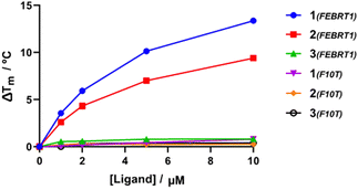
To assess the ligand selectivity for G4 DNA, Förster resonance energy transfer (FRET) melting assays,35 which measure ligand-induced stabilisation of the secondary DNA structure as observed by the change in apparent melting temperature (ΔTm) of the folded species, were conducted at a range of concentrations (1–10 μM) against fluorophore-labelled G4 sequences at 200 nM: polymorphic G4 found in T. brucei (Febr1T-K+, a mixed G4 topology),7 human telomeric G4 in potassium buffer (FhtelT-K+, mixed parallel/hybrid G4)36 and sodium buffer (FhtelT-Na+, antiparallel G4),37 the c-Myc promoter G-quadruplex (FmycT-K+, parallel G4)38 and a hairpin duplex DNA sequence (F10T-K+) (see ESI† for full details). Our results show that 4-Py 1 exhibited higher binding affinity towards G4 sequences when compared to 3-Py 2 and 2-Py 3 (ΔTm for 1 was higher by 4 °C and 12 °C than those for compounds 2 and 3, respectively, at 10 μM, Fig. 2 and Table S2, ESI†), with 3 showing minimal stabilization to all DNA sequences. No clear preference towards stabilization of a specific G4 topology was observed for 1 and 2, but with a notable selectivity with respect to duplex DNA since a negligible stabilisation on the duplex DNA model F10T is seen for all compounds at all concentrations tested.
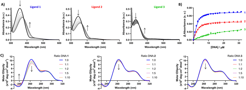
Having established G4 selectivity for our azobenzene ligands, we then further examined the binding affinity and binding mode under physiologically-relevant conditions of ligands 1–3 with the unlabelled polymorphic EBR1 G4 specific to T. brucei in K+ buffer, using a combined approach involving ultraviolet-visible absorbance (UV/Vis) and circular dichroism (CD) spectroscopy titration studies.39 UV/Vis observed binding isotherms were fitted to an independent-and-equivalent-sites binding model, and the binding constants (Ka) and stoichiometries were determined. Results from the titration of EBR1-K+ revealed hypochromicity, and a striking bathochromic shift for 1 and 2 (ca. >15 nm) in comparison with the lower red-shift in the absorbance for 3 (ca. <10 nm) (Fig. 3A). This effect is indicative of end-stacking ligand binding modes, where the energy of the π–π* transition responsible for the Soret band is lowered by the interactions of the ligand chromophores with the G-tetrad.40 The titration with EBR1-K+ yielded Ka values of 0.7 ± 0.05, 0.4 ± 0.04 and 0.02 ± 0.002 × 10⁶ M−1 for 1, 2 and 3, respectively (Fig. 3B), whereby 1 shows ≈2- and 35-fold selectivity over ligands 2 and 3. The observed binding isotherms were successfully fitted to a 2![[thin space (1/6-em)]](https://www.rsc.org/images/entities/char_2009.gif) :
:![[thin space (1/6-em)]](https://www.rsc.org/images/entities/char_2009.gif) 1 binding model, which is also in agreement with the potential end-stacking of the ligand on terminal G-tetrads. 2-Py azobenzene 3, which displayed negligible stabilization of EBR1 on FRET, exhibited only subtle perturbations (Fig. 3B), indicative of a weak interaction. Notably, these observed affinities mirror the trends observed in the thermal melting assay, with 4-Py azobenzene 1 emerging as the most potent G4 ligand of the series.
1 binding model, which is also in agreement with the potential end-stacking of the ligand on terminal G-tetrads. 2-Py azobenzene 3, which displayed negligible stabilization of EBR1 on FRET, exhibited only subtle perturbations (Fig. 3B), indicative of a weak interaction. Notably, these observed affinities mirror the trends observed in the thermal melting assay, with 4-Py azobenzene 1 emerging as the most potent G4 ligand of the series.
These results suggest that the spatial positioning of the pyridinium N is key for the inherent selectivity observed towards four-stranded structures over duplex sequences and it is also crucial for optimal binding with 4-Py 1 and 3-Py 2 exhibiting micromolar G4 affinity, whilst 2-Py displays affinity 1 order of magnitude lower. The lack of G4 stabilization by 3 might be attributed to the shorter distance between the N atoms of both pyridinium rings, which do not facilitate the correct orientation for G4 groove binding.
To further probe the different G4 stabilization modes and potential topology changes induced by 1–3, circular dichroism (CD) experiments were also conducted on EBR1-K+. The CD spectrum of EBR1 is characterized by two positive bands at 260 and 295 nm and a negative band at 240 nm indicative of a predominant parallel G-quadruplex topology.7 Although no conformational change was observed in K+ conditions (Fig. 3C), binding of all ligands with the EBR1 sequence is evidenced by perturbation of the positive (260 nm) and negative (240 nm) bands. The effect is most striking for azobenzene 1, which is consistent with this compound being the more potent of the three pyridinium ligands investigated in the current study. Indeed, 1 induces hypochromicity in the positive band at 260 nm and the negative band at 240 nm. These effects suggest that the ligand induces a disruption of the folded topology, possibly arising from an intercalative binding mechanism at higher concentration. Lesser spectral perturbations were observed for azobenzene 3, corroborating the results from the FRET and UV-Vis assays, where weaker stabilization of G4 was observed over the range of concentrations studied.
Next, we examined the cytotoxicity and antiparasitic activity of ligands 1–3 against T. brucei and L. major strains and MRC5 fibroblast cells as a healthy control (Table 1). Interestingly, 4-Py azobenzene 1 shows submicromolar efficiency against L. major and subnanomolar efficiency against T. brucei. In fact, the antitrypanosomal activity observed within the series follows the same tendency observed in binding to quadruplexes, with 1 being the most efficient, then 2 and finally 3 with the lowest activity. Remarkably, the selectivity index (IC50 MRC-5/IC50T. brucei) was 2285 fold in the range of that obtained for suramin.
| Ligand | MRC5 | L. major | T. brucei | SI MRC5/L. major | SI MRC5/T. brucei |
|---|---|---|---|---|---|
| 1 | 1.6 ± 0.5 | 0.7 ± 0.2 | 0.0007 ± 0.00008 | 2.3 | 2285.7 |
| 2 | >100 | 10.5 ± 1 | 0.37 ± 0.10 | >9.5 | >270 |
| 3 | 30.5 ± 9.8 | 53.7 ± 4.7 | 18.8 ± 0.47 | <1 | 1.6 |
| Suramin | 350 | — | 0.038 ± 0.003 | — | 9210 |
In conclusion, we describe three G-quadruplex ligands based on an azobenzene scaffold featuring methyl pyridinium side chains with 2-, 3- or 4-substitution pattern with regards to the azobenzene core, which varies the overall spatial presentation of the cationic head. Our study reveals that although the structural changes are relatively small, a significant effect is seen on G4 binding affinity as demonstrated by FRET, UV-Vis and CD experiments. We found that 4-Py 1 exhibited higher binding affinity and selectivity towards G4 sequences of mixed topology (e.g. Febr1T-K+ and F21T-K+) when compared to 3-Py 2 and 2-Py 3, with 3, which features the N-methyl group closer to the azobenzene core, showing minimal stabilization of all DNA sequences. These results suggest that the position of the positively charged N in the pyridinium ring is a key driving force for G4 stabilization and selectivity, and should be considered as an important factor when designing or tuning G4 interactive compounds. Furthermore, we were able to correlate G4 binding affinity with antiparasitic activity and found that 4-Py azobenzene 1 exhibited submicromolar efficiency against L. major and subnanomolar efficiency against T. brucei and a superb selectivity index against MRC5 fibroblast cells. Although there is no preference for specific G4 topologies when we compare all the topologies screened, the ligand is very selective towards G4 over duplex DNA. The observed antiparasitic activity and selectivity index may come from a variety of reasons, such as differences in cellular uptake between the parasites and mammalian cells, differences in cell cycle rate (human typical cell cycle is 24 h, whereas the T. brucei divides every 2 h) or differential nucleus entry due to the dissimilar nuclear membrane composition.41 Our study provides insights into key structural features required for G4 binding and target selectivity and paves the way for the development of novel antiparasitic strategies.
MCG thanks ERC-COG: 648239, J. R.-S. thanks MSCA fellowship (project 843720-BioNanoProbes). E. H. thanks EPSRC EP/L015366/1 and J. Y. J. (EPSRC EP/L015366/1 and EP/S026215/1) for their PhD studentships. JCM thanks the Spanish Ministerio de Ciencia, Innovación y Universidades (Grant PID2021-127109OB-I00).
Data availability
The data supporting this article have been included as part of the ESI.† This includes synthetic protocols and characterization data for all compounds, all biophysical characterization using FRET, CD and UV-Vis and details on antiparasitic assays.Conflicts of interest
There are no conflicts to declare.Notes and references
- N. Saranathan and P. Vivekanandan, Trends Microbiol., 2019, 27, 148–163 CrossRef CAS PubMed.
- M. Gellert, M. N. Lipsett and D. R. Davies, Proc. Natl. Acad. Sci. U. S. A., 1962, 48, 2013–2018 CrossRef CAS.
- P. Alberti and J.-L. Mergny, Proc. Natl. Acad. Sci. U. S. A., 2003, 100, 1569–1573 Search PubMed.
- P. Alberti, A. Bourdoncle, B. Saccà, L. Lacroix and J.-L. Mergny, Org. Biomol. Chem., 2006, 4, 3383–3391 RSC.
- S. Neidle, J. Med. Chem., 2016, 59, 5987–6011 CrossRef CAS PubMed.
- E. H. Blackburn and P. B. Challoner, Cell, 1984, 36, 447–457 Search PubMed.
- E. Belmonte-Reche, M. Martínez-García, A. Guédin, M. Zuffo, M. Arévalo-Ruiz, F. Doria, J. Campos-Salinas, M. Maynadier, J. J. López-Rubio, M. Freccero, J.-L. Mergny, J. M. Pérez-Victoria and J. C. Morales, J. Med. Chem., 2018, 61, 1231–1240 CrossRef CAS PubMed.
- J. R. Franco, G. Cecchi, G. Priotto, M. Paone, A. Diarra, L. Grout, R. C. Mattioli and D. Argaw, PLoS Neglected Trop. Dis., 2017, 11, e0005585 Search PubMed.
- A. Luscher, H. P. de Koning and P. Maser, Curr. Pharm. Des., 2007, 13, 555–567 Search PubMed.
- N. Baker, H. P. de Koning, P. Maser and D. Horn, Trends Parasitol., 2013, 29, 110–118 Search PubMed.
- M. C. Field, D. Horn, A. H. Fairlamb, M. A. J. Ferguson, D. W. Gray, K. D. Read, M. De Rycker, L. S. Torrie, P. G. Wyatt, S. Wyllie and I. H. Gilbert, Nat. Rev. Microbiol., 2017, 15, 447 CrossRef CAS.
- J. Ramos-Soriano and M. C. Galan, JACS Au, 2021, 1, 1516–1526 CrossRef CAS PubMed.
- S. Balasubramanian, L. H. Hurley and S. Neidle, Nat. Rev. Drug Discovery, 2011, 10, 261–275 CrossRef CAS PubMed.
- M. Nadai, F. Doria, I. Frasson, R. Perrone, V. Pirota, G. Bergamaschi, M. Freccero and S. N. Richter, ACS Infect. Dis., 2024, 10, 489–499 CrossRef CAS.
- M. Zuffo, A. Stucchi, J. Campos-Salinas, M. Cabello-Donayre, M. Martinez-Garcia, E. Belmonte-Reche, J. M. Perez-Victoria, J. L. Mergny, M. Freccero, J. C. Morales and F. Doria, Eur. J. Med. Chem., 2019, 163, 54–66 Search PubMed.
- L. Monti and M. D. Antonio, ChemBioChem, 2023, 24, e202300265 Search PubMed.
- M. P. O'Hagan, P. Peñalver, R. S. L. Gibson, J. C. Morales and M. C. Galan, Chem. – Eur. J., 2020, 26, 6224–6233 Search PubMed.
- M. Perez-Soto, P. Penalver, S. T. G. Street, D. Weenink, M. P. O'Hagan, J. Ramos-Soriano, Y. J. Jiang, G. J. Hollingworth, M. C. Galan and J. C. Morales, Bioorg. Med. Chem., 2022, 71, 116946 Search PubMed.
- M. Arevalo-Ruiz, F. Doria, E. Belmonte-Reche, A. De Rache, J. Campos-Salinas, R. Lucas, E. Falomir, M. Carda, J. M. Perez-Victoria, J. L. Mergny, M. Freccero and J. C. Morales, Chem. – Eur. J., 2017, 23, 2157–2164 Search PubMed.
- E. Belmonte-Reche, A. Benassi, P. Penalver, A. Cucchiarini, A. Guedin, J. L. Mergny, F. Rosu, V. Gabelica, M. Freccero, F. Doria and J. C. Morales, Eur. J. Med. Chem., 2022, 232, 114183 CrossRef CAS.
- S. T. G. Street, P. Peñalver, M. P. O'Hagan, G. J. Hollingworth, J. C. Morales and M. C. Galan, Chem. Eur. J., 2021, 27, 7712–7721 CrossRef CAS.
- J. Guillon, A. Cohen, C. Boudot, S. Monic, S. Savrimoutou, S. Moreau, S. Albenque-Rubio, C. Lafon-Schmaltz, A. Dassonville-Klimpt, J. L. Mergny, L. Ronga, M. Bernabeu de Maria, J. Lamarche, C. D. Lago, E. Largy, V. Gabelica, S. Moukha, P. Dozolme, P. Agnamey, N. Azas, C. Mullie, B. Courtioux and P. Sonnet, Pathogens, 2022, 11, 1339 Search PubMed.
- J. Guillon, A. Cohen, N. M. Gueddouda, R. N. Das, S. Moreau, L. Ronga, S. Savrimoutou, L. Basmaciyan, A. Monnier, M. Monget, S. Rubio, T. Garnerin, N. Azas, J. L. Mergny, C. Mullie and P. Sonnet, J. Enzyme Inhib. Med. Chem., 2017, 32, 547–563 CrossRef PubMed.
- J. Guillon, A. Cohen, C. Boudot, A. Valle, V. Milano, R. N. Das, A. Guedin, S. Moreau, L. Ronga, S. Savrimoutou, M. Demourgues, E. Reviriego, S. Rubio, S. Ferriez, P. Agnamey, C. Pauc, S. Moukha, P. Dozolme, S. D. Nascimento, P. Laumaille, A. Bouchut, N. Azas, J. L. Mergny, C. Mullie, P. Sonnet and B. Courtioux, J. Enzyme Inhib. Med. Chem., 2020, 35, 432–459 CrossRef PubMed.
- H. M. Craven, G. Nettesheim, P. Cicuta, A. M. Blagborough and C. J. Merrick, Int. J. Parasitol.: Drugs Drug Resist., 2023, 23, 106–119 Search PubMed.
- M. P. O'Hagan, J. Ramos-Soriano, S. Haldar, S. Sheikh, J. C. Morales, A. J. Mulholland and M. C. Galan, Chem. Commun., 2020, 56, 5186–5189 RSC.
- M. Pérez-Soto, J. Ramos-Soriano, P. Peñalver, E. Belmonte-Reche, M. P. O’Hagan, A. Cucchiarini, J. L. Mergny, M. C. Galán, M. C. López-López, M. C. Thomas and J. C. Morales, Eur. J. Med. Chem., 2024, 276, 116641 CrossRef PubMed.
- X. Wang, J. Huang, Y. Zhou, S. Yan, X. Weng, X. Wu, M. Deng and X. Zhou, Angew. Chem., Int. Ed., 2010, 49, 5305–5309 CrossRef PubMed.
- X. Xing, X. Wang, L. Xu, Y. Tai, L. Dai, X. Zheng, W. Mao, X. Xu and X. Zhou, Org. Biomol. Chem., 2011, 9, 6639–6645 RSC.
- T. Tian, Y. Song, J. Wang, B. Fu, Z. He, X. Xu, A. Li, X. Zhou, S. Wang and X. Zhou, J. Am. Chem. Soc., 2016, 138, 955–961 CrossRef PubMed.
- Y. Takebayashi, J. Ramos-Soriano, Y. J. Jiang, J. Samphire, E. Belmonte-Reche, M. P. O’Hagan, C. Gurr, K. J. Heesom, P. A. Lewis, T. Samernate, P. Nonejuie, J. Spencer and M. C. Galan, bioRxiv, 2024, preprint DOI:10.1101/2022.09.01.506212.
- F. A. Jerca, V. V. Jerca and R. Hoogenboom, Nat. Rev. Chem., 2022, 6, 51–69 CrossRef PubMed.
- M. P. O'Hagan, S. Haldar, M. Duchi, T. A. A. Oliver, A. J. Mulholland, J. C. Morales and M. C. Galan, Angew. Chem., Int. Ed., 2019, 58, 4334–4338 CrossRef PubMed.
- M. P. O'Hagan, S. Haldar, J. C. Morales, A. J. Mulholland and M. C. Galan, Chem. Sci., 2021, 12, 1415–1426 RSC.
- A. De Cian, L. Guittat, M. Kaiser, B. Saccà, S. Amrane, A. Bourdoncle, P. Alberti, M.-P. Teulade-Fichou, L. Lacroix and J.-L. Mergny, Methods, 2007, 42, 183–195 CrossRef CAS PubMed.
- K. N. Luu, A. T. Phan, V. Kuryavyi, L. Lacroix and D. J. Patel, J. Am. Chem. Soc., 2006, 128, 9963–9970 CrossRef CAS PubMed.
- Y. Wang and D. J. Patel, Structure, 1993, 1, 263–282 CrossRef CAS PubMed.
- A. Ambrus, D. Chen, J. Dai, R. A. Jones and D. Yang, Biochemistry, 2005, 44, 2048–2058 CrossRef CAS.
- J. M. Nicoludis, S. P. Barrett, J. L. Mergny and L. A. Yatsunyk, Nucleic Acids Res., 2012, 40, 5432–5447 CrossRef CAS.
- A. P. Falanga, A. D'Urso, G. Travagliante, C. M. A. Gangemi, M. Marzano, S. D'Errico, M. Terracciano, F. Greco, L. De Stefano, P. Dardano, I. Rea, G. Piccialli, G. Oliviero and N. Borbone, Int. J. Biol. Macromol., 2024, 268, 131801 CrossRef CAS.
- J. P. Daniels, K. Gull and B. Wickstead, Cell biology of the trypanosome genome, Microbiol. Mol. Biol. Rev., 2010, 74(4), 552–569 CrossRef CAS PubMed.
Footnotes |
| † Electronic supplementary information (ESI) available. See DOI: https://doi.org/10.1039/d4cc03106g |
| ‡ Present address: Instituto de Investigaciones Químicas (IIQ), CSIC – Universidad de Sevilla, Av. Américo Vespucio 49, Seville, 41092, Spain. |
| This journal is © The Royal Society of Chemistry 2024 |


