DOI:
10.1039/C3RA46093B
(Paper)
RSC Adv., 2014,
4, 9012-9020
Silymarin coated gold nanoparticles ameliorates CCl4-induced hepatic injury and cirrhosis through down regulation of hepatic stellate cells and attenuation of Kupffer cells†
Received
24th October 2013
, Accepted 21st January 2014
First published on 21st January 2014
Abstract
Silymarin coated gold nanoparticles with a mean size of 20 nm were synthesized and functionalized in one pot using silymarin as a reducing and stabilizing agent. Conjugation of gold with silymarin was confirmed with FT-IR and UV-visible techniques. The aim of this study was to investigate the hepatoprotective and antifibrotic potential of Silymarin coated gold nanoparticles. For this purpose oxidative liver damage was induced in Wistar rats by intraperitoneal injection of CCl4 dissolved in olive oil (1![[thin space (1/6-em)]](https://www.rsc.org/images/entities/char_2009.gif) :1 v/v, 1 ml kg−1). Silymarin coated gold nanoparticles were administered intragastrically once per day for 14 weeks in a dose of 30 mg kg−1 of body weight. Hepatoprotective and antifibrotic activities of silymarin coated gold nanoparticles were assessed in terms of reduction in serum enzymes (ALT, AST, ALP), through histopathology and immunohistochemistry techniques. It also reduced the CCl4-induced damaged area as well as fibrotic area to 0% as assessed by histopathology. The Alpha SMA and Kupffer cells were also reduced in number around the portal traid area by the silymarin coated gold nanoparticles. These hepatoprotective and antifibrotic effects were better than the positive control silymarin. Our results suggest the therapeutic effect of silymarin coated gold nanoparticles in CCl4-induced liver injury and cirrhosis by promoting extracellular matrix degradation, hepatic stellate cells inactivation with strong enhancement of hepatic regenerative capacity. Silymarin coated gold nanoparticles could be administered for up to 14 weeks without inducing side effects or alterations of the histological structure of kidneys, heart, pancreas and lungs.
:1 v/v, 1 ml kg−1). Silymarin coated gold nanoparticles were administered intragastrically once per day for 14 weeks in a dose of 30 mg kg−1 of body weight. Hepatoprotective and antifibrotic activities of silymarin coated gold nanoparticles were assessed in terms of reduction in serum enzymes (ALT, AST, ALP), through histopathology and immunohistochemistry techniques. It also reduced the CCl4-induced damaged area as well as fibrotic area to 0% as assessed by histopathology. The Alpha SMA and Kupffer cells were also reduced in number around the portal traid area by the silymarin coated gold nanoparticles. These hepatoprotective and antifibrotic effects were better than the positive control silymarin. Our results suggest the therapeutic effect of silymarin coated gold nanoparticles in CCl4-induced liver injury and cirrhosis by promoting extracellular matrix degradation, hepatic stellate cells inactivation with strong enhancement of hepatic regenerative capacity. Silymarin coated gold nanoparticles could be administered for up to 14 weeks without inducing side effects or alterations of the histological structure of kidneys, heart, pancreas and lungs.
1. Introduction
Metal and oxide nanoparticles are currently being considered for a wide array of medical applications, including sensing,1–5 photodynamic therapy,6 drug delivery,7 imaging8 and hyperthermia.9 In most cases, chemically inert nanoparticles are employed to minimize toxicity and side effects. For example, Au nanoparticles are often employed for drug delivery10 and photothermal therapy,11 and Fe oxide nanoparticles are employed for MRI12 and hyperthermia.13 Chemically reactive materials are seldom employed, typically only as biocides for external application. For example, Ag nanoparticles have been employed for millennia as antibacterials and can now be found in commercial detergents14 and in clothing.15 Cu nanoparticles, another well-known biocide, are being employed as antifungals and antibacterials for clothing.16 Several reports have also shown17–21 that the antibacterial activity of several biocides can be amplified by conjugation to Ag nanoparticles. These results are not completely surprising, since the conjugates couple two biocides, one organic (the capping agents) and one inorganic (the Ag nanoparticles). These two biocides can affect different metabolic pathways of the targeted organisms and can thus be more effective than each component taken individually. Recent reports, however, have suggested that nanoparticles of chemically inert materials can also enhance the potency of certain molecules. For example, the group of Kotov showed that leukemia cells were killed more efficiently by conjugation of 6-mercaptopurine to Au nanoparticles.22 Jin and He23 showed that the antibacterial activity of nisin increased when MgO was added to a culture of food borne pathogens such as E. coli, and our own group24 showed that conjugation to Au nanoparticles increased by up to three times the antibacterial, antifungal, insecticidal and cytotoxic activity of the biocide 2–4, dihydroxybenzene carbodithioic acid (DHT). The latter results, they showed that conjugation to Au nanoparticles increased the activity of drugs not only against unicellular organisms but also against more complex organisms such as brine shrimp larvae and insects such as fruit parasites. We therefore wondered if the enhanced activity of Au bioconjugates could be a more general phenomenon, and, specifically, if Au nanoparticle conjugates could improve the therapeutic activity of drugs used to treat complex pathologies. As a model system, we focused on oxidative liver damage. Liver is a convenient target organ since it removes bioconjugates very efficiently from the blood stream.25 By choosing a liver pathology, the conjugates do not need to be protected from liver absorption, which simplifies synthesis and data interpretation. In addition, oxidative liver damage, which ultimately leads to cirrhosis, is easy to induce in animals and it is also a clinically relevant and widespread pathology. Liver cirrhosis is a relatively common disease which in the industrialized world is often associated to excessive alcohol consumption. Cirrhosis, however, can be also caused by the Hepatitis B and C viruses, which are estimated to affect 500 million people worldwide and are especially widespread in developing countries. In Pakistan alone, with a population of about 150 million, data from the last 10 years show a prevalence of 3–4% for hepatitis B26 and 5–7% for hepatitis C.27
Here, we show in vivo that conjugation to Au nanoparticles enhances the therapeutic effect of silymarin, a flavonoid that is being considered for the treatment of several hepatic pathologies. Au–silymarin conjugates induced complete recovery from oxidative liver damage and protected from cirrhosis. Administration of silymarin, our positive control, leads to a partial recovery of hepatic structure and enzymatic levels, while Au nanoparticles had no therapeutic effect. Most importantly, Au–silymarin conjugates could be administered for up to 14 weeks without any noticeable side effects and without inducing any discernible changes in the histological structure of heart, kidney and lungs of the animals.
2. Results and discussion
2.A. Nanoparticle synthesis
Silymarin is a mild reducing agent, and during addition the color of the reaction mixture changed from yellow to violet, indicating reduction of the Au ions and formation of Au nanoparticles. UV-vis spectra (Fig. S1†) of the suspensions revealed an absorption maximum at 520 nm, which is within the range expected for non-coagulated suspensions of Au nanoparticles.28 Conjugation of silymarin to the gold nanoparticles was supported by FTIR spectroscopy,38 reported in Fig. S.2.† Transmission electron microscopy (TEM) showed that the nanoparticles had a narrow size distribution with a mean size of 20 nm, as shown in Fig. S3.†
2.B. Biological studies
Although viral hepatitis is the most common cause of liver injury and inflammation, in this study we used the widely accepted CCl4-induced model because the rats are naturally immune to the hepatitis B and C virus. Carbon tetrachloride (CCl4)-induced liver injury is a widely used and well-characterized animal model of xenobiotic-induced, oxidative stress-mediated hepatotoxicity.29 CCl4 induces the production of several types of reactive oxygen species (ROS) via cytochrome P450. The ensuing liver injury presents symptoms which are similar to those of chronic liver injury in humans.30,31
Acute liver injury.
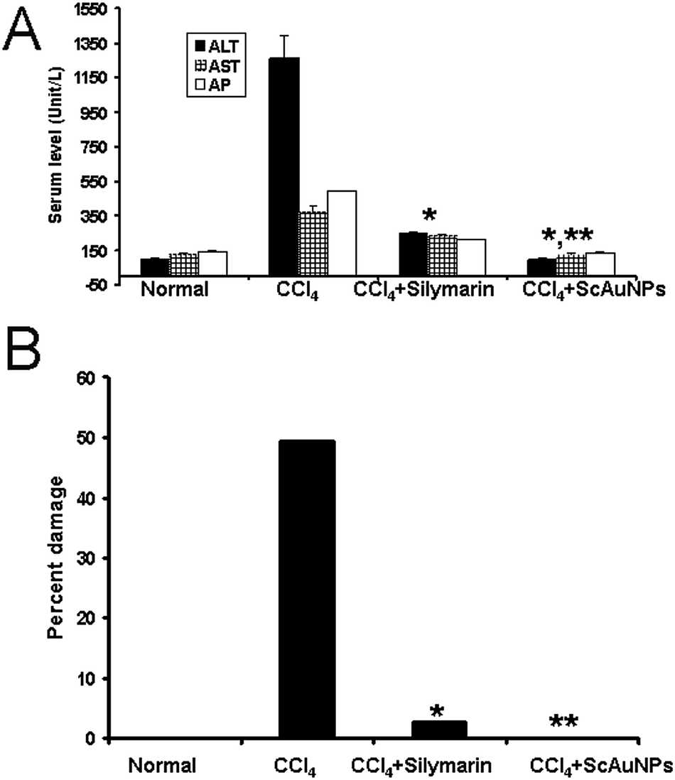
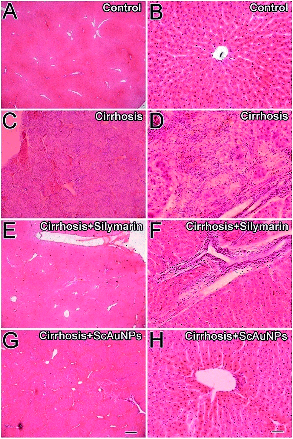
Acute liver injury was induced by intraperitoneal injection of CCl4 as illustrated in the Experimental section. Liver sections from control rats stained with H/E showed normal hepatic architecture (Fig. 1A and B) and normal levels of liver enzymes in the serum (Fig. 2A). The area surrounding the central vein showed hepatic cords and sinusoids typical of liver architecture. Liver sections from the CCl4 model group (Fig. 1C and D) exhibited necrotic areas (indicated by red arrows) which were characterized by a pale eosin staining. The necrotic areas presented necrotic hepatocytes without nucleus. Cells other than the hepatocytes, however, had a normal appearance. In the more heavily stained regions (indicated by green arrow), all cells appeared to be healthy. There was mixed inflammatory infiltrate around the central vein (Fig. 1D). Often, times hepatocytes in the vicinity of hepatic lesions, particularly those situated immediately alongside the lesion border, showed hydropic degeneration. Sinusoidal spaces filled with erythrocytes were also observed. The necrotic area was calculated by the NIS-elements software (Nikon) and the results are reported in Fig. 2B. Enzymatic levels confirmed the histological analysis. Carbon tetrachloride causes acute liver injury through its bioactivation yields carbon trichloride free radicals (CCl3).32 These free radicals cause lipid peroxidation (LP), which produces hepatocellular damage and enhanced production of fibrotic tissue.33 As a result of damage to cell membrane integrity cellular plasma enzymes such as ALT, AST and AP leak out34 raising their blood level. Not surprisingly, enzymatic levels in the CCl4 group were 3 to 7 times higher than in the control group. In the positive control group, which received 200 mg kg−1 body weight of silymarin, hepatic lesions were significantly reduced in extent and less frequent (Fig. 1E and F). However, both the enzyme level (Fig. 2A) and the histopathological analysis (Fig. 2B) indicated ∼3% damage with some mixed inflammatory infiltrate around the central vein area. In contrast, the liver of rats receiving 30 mg kg−1 of ScAuNPs showed normal hepatic architecture with no sign of hepatic injury or inflammation (Fig 1G and H). Enzymatic levels (**, P < 0.001, Fig. 2A) and histopathological analysis (**, P < 0.001, Fig 2B) showed that this protective effect was higher than the positive control silymarin. After the 48 hours of treatment, serum total and direct bilirubin did not show any changes in any of the groups mentioned above.
 |
| | Fig. 1 Histological analysis of CCl4-induced acute liver injury. (A and B) Control group. (C and D) Group injected with CCl4. (E and F) Positive control group (CCl4 and silymarin). (G and H) Group injected with CCl4 and treated with ScAuNPs. Necrotic or injured areas appear as pale regions and are marked by red arrows. Healthy areas are marked by green arrows. Scale bar for A, C, E and G is 250 μm and is shown in G. Scale bar for B, D, F and H is 25 μm and is shown in H. | |
 |
| | Fig. 2 (A) Serum AST, ALT and AP levels as markers of acute liver injury. The ScAuNPs showed complete protection against CCl4-induced liver injury as compared to CCl4 (*, P < 0.001) or CCl4+ silymarin (**, P < 0.001). (B). Histopatological analysis of stained sections, obtained using NIS-elements software (Nikon). | |
Chronic liver injury (cirrhosis).
Liver specimens obtained from control animals did not show any histological abnormalities (Fig. 3A and B). The CCl4-induced liver cirrhosis group was characterized by abnormal hepatocellular degeneration with formation of fibrous tissue infiltrated with inflammatory cells. There was such a vast rearrangement of the hepatic structure that hepatic cords, sinusoids and hepatic lobules were not recognizable. Septal fibrosis and micronodule formation was very prominent (Fig. 3C and D). Liver specimens obtained from the silymarin-treated group (positive control) showed infiltration of inflammatory cells around the portal triad area. However, the parenchymal cells had normal morphological structure (Fig. 3E and F). The liver treated with ScAuNPs showed complete protection from cirrhosis with normal-looking hepatocytes, sinusoids and hepatic lobules (Fig. 3G and H). Treatment with ScAuNPs also completely resolved the inflammatory infiltrate around the portal area as well as fibrous nodular formation.
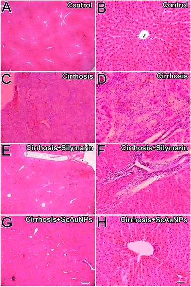 |
| | Fig. 3 Histological analysis of CCl4-induced chronic (14 weeks) liver injury (cirrhosis). (A and B) Control group. (C and D) Group injected with CCl4. (E and F) Positive control group (CCl4 and silymarin). (G and H) CCl4 and ScAuNPs. Scale bar for A, C, E and G is 250 μm and is shown in G. Scale bar for B, D, F and H is 25 μm and is shown in H. | |
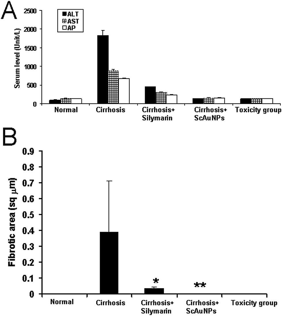
Masson's trichrome stain was used for the detection of fibrosis of liver which is easily identified by a blue coloration. The livers from control rat stained with Masson's trichrome stain showed traces of fibrosis only in the vascular walls (Fig. 4A and B). Liver section from the cirrhosis model group showed extensive multiple nodular fibrosis predominantly in the portal triad areas (Fig. 4C and D). Some fibrotic lesions were still present in the livers of rats treated with 500 mg kg−1 per day of standard silymarin for 14 weeks (Fig. 4E and F), but the fibrosis was reduced and less frequent compared to the cirrhosis model group, whereas pericellular fibrosis was observed in the surrounding liver parenchyma (Fig. 4E and F). The livers of rat treated with ScAuNPs at a dose of 30 mg kg−1 per day for 14 weeks showed only minor, markedly small fibrotic deposits in the periportal areas (Fig. 4G and H). The hepatocytes were looking undamaged with distinct hepatocyte cell morphology and sinusoidal spaces (Fig. 4H). However, both the enzyme level (Fig. 5A) and the calculation of fibrotic area (Fig. 5B) indicated liver damage with ∼3.5% fibrosis with some mixed inflammatory infiltrate around the portal tracts area in the silymarin treated group. In contrast, the liver of rats receiving 30 mg kg−1 of (ScAuNPs) showed normal hepatic architecture with almost no sign of hepatic fibrosis or inflammation (Fig. 4G and H and 5A and B). Both the enzyme level (**, P < 0.001, Fig. 5A) and histopathological analysis of fibrosis (**, P < 0.001, Fig. 5B) showed that this protective effect was more than the positive control silymarin.
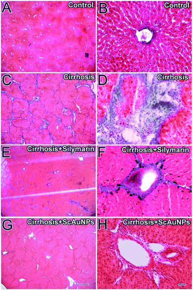 |
| | Fig. 4 Effect of ScAuNPs on CCl4-induced liver cirrhosis (Masson's trichrome staining): animals were sacrificed after treating without (A and B) or with CCL4 (C and D), or with CCl4 and silymarin (E and F) or with CCl4 and ScAuNPs (G and H) for 14 weeks. Note that after 3 months of treatment the liver architecture was changed (C and D) and showed prominent fibrosis (C and D, blue stain). These changes were decreased with silymarin (E and F) and were completely absent with the ScAuNPs (G and H). Scale bar for A, C, E and G is 250 μm and is shown in G. Scale bar for B, D, F and H is 25 μm and is shown in H. | |
 |
| | Fig. 5 Quantification of the effects of ScAuNPs on CCl4-induced liver cirrhosis: (A) serum AST, ALT and AP levels as markers of liver cirrhosis under various conditions. Note that the ScAuNPs showed complete protection against CCl4-induced liver cirrhosis as compared to CCl4 (*, P < 0.001) or CCl4+ silymarin (**, P < 0.001). B) Fibrotic area of the liver as assessed by Masson's Trichrome staining under various conditions. Note the slight fibrotic area (3.5%) in the silymarin group while ScAuNPs showed no fibrosis at all (0%). The toxicity group (treatment with only ScAuNPs also showed no difference in either the enzyme level or percent damage compared to control. | |
To further confirm the enhanced protective action of the Au nanoparticle conjugates, a detailed immunohistochemical characterization of the rats treated for 14 weeks was carried out. Hepatic stellate cells (HSCs) were identified using the α-SMA antibody. In the livers of control animals the distribution of α-SMA-positive fibrogenic cells was restricted to the smooth musculature and to the portal and central veins, while other liver areas remained negative (Fig. 6A–D). Immunohistochemical detection of α-SMA as a marker for hepatic stellate cells (HSCs) activation revealed minimal staining in the blood vessels (Fig. 6A–D). CCl4-induced liver cirrhosis strongly induced perisinusoidal α-SMA expression, which are activated HSCs (Fig. 6E). α-SMA positive cells were located at the scar–parenchyma interface (Fig. 6H). The HSCs were mostly present in the periportal and in the central lobular area along the sinusoidal walls (Fig. 6E and H). Rats treated with Silymarin as a positive control showed marked positive hepatic stellate cells along the side of sinusoidal spaces and in the portal triad areas (Fig. 6I and L). Rats treated with ScAuNPs showed complete reversion of α-SMA positive HSCs (Fig. 6M). The livers of these rats showed a staining pattern similar to the control group's α-SMA positive cells in the vicinity of portal triad (Fig. 6P and D). The liver macrophage cells (Kupffer cells, KCs) were identified using the CD68 antibody. The CD68+ KCs, in the livers of control animals, were present in hepatic sinusoids and were at low number (Fig. 7A and D). Immunohistochemical detection of CD68+ KCs revealed minimal staining in the sinusoid of the normal control group (Fig. 7D). In liver cirrhosis model group, CD68+ KCs with strong staining appeared not only in hepatic sinusoids, but also in portal areas and adjacent to fibrotic septa (Fig. 7E and H). The rats of the positive control group showed marked positive KCs along the side of sinusoidal spaces as well as in the portal triad areas (Fig. 7I and L). Rats treated with ScAuNPs showed CD68+ KCs picture that was very similar to the control (Fig. 7M).
 |
| | Fig. 6 Effect of ScAuNPs on the expression of α-SMA in CCl4-induced liver cirrhosis: animals were sacrificed after treating without (A–D) or with CCl4 (E–H), or with CCl4 and silymarin (I–L) or with CCl4 and ScAuNPs (M–P), and the expression of α-SMA (A, E, I and M) was analyzed by immunohistochemistry. The nucleus was stained with DAPI (B, F, J and N). Note that DAPI is shown in the green channel because of easy visualization when merged with the red channel. DIC pictures of the sections are shown in C, G, K and O. Merges of the α-SMA immunohistochemistry with DAPI and DIC are shown in D, H, L and P. Note the considerable increase in the expression of α-SMA when the animals are cirrhotic (E and H) which is decreased by the treatment of silymarin (I and L) and is similar to control levels by the treatment with ScAuNPs (M and P). Scale bar is 50 μm. | |
 |
| | Fig. 7 Effect of ScAuNPs on the expression of CD68, a macrophage marker, in CCl4-induced liver cirrhosis: animals were sacrificed after treating without (A–D) or with CCl4 (E–H), or with CCl4 and silymarin (I–L) or with CCl4 and ScAuNPs (M–P), and the expression of CD68 (A, E, I and M) was analyzed by immunohistochemistry. The nucleus was stained with DAPI (B, F, J and N). Note that DAPI is shown in the green channel because of easy visualization when merged with the red channel. DIC pictures of the sections are shown in C, G, K and O. Merges of the CD68 immunohistochemistry with DAPI and DIC are shown in D, H, L and P. Note the considerable increase in the expression of CD68 when the animals are cirrhotic (E and H) which is decreased by the treatment of silymarin (I and L) and is similar to control levels by the treatment with ScAuNPs (M and P). Scale bar is 50 μm. | |
The number of α-SMA+ cells in the portal triad area is reported in Fig. 8A. A decreased (yet elevated from the control group) number of α-SMA+ cells were noticed in the positive control group compared to the cirrhosis model group. In contrast, the liver of rats treated with ScAuNPs showed a number of α-SMA+ cells similar to the control group. The number of CD68+ KCs in the portal triad area is reported in Fig. 8B. CD68+ KCs revealed an increase in their number in the cirrhosis group compared to the control. In the positive control group the number of CD68+ KCs decreased compared to the cirrhosis group but were still higher than the control group. In contrast, the liver of rats treated with ScAuNPs showed a number of CD68+ KCs which was lower than the positive control and quite comparable to the positive control group.
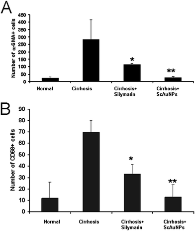 |
| | Fig. 8 Quantification of the α-SMA+ and CD68+ cells after ScAuNPs treatment on CCl4-induced liver cirrhosis: (A) quantification of the number of α-SMA+ cells under various conditions. CCl4-induced liver cirrhosis showed increase in the number of α-SMA+ cells as compared to control in the portal tract region. Silymarin treatment somewhat reduced this number while ScAuNPs completely reduced this number to control levels. (B) Quantification of the number of CD68+ cells under various conditions. CCl4-induced liver cirrhosis showed increase in the number of CD68+ cells as compared to control in the portal tract region. Silymarin treatment somewhat reduced this number while ScAuNPs completely reduced this number to control levels. | |
Most important, treatment with ScAuNPs showed no toxicity in a 14 weeks study. No symptoms of toxicity such as weight loss, piloerection, immobility etc. were observed. A post-mortem study was performed which showed no sign of damage to liver, kidney, heart, pancreas, stomach, intestine, lungs spleen etc. Histopathological analysis was performed of liver, kidney, heart, and pancreas tissue which all showed a normal histological picture (Fig. 9).
 |
| | Fig. 9 Histopatological analysis of organs of animals injected for 14 weeks with Au–silymarin conjugates. Liver (A), kidney (B), heart (C) and pancreas (D). No abnormalities were observed in these tissues. | |
Thus, our results show that ScAuNPs have a superior hepatoprotective activity than pure silymarin. Now, one of the main issues of silymarin is its poor solubility, which leads to a bioavailability of between 20 and 50% of a dose administered orally.35 The solubility of ScAuNPs, on the other hand, is very high. Our conjugates could be dissolved in water at concentrations as high as 200 mg ml−1. For this reason, rats were fed with different amounts of drugs. The positive control group was given 200 mg kg−1 of pure silymarin in the acute toxicity experiments and 500 mg kg−1 in the chronic liver injury experiments. This corresponded to an effective dose of 40 mg kg−1 for the worst-case scenario of 20% bioavailability (100 mg kg−1 for the chronic study) and of 100 mg kg−1 in the best-case scenario of 50% bioavailability (250 mg kg−1 for the chronic study). The group treated with ScAuNPs was given a dose of 30 mg kg−1 in all studies. Given the high solubility of ScAuNPs, we can assume that the full amount will become bioavailable. However, one must remember that ScAuNPs are, for the most part, Au, and that silymarin represents only a minor weight fraction of the conjugates. In recent experiments,24 for example, we determined that carbothiamate surfactants represented only about 7% of the weight of 20 nm diameter Au nanoparticle conjugates. Kotov et al.22 synthesized Au-mercaptopurine conjugates with a nanoparticle diameter of about 4 nm and a 50% weight of organics. The %weight of the organics scales with the surface-to-volume ratio, which in turn depends on the radius of the nanoparticles. For our ∼10 nm nanoparticles we therefore estimate a silymarin content between 15 and 20 wt%. Thus, the groups treated with ScAuNPs received an effective silymarin dose between 4.5 and 6 mg kg−1, which is about an order of magnitude lower than the silymarin received by the positive control group. We conclude that the superior protective effect of ScAuNPs is not due to solubility but, rather, to conjugation. Our conclusion is corroborated by recent work by Parveen et al.36,37 In this work, silymarin solubility and bioavailability were increased 4 to 6 times by a nanoemulsification technique. Liver damage was induced chemically in rats, and nanoemulsions were found to improve the protective action of silymarin. However, the increased solubility did not lead to complete recovery of the hepatic functions. Histological analysis showed that the liver of rats treated with nanoemulsified silymarin exhibited an abnormal number of fat vacuoles. Enzymatic levels were also improved when compared to the untreated group, but they were higher than the control group and comparable to the levels of the positive control group. In our experiments, instead, histology and enzymatic levels of the group treated with ScAuNPs were undistinguishable from the control group of healthy animals.
3. Conclusion
In conclusion, we have shown that conjugates of silymarin and Au nanoparticles are more effective than the positive control silymarin in preventing and repairing acute and chronic liver damage. Animals treated with the Au conjugates showed a normal liver structure and hepatic functions. In contrast, animals treated with the positive control silymarin showed residual damage and inflammation and a slightly abnormal liver function. The nanoparticles conjugates did not show any toxic effects or alterations of the liver enzymatic levels even after 14 weeks of treatment.
4. Experimental details
4.A. Materials synthesis
Materials and instruments.
Tetrachloroauric(III) acid trihydrate (HAuCl4) and NaOH were purchased from TCI. Silymarin was purchased from MP biomedicals, France. UV-vis spectra were recorded with a Shimadzu UV-240, Hitachi U-3200 spectrometer with a path length of 1 cm. IR spectra were recorded using a IR spectrophotometer (IR-460 Shimadzu) by preparing a pellet of a 1:1 mixture of AuNPs (after centrifugation) and KBr. Transmission electron spectroscopy (TEM) was carried out with a Zeiss Libra operated at 120 keV.
Synthesis of silymarin (SM) stabilized gold nanoparticles.
Since silymarin has low solubility in water, an alkaline solution of silymarin was prepared in 0.1 M sodium hydroxide (NaOH). 1 mL of this silymarin solution (1 mM) was then added drop wise to an aqueous solution of tetrachloroauric(III) acid (5 mL, 1 mM). The resulting mixture was stirred for four hours at room temperature. Silymarin-stabilized gold nanoparticles were collected in the form of precipitate after centrifugation.
4.B. Biology experiments
Animals.
Male wistar rats weighing 190 to 200 g were housed in individual cages kept at 22 to 26 °C under 12 hours light/dark cycles, with free access to standard laboratory chow and tap water ad libitum. All procedures were conducted according to the guidelines for the care and use of laboratory animals approved by our Institution ethical committee.
Chemicals and antibodies.
Serum amino–transferases activity assay kits were purchased from Roche Diagnostic GmbH, Mannheim, Germany. Carbon tetrachloride (CCl4) was obtained from Sigma Chemical Co., St Louis, MO, USA. Silymarin purchased from MP Biomedicals Rue Geiler de Kaysersberg lllkirch Cedex 67402, France Monoclonal mouse anti-α-SMA antibody and monoclonal mouse anti-CD68 antibodies were purchased from Sigma, Saint Louis, USA and abcam®, Kendall squre Suite B2304 Cambridge, MA 02139-1517 USA respectively. All other chemicals were obtained from local commercial suppliers.
Animal models of hepatitis.
Although viral hepatitis is the most common cause of liver injury and inflammation, in this study we used the widely accepted CCl4-induced model because the rats are naturally immune to the hepatitis B and C virus. Carbon tetrachloride (CCl4)-induced liver injury is a widely used and well-characterized animal model of xenobiotic-induced, oxidative stress-mediated hepatotoxicity.29 The symptoms of CCl4-induced chronic liver injury are similar to those of chronic liver injury in humans.30 CCl4 induces the production of several types of reactive oxygen species (ROS) via cytochrome P450, thereby causing liver injury.31
Acute liver injury model.
Male wistar rats were divided into four groups consisting of six animals in each group. Group I served as normal control received vehicle, 1 ml kg−1 body weight for 5 days. Group II acute hepatitis group received (CCl4), (1 ml kg−1 body weight, i.p.) in a volume of olive oil equivalent to 1:1 v/v. Group III was treated with the standard drug silymarin (200 mg/kg body weight, p.o.) daily for 3 days, and also received CCl4–olive oil (1:1 v/v, 1 ml kg−1 body weight, i.p.) on 3rd day 1 hour after administration of silymarin. Groups IV (treated group) were administered orally a dose of 30 mg kg−1 body weight of gold nano-particles (p.o.), for 3 days in addition received CCl4–olive oil (1:1 v/v, 1 ml kg−1 body weight, i.p.) on 3rd day 1 hour after intraperitoneal injection of CCl4. After 48 hours of treatment the animals were sacrificed, blood was collected and allowed to clot, and serum was separated for assessment of biochemical parameters.
Chronic liver injury (Cirrhosis) model.
Male Wistar rats (200 g body weight) were obtained from the Animal House of ICCBS, University of Karachi. Animals were randomly divided into four groups (n = 6) according to experimental plan. Group I (Normal control group): rats received vehicle only for 14 weeks at a dose of 1 ml kg−1 body weight daily. Group II (Cirrhosis model group): rats received 1 ml kg−1 body weight of carbon tetrachloride in olive oil at a volume of 1:1 v/v, i.p. twice a week for 14 weeks. Group III (Silymarin treated group) was treated for 14 weeks with the standard drug silymarin (500 mg/kg per day body weight, i.g.) after 3 days of CCl4–olive oil (1:1 v/v, 1 ml kg−1 body weight, i.p) single injection. Group IV (silymarin–Au nanoparticle conjugates treated group) received a single intraperitoneal injection of carbon tetrachloride (1:1 v/v, 1 ml kg−1 body weight, i.p. twice a week) before 3 days followed by daily treatment with silymarin–Au nanoparticles at a dose of (30 mg kg−1 per day, i.g) for 14 weeks. After 3 days of the final injection of CCl4, rats were anesthetized with sodium pentothal. Animals were starved overnight before sacrifice. The serum was stored at −26 °C after separation until assayed. The liver and kidney were carefully dissected and cleaned of extraneous tissue, and part of the liver tissue was immediately transferred into 10% formalin for histopathological investigation.
Liver function tests.
For the acute liver injury, after 48 hours of treatment with CCl4 the animals were sacrificed, blood was collected and allowed to clot, and serum was separated for assessment of biochemical parameters. Serum levels of alanine aminotranferase (ALT), aspartate transaminase (AST), and alkaline phosphatase (AP) were measured by standard techniques using the Reflotron® plus dry chemistry analyzer (Roche Diagnostic). Similarly, for liver cirrhosis models AST, ALT and AP were measured after 14 weeks of experiments.
Histological techniques.
Liver samples from all animals groups were processed for 6 μm thick paraffin sections and stained with hematoxylin and eosin (H/E) using standard procedures. Briefly, liver tissues were fixed in 4% neutral buffered formalin and embedded in paraffin after dehydration. For H&E staining, liver sections were deparaffinized in xylene followed by rehydration and washed in water for 15 minutes and stained with H&E. Liver sections were visualized using a Nikon 90i microscope (Nikon) and the images were acquired with a Nikon DXM 1200C camera using NIS-Elements image analysis software AR 3.0 (Nikon). Image analysis was performed with NIS-Elements and Adobe photoshop software.
Masson's trichrome staining.
For identification of collagen fibers Masson's trichrome staining was used. First liver sections were deparaffinized in xylene, rehydrated followed by using staining techniques according to manufacturer instructions (Carl Roth, Germany). Liver sections were then analyzed using Nikon 90i microscope.
Immunohistochemistry.
For immunohistochemistry, the liver sections were deparaffinised, rehydrated, washed in water for 15 minutes. Each section was incubated with a blocking solution (Roti-immunoblock, Carl Roth, Karlsruhe, Germany) for 20 min at room temperature. Then, the sections were incubated with a primary antibody (1:100) for 1 hour at 37 °C. For hepatic stellate cells (HSCs) monoclonal α-SMA antibody (clone 1A4, Sigma) and for liver macrophages monoclonal CD68 (clone ED1, abcam) antibody was used. After thoroughly washing with PBS, the sections were incubated with a mixture of Texas Red-conjugated goat anti-mouse IgG (1:100) for 40 min. After washing extensively with PBS the nuclei were stained with DAPI (1:10000 fold dilution of a 1 mg ml−1 stock solution) for 1 min, washed with PBS and mounted in mounting media. The fluorescent images were acquired and analyzed using a Nikon 90i multichannel fluorescence microscope and Nikon DXM 1200C camera with NIS-Elements image analysis software AR 3.0 (Nikon). Finally, image processing was performed with Adobe Photoshop software.
References
- J. H. W. Leuvering, P. J. H. M. Thal, D. D. White and A. H. W. M. Schuurs, J. Immunol. Methods, 1983, 62, 163–174 CrossRef.
- T. Gribnau, J. Leuvering and H. Van Hell, J. Chromatogr. B: Biomed. Sci. Appl., 1986, 376, 175–189 CrossRef.
- P. Englebienne, A. V. Hoonacker and M. Verhas, J. Spectrosc., 2003, 17, 255–273 CrossRef PubMed.
- R. Elghanian, J. J. Storhoff, R. C. Mucic, R. L. Letsinger and C. A. Mirkin, Science, 1997, 277, 1078–1081 CrossRef.
- V. Bogatyrev, L. Dykman, Y. M. Krasnov, V. Plotnikov and N. Khlebtsov, Colloid J., 2002, 64, 671–680 CrossRef.
- F. Lu, T. L. Doane, J.-J. Zhu and C. Burda, Inorg. Chim. Acta, 2012 DOI:10.1016/j.ica.2012.05.038.
- P. Ghosh, G. Han, M. De, C. K. Kim and V. M. Rotello, Adv. Drug Delivery Rev., 2008, 60, 1307–1315 CrossRef PubMed.
- P. K. Jain, K. S. Lee, I. H. El-Sayed and M. A. El-Sayed, J. Phys. Chem. B, 2006, 110, 7238–7248 CrossRef PubMed.
- P. Diagaradjane, A. Shetty, J. C. Wang, A. M. Elliott, J. Schwartz, S. Shentu, H. C. Park, A. Deorukhkar, R. J. Stafford and S. H. Cho, Nano Lett., 2008, 8, 1492–1500 CrossRef PubMed.
- G. Han, P. Ghosh and V. M. Rotello, Nanomedicine, 2007, 2, 113–123 CrossRef PubMed.
- X. Huang, P. K. Jain, I. H. El-Sayed and M. A. El-Sayed, Lasers Med. Sci., 2008, 23, 217–228 CrossRef PubMed.
- B. Chertok, B. A. Moffat, A. E. David, F. Yu, C. Bergemann, B. D. Ross and V. C. Yang, Biomaterials, 2008, 29, 487–496 CrossRef PubMed.
- F. Sonvico, S. Mornet, S. Vasseur, C. Dubernet, D. Jaillard, J. Degrouard, J. Hoebeke, E. Duguet, P. Colombo and P. Couvreur, Bioconjugate Chem., 2005, 16, 1181–1188 CrossRef PubMed.
- C. A. Impellitteri, T. M. Tolaymat and K. G. Scheckel, J. Environ. Qual., 2009, 38, 1528–1530 CrossRef PubMed.
- T. M. Benn and P. Westerhoff, Environ. Sci. Technol., 2008, 42, 4133–4139 CrossRef.
-
http://www.cupron.com/
.
- A. Panáček, L. Kvitek, R. Prucek, M. Kolar, R. Vecerova, N. Pizurova, V. K. Sharma, T. j. Nevečná and R. Zboril, J. Phys. Chem. B, 2006, 110, 16248–16253 CrossRef PubMed.
- C. Baker, A. Pradhan, L. Pakstis, D. J. Pochan and S. I. Shah, J. Nanosci. Nanotechnol., 2005, 5, 244–249 CrossRef PubMed.
- H. Kong and J. Jang, Langmuir, 2008, 24, 2051–2056 CrossRef PubMed.
- M. Singh, S. Singh, S. Prasad and I. Gambhir, Digest J. Nanomater. Biostructures, 2008, 3, 115–122 Search PubMed.
- X. Xu, Q. Yang, Y. Wang, H. Yu, X. Chen and X. Jing, Eur. Polym. J., 2006, 42, 2081–2087 CrossRef PubMed.
- P. Podsiadlo, V. A. Sinani, J. H. Bahng, N. W. S. Kam, J. Lee and N. A. Kotov, Langmuir, 2008, 24, 568–574 CrossRef PubMed.
- T. Jin and Y. He, J. Nanopart. Res., 2011, 13, 6877–6885 CrossRef.
- S. S. Naz, N. U. Islam, M. R. Shah, S. S. Alam, Z. Iqbal, M. Bertino, L. Franzel and A. Ahmed, J. Nanobiotechnol., 2013, 11, 13 CrossRef PubMed.
- S. Stolnik, L. Illum and S. Davis, Adv. Drug Delivery Rev., 1995, 16, 195–214 CrossRef.
- F. André, Vaccine, 2000, 18, S20–S22 CrossRef.
- S. J. Zuberi, International Hepatology Communications, 1996, 5, 19–26 CrossRef.
-
U. Kreibig and M. Vollmer, Optical properties of metal clusters, Springer Verlag, 1995 Search PubMed.
- R. O. Rechnagel, E. A. Glende and G. L. Plaa, Crit. Rev. Toxicol., 1973, 2, 263–297 CrossRef PubMed.
- S. Basu, Toxicology, 2003, 189, 113–127 CrossRef.
- L. W. Weber, M. Boll and A. Stampfl, Crit. Rev. Toxicol., 2003, 33, 105–136 CrossRef PubMed.
- G. Poli, Mol. Aspects Med., 2000, 21, 49–98 CrossRef.
-
H. Tsukamoto, M. Matsuoka and S. French, Experimental models of hepatic fibrosis: a review, 1990 Search PubMed.
- G. K. Marathe, K. A. Harrison, L. J. Roberts, J. D. Morrow, R. C. Murphy, L. W. Tjoelker, S. M. Prescott, G. A. Zimmerman and T. M. McIntyre, J. Lipid Res., 2001, 42, 587–596 Search PubMed.
- S. Javed, K. Kohli and M. Ali, Alternative Med. Rev., 2011, 16, 239–249 Search PubMed.
- R. Parveen, S. Baboota, J. Ali, A. Ahuja, S. S. Vasudev and S. Ahmad, Int. J. Pharm., 2011, 413, 245–253 CrossRef PubMed.
- R. Parveen, S. Baboota, J. Ali, A. Ahuja, S. S. Vasudev and S. Ahmad, Arch. Pharmacal Res., 2011, 34, 767–774 CrossRef PubMed.
- N. A. Begum, S. Mondal, S. Basu, R. A. Laskar and D. Mandal, Colloids Surf., B, 2009, 71, 113–118 CrossRef PubMed.
Footnote |
| † Electronic supplementary information (ESI) available. See DOI: 10.1039/c3ra46093b |
|
| This journal is © The Royal Society of Chemistry 2014 |
Click here to see how this site uses Cookies. View our privacy policy here.