Additive-free graphene-based inks for 3D printing functional conductive aerogels†
Elnaz
Erfanian
ab,
Milad
Goodarzi
c,
Gabriel
Banvillet
b,
Farbod
Sharif
a,
Mohammad
Arjmand
 c,
Orlando J.
Rojas
 bde,
Milad
Kamkar
 *f and
Uttandaraman
Sundararaj
*a
aDepartment of Chemical and Petroleum Engineering, University of Calgary, Calgary, AB T2N 1N4, Canada. E-mail: u.sundararaj@ucalgary.ca
bDepartment of Chemical and Biological Engineering, Bioproducts Institute, University of British Columbia, Vancouver, BC V6T 1Z4, Canada
cNanomaterials and Polymer Nanocomposites Laboratory, School of Engineering, University of British Columbia, Kelowna, BC V1V 1V7, Canada
dDepartment of Chemistry, Department of Wood Science, University of British Columbia, Vancouver, BC V6T 1Z4, Canada
eDepartment of Bioproducts and Biosystems, School of Chemical Engineering, Aalto University, P. O. Box 16300, FIN-00076, Espoo, Finland
fDepartment of Chemical Engineering, The Waterloo Institute for Nanotechnology University of Waterloo, 200 University Avenue West Waterloo, ON N2L 3G1, Canada. E-mail: milad.kamkar@uwaterloo.ca
First published on 18th September 2024
Abstract
This study demonstrates an all-graphene, additive-free, aqueous-based ink for direct ink writing (DIW) to 3D-print functional aerogels for applications in electronics and electromagnetic interference (EMI) shields. We employ a two-step electrochemical method with a specially designed intercalation step that controls the surface functionality of graphene nanosheets. Comprehensive characterization reveals the significant impact of the physicochemical properties of graphene nanosheets on homogeneity, rheology, electrical conductivity, and EMI shielding effectiveness (SE). A critical observation is that rheology alone is insufficient to predict the printability of two-dimensional particulate systems, while ink homogeneity, dictated by inter-sheet interactions, plays a vital role. By focusing on optimizing intercalation conditions, we find that phosphoric acid treatment is most effective in enhancing both printability and conductivity, achieving an electrical conductivity of 158 S cm−1 and an EMI SE of 50 dB (at 50 μm thickness) without requiring any post-processing reduction. Systematic experiments with varying durations of phosphoric acid intercalation establish that a 10-minutes treatment produces inks with superior 3D printing fidelity. This innovative approach to graphene ink production enables rapid, continuous, and large-scale manufacturing of lightweight, porous materials, avoiding the need for environmentally harmful reductant chemistries or high-temperature processing. Furthermore, eliminating the reduction step in the fabrication process aligns with industrial demands for energy-efficient production processes and high output rates, marking a significant advancement in the field of materials science and offering promising prospects for applying graphene-based inks in advanced manufacturing technologies.
1. Introduction
The potential of graphite and its derivatives, such as graphene, has been widely acknowledged for its remarkable properties and wide-scale applicability in various domains, from advanced electronics to biomedical applications.1,2 It holds the potential to significantly impact our everyday lives if it can be processed as readily as polymers and metals, which can be shaped by well-established thermochemical transformations.3 Hence, the emergence of next-generation transformation techniques using water and environmentally friendly solvents as primary agents for facilitating flowability becomes highly relevant for processing and shaping graphitic structures with complex architectures.4 This contrasts with traditional processing methods reliant on heat, signifying a shift towards more sustainable practices.Traditional graphite forms consist of powder, flake, or foil. Indeed, these conventional forms of synthetic and natural graphite have restricted the full application potential of graphitic structures. Thus, in the evolving landscape of materials science, the transition from traditional graphite forms to intricate three-dimensional (3D) graphitic structures marks a significant milestone.5,6 Recent advancements in additive manufacturing, particularly direct ink writing (DIW), have significantly improved the processing and shaping of nanomaterials in their hydrogel state.5,7 However, this field still faces substantial challenges in handling graphitic structures, especially graphene.8 The hydrophobic nature of graphene nanosheets and the strong van der Waals forces among them hinder their dispersibility in aqueous media, which is otherwise preferred for its cost-effectiveness and minimal environmental impact.9,10 As a result, much of the current research focuses on graphene oxide (GO) for its enhanced water dispersibility due to its high polarity.
Nevertheless, common GO production methods, such as the Hummers' method, involve hazardous oxidants in acidic conditions, posing environmental and safety concerns and risking the introduction of impurities.11–14 These processes also compromise the electrical properties of GO due to the high density of oxygen-containing functional groups, necessitating further post-processing like chemical, thermal, and microwave reduction. This further hinders the scalable production of graphene-based printed electronics, as they are time-, cost- and energy-intensive and introduce contaminants from reducing agents.15–17 For instance, GO-based binder-free inks have been successfully formulated.18 However, thermal reduction at 1000 °C in an argon-filled tube furnace was required for their utilization as Li–O2 cathodes.
The second challenge of fabrication of graphene-based inks lies in their poor rheological properties, e.g., low storage modulus and relatively high loss modulus (liquid-like behavior), making them unsuitable for direct printing.3,19,20 To enable a smooth flow under pressure, the inks' rheological properties must fulfill printability criteria, such as shear-thinning behavior. This should be conjugated with suitable viscoelasticity to maintain the shape after printing and filament deposition on the bed.21 To this end, approaches like incorporating additives, such as surfactants, binders, and crosslinkers, to fine-tune the rheological features have been suggested to enable desired flow behavior and printability.3,20 In our recent work,22 we implemented the electrochemical method to produce an ink featuring superior electrical conductivity. Although the pure graphene-based ink lacked colloidal stability and thus was not printable, integrating it with TEMPO-oxidized cellulose nanofibrils improved its colloidal dispersion and rheological properties, paving the way for high-resolution 3D printing via DIW technique. However, these additives significantly compromised the final properties of the products, e.g., reducing the electrical conductivity and increasing the density of the resulting 3D-printed structures.
Additionally, incorporating additives might introduce complex post-processing steps to remove these additives. For instance, Zhu et al.23 employed silica as a rheological modifier to adjust the necessary rheological properties of GO-based ink for 3D printing. However, this necessitated the removal of silica through chemical etching with hydrofluoric acid. Such aggressive removal methods can potentially compromise the physiochemical properties of the printed structure. Tran et al.24 resolved this issue using a conductive additive. They developed a 3D printable, aqueous, electrochemically synthesized graphene-based ink dispersed within a recrystallized PEDOT:PSS nanofibrils network. Despite the high conductivity of all components, achieving superior printability necessitated careful engineering of the interfacial interactions between exfoliated graphene flakes and PEDOT:PSS nanofibrils, along with controlling the concentration of the entangled nanofibrillar network to attain desirable rheological properties. Furthermore, multicomponent inks are highly susceptible to inhomogeneity. Consequently, achieving pure graphene-based dispersions with proper rheological and colloidal behavior toward high-fidelity 3D printing is still a considerable challenge.
In our study, we address these critical challenges in the field of materials science by creating additive-free, highly conductive all-graphene aqueous inks. Our goal is to harmonize conductivity with processability without relying on external additives, achieved through meticulous engineering of the nanoscale chemistry and microscale assembly of graphene nanosheets. This was accomplished using a novel two-step electrochemical synthesis method, which allows precise control over the surface functionality of the nanosheets, leading to the formulation of inks that exhibit exceptional 3D printing fidelity at room temperature. Consequently, our efforts have facilitated the creation of 3D-printed, additive-free, and highly conductive graphitic aerogels. This achievement marks a substantial broadening of the application spectrum for graphitic structures, allowing for the seamless integration of graphene-based 3D structures with meticulously designed configurations and controlled fabrication processes. This advancement underscores the importance of addressing the challenges in balancing intrinsic properties with new forms in material science.
2. Results and discussion
2.1. Electrochemically synthesized graphene nanosheets
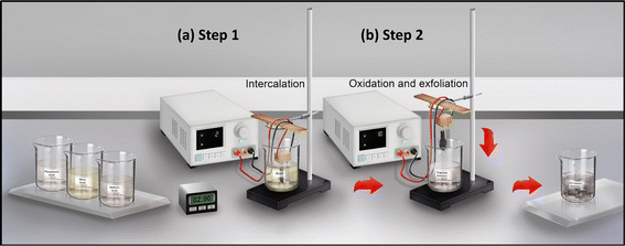
Electrochemically synthesized graphene nanosheets (ECGs) were derived from graphite foil employing a two-step electrochemical technique, which encompasses: (1) intercalation; and (2) oxidation followed by exfoliation, as depicted in Fig. 1. An outlier in this process involved the synthesis of one sample through a conventional, single-step electrochemical (EC) method, designated as El, for reference purposes.During the intercalation phase, graphite intercalated compounds (GICs) were generated by incorporating various acids over fixed durations. For the exfoliation phase, uniform conditions were applied across all samples. Table 1 compiles these conditions alongside the corresponding ECG labels, which are designed to reflect both the type of acid used and the duration of intercalation.
| Step 1 electrolyte | Name | Intercalation voltage (V) | Measured intercalation current Im (A) | Exfoliation voltage (V) | Measured exfoliation current Im (A) | Step 2 electrolyte | Area of graphite (cm2) | Synthesis yield [%] |
|---|---|---|---|---|---|---|---|---|
| Nitric acid | N-2 min | 2 | 1.5 | 10 | 1.8 | (NH4)2SO4 | 1 × 5 | 30 |
| Sulfuric acid | S-2 min | 2 | 0.3 | 10 | 0.96 | (NH4)2SO4 | 1 × 5 | 65 |
| Phosphoric acid | P-2 min | 2 | 0.02 | 10 | 0.75 | (NH4)2SO4 | 1 × 5 | 85 |
| (NH4)2SO4 | El | — | — | 10 | 0.75 | — | 1 × 5 | 90 |
In Step I, the acids act as intercalating agents, providing ions that facilitate the flow of electrical current through the solution and the graphite layers.25 Among the acids used, nitric acid, the strongest, exhibits the highest intercalation current (1.5 A), while phosphoric acid shows the lowest current (0.02 A). Upon reaching the appropriate potential, intercalating agents from different acids, such as HSO4−/SO42− ions in the sulfuric acid,26–28 NO3− ions in the nitric acid,29 and H2PO4− ions in the phosphoric acid30,31 are intercalated into the graphene layers.32 The GIC phase was named based on the acid type, namely, H2SO4-GIC, HNO3-GIC, and H3PO4-GIC for sulfuric, nitric, and phosphoric acid intercalation, respectively. During intercalation, a reduction of the acid molecules occurred at the graphite surface. These reduced acid molecules (protons and anions) then intercalated between the graphene layers in the graphite, weakening the van der Waals forces and disintegrating the graphene layers.33 After intercalation (Step I), the fabricated GIC was subjected to a constant electrical potential in Step II, inducing the exfoliation of GIC into graphene sheets. This process is facilitated by producing gas (usually oxygen, and others) at the electrode surface, generating mechanical stress that forces the graphite layers apart.34 Furthermore, in Step II, water molecules co-intercalated into the interlayer spaces previously occupied by ions. These water molecules react with sp2-bonded carbon atoms, forming C–OH and alkoxy groups on the graphene surface. Subsequently, alkoxy groups partially transform into carbonyl groups during oxidation.34 After exfoliation, the graphene yield was determined by comparing the mass difference between the disintegrated/non-oxidized graphite from Step I (collected at the bottom of the beaker), the unexfoliated graphite residue (which settles during the process and is separated by centrifugation), and the initial mass of the graphite foil used at the start, which was in contact with the electrolyte. According to Table 1, the synthesis yield was the highest (85%) for the P-2 min process, whereas the N-2 min process yielded the lowest value (30%) among all samples. The strong nitric acid acidity and the electrolyte's higher electrical properties in the N-2 min sample lead to significant graphite loss in the intercalation step, owing to its direct penetration, oxidation, and breaking (unzipping) of the graphitic layers. In contrast, the phosphoric acid electrolyte left minimal residues in the first step, thereby facilitating its reuse and enhancing the scalability of the process with limited chemical consumption. Table 1 presents comprehensive data on yield, intercalation, and exfoliation of ECGs.
2.2. Chemical and physical characterization of synthesized graphene
Fig. 2(a) displays the X-ray diffraction (XRD) spectra of the synthesized ECGs and graphite. The graphite shows a sharp peak at 2θ = 26.48°, originating from the (002) planes of its crystalline structure.35 This peak's high intensity and sharpness indicate the high purity of graphite.35 The one-step EC synthesis procedure leads to a broad and more asymmetric peak in the El sample, signifying the formation of high-quality graphene.36 Incorporating an acid intercalation step into the procedure significantly impacted the XRD spectrum, with variations observed for different acids. Intercalation with phosphoric acid did not introduce any changes to the XRD spectrum compared to the El sample, likely due to the lower acidic strength of phosphoric acid, resulting in less oxidation. Among the ECGs, the S-2 min sample exhibited moderate features, with a low-intensity peak at 2θ = 13.2° corresponding to the (002) layers in the GO crystalline phase. A similar trend was observed in the N-2 min sample, except that the peak of the GO phase was sharper and more intense, and the peak for graphite was weakened. To calculate the difference in interlayer spacing before and after intercalation, we utilized Bragg's Law, which allowed us to calculate the spacing between ECG planes using the diffracted X-rays angles. Detailed calculations and results, demonstrating significant increases in the interlayer spacing due to intercalation, are provided in the ESI.† These results indicate that intercalation significantly increases the interlayer spacing of graphene, suggesting successful insertion of ions between the graphene layers. The differences in spacing also imply variations in the degree of intercalation depending on the used acid in the first step.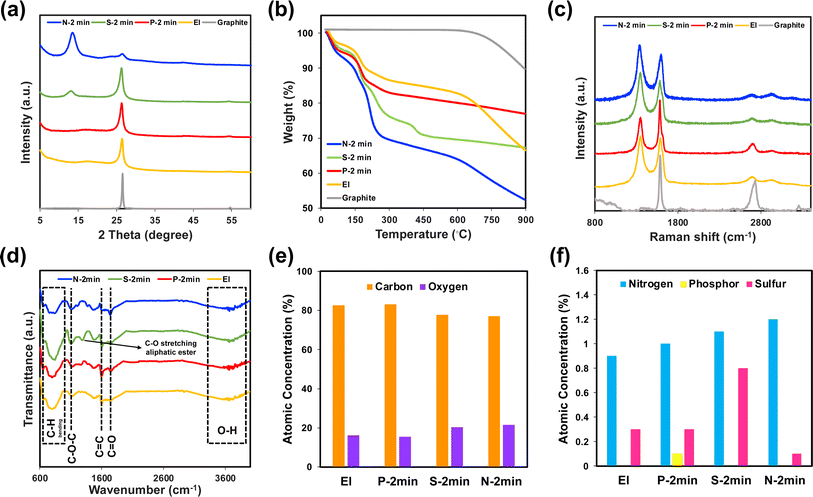 | ||
| Fig. 2 (a) XRD spectra, (b) TGA curves, (c) Raman spectra (data for El and graphite reproduced from ref. 22 with permission from Elsevier, Copyright 2023), and (d) FTIR spectra of graphite and El, P-2 min, N-2 min, and S-2 min samples. (e and f) Elemental analysis for the ECGs obtained from XPS surveys. | ||
Graphite exhibits no weight loss, even at higher temperatures, demonstrating its high quality. Graphite degradation commences at approximately 650 °C under an inert atmosphere, nitrogen. All samples demonstrated a uniform pattern of weight loss attributed to water desorption in the initial phase, marked by a pronounced decrease in the TGA curves. The extent of OCGs content, as suggested by the TGA curves, aligns closely with observations from the XRD spectra. XRD shows crystallinity and OCG presence, while TGA indicates weight loss from OCG decomposition. The N-2 min sample shows significant weight loss in the second phase due to high OCG density, corresponding to high oxidation and defects observed in XRD. In contrast, the P-2 min sample shows moderate weight loss in the second phase and fewer OCGs, which aligns with fewer defects and less oxidation observed in XRD. The TGA curve for the S-2 min sample was notable for displaying multiple steps, each representing different types of OCGs. Moreover, TGA analysis was able to differentiate the polarity between the El and P-2 min samples—a detail that XRD spectra could not elucidate. The P-2 min sample, in particular, showed considerable weight loss in the second stage, suggesting a higher content of OCGs and thus higher polarity compared to the El sample.
An additional noteworthy finding is the superior thermal stability of the P-2 min and S-2 min samples, which was preserved up to 900 °C, exceeding the resilience of the original graphite. This enhanced thermal stability is paramount for the longevity of graphene-based materials, especially for applications subjected to high temperatures. Enhancing this property is essential for expanding the potential uses of these materials. In this context, the incorporation of phosphate and sulfate functional groups within the P-2 min and S-2 min samples, respectively, plays a vital role in impeding the decomposition of the graphene nanosheets, thereby contributing to their improved thermal stability. For instance, Dai et al.38 found that phosphate groups in functionalized graphene oxide improve the thermal stability and flame retardancy of polystyrene composites. Zhong et al.39 demonstrated that phosphate groups on graphene oxide form a protective barrier, reducing oxidation and defect formation. These studies collectively highlight the role of these functional groups in improving the thermal properties and durability of graphene-based materials.
Fourier Transform Infrared Spectroscopy (FTIR) revealed the types of bonds on the carbonaceous nanomaterials. The FTIR spectra aligned well with the aforementioned characterizations (XRD, TGA, and Raman). As anticipated, the N-2 min and S-2 min samples display a broader range of OCGs. Moreover, in line with the expectations, the S-2 min sample exhibits more intense peaks. Distinct, sharp peaks at 1630 cm−1 and 1730 cm−1 are attributable to C![[double bond, length as m-dash]](https://www.rsc.org/images/entities/char_e001.gif) C and C
C and C![[double bond, length as m-dash]](https://www.rsc.org/images/entities/char_e001.gif) O bonds, respectively. Various forms of C–O bonds are also present, indicating the stretching of aliphatic ethers and esters. However, the presence of aliphatic esters is uniquely observed in the S-2 min sample.
O bonds, respectively. Various forms of C–O bonds are also present, indicating the stretching of aliphatic ethers and esters. However, the presence of aliphatic esters is uniquely observed in the S-2 min sample.
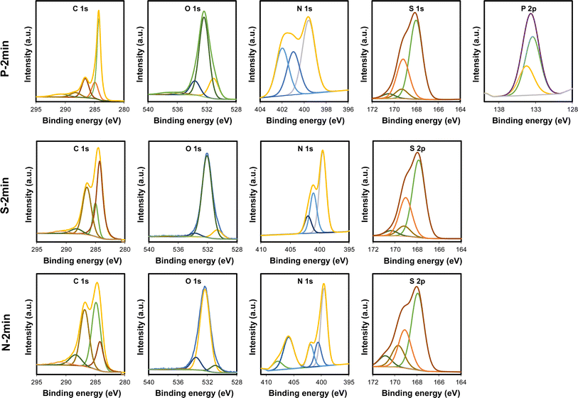
X-ray photoelectron spectroscopy (XPS) was employed to further investigate the chemical nature, composition, and heteroatom doping species of the ECGs (Fig. 3). The survey scans of all exfoliated graphene samples confirm the presence of oxygen, suggesting its presence, along with nitrogen and sulfur. The quantified atomic concentrations of all ECGs were calculated using XPS analysis and are shown in Table 2. High-resolution C 1s, N 1s, S 2p, and P 2p were used to provide further insight into the chemical characteristics of the ECGs. All samples' C 1s XPS spectra reveal five peaks at average binding energies corresponding to various carbonic bonds. For the P-2 min sample, the main component of the C 1s peak is C–C graphitic (sp2) carbon bonding, which is significantly higher than other peaks. However, for the N-2 min and S-2 min samples, a peak at 286.5 eV was also notable, corresponding to carbon atoms bound to oxygen atoms in hydroxyl and epoxy groups.42 The relative intensity of –C(![[double bond, length as m-dash]](https://www.rsc.org/images/entities/char_e001.gif) O)–OH, typically observed at the edges, was the highest for the N-2 min and S-2 min samples. This is because stronger acids, like nitric and sulfuric acids, led to unzipping and fragmentation of the graphitic layers, producing more available edge sites for carbonylation and carboxylation. Deconvolution of O 1s spectra confirmed the presence of different OCGs in all samples. The atomic ratio of carbon to oxygen (C/O) quantified by XPS ranges from 25.67 for graphite to 3.56 for the N-2 min sample. The high atomic percentage ratio of C/O (>5) for the El and P-2 min reflects their low degree of oxidation, resulting in high conductivity for these two samples.
O)–OH, typically observed at the edges, was the highest for the N-2 min and S-2 min samples. This is because stronger acids, like nitric and sulfuric acids, led to unzipping and fragmentation of the graphitic layers, producing more available edge sites for carbonylation and carboxylation. Deconvolution of O 1s spectra confirmed the presence of different OCGs in all samples. The atomic ratio of carbon to oxygen (C/O) quantified by XPS ranges from 25.67 for graphite to 3.56 for the N-2 min sample. The high atomic percentage ratio of C/O (>5) for the El and P-2 min reflects their low degree of oxidation, resulting in high conductivity for these two samples.
| C 1s (at%) | O 1s (at%) | N 1s (at%) | P 2p (at%) | S 2p (at%) | |
|---|---|---|---|---|---|
| P-2 min | 83.3 | 15.3 | 1 | 0.1 | 0.3 |
| S-2 min | 77.8 | 20.3 | 1.1 | 0 | 0.8 |
| N-2 min | 77.1 | 21.6 | 1.2 | 0 | 0.1 |
| El | 82.6 | 16.2 | 0.9 | 0 | 0.3 |
In the high-resolution XPS spectrum of N 1s, three peaks were identified, corresponding to pyridinic N, pyrrolic N, and graphitic N. Pyridinic is the most notable peak for all samples. P-2 min possesses a higher graphitic-nitrogen content than other samples. For N-2 min, two additional peaks appear, attributed to oxidized-like nitrogen bonding configurations, such as –NO2 and –NO3.43,44 The presence of sulfur in all samples was confirmed by deconvoluting the high-resolution S 2p spectra. This is attributed to the ammonium sulfate used in the second step of each synthesis. In P-2 min, the XPS spectra confirm phosphonation by the presence of P 2p peaks. Two peaks can be fitted for P-2 min, corresponding to P–C and C–P–O bonding configurations.45 The main component of the P 2p peak is P–C bonding, suggesting the successful incorporation of phosphorus into the P-2 min sample.
In conclusion, the polarity of the samples increased in the following order: N-2 min > S-2 min > P-2 min > El. The innovative two-step EC synthesis offers a novel approach to producing functionalized graphene nanosheets. During the intercalation process, HSO4−/SO42− ions in sulfuric acid, NO3− ions in nitric acid, and H2PO4−/HPO42−/PO43− ions in phosphoric acid can be intercalated into the layers of graphite, expanding the interlayer spacing and facilitating the exfoliation of individual graphene sheets. This process can also lead to the functionalization of graphene, where the intercalated ions or molecules become chemically bonded to the graphene surface. This process can be tailored to produce graphene with specific properties desired for certain applications.
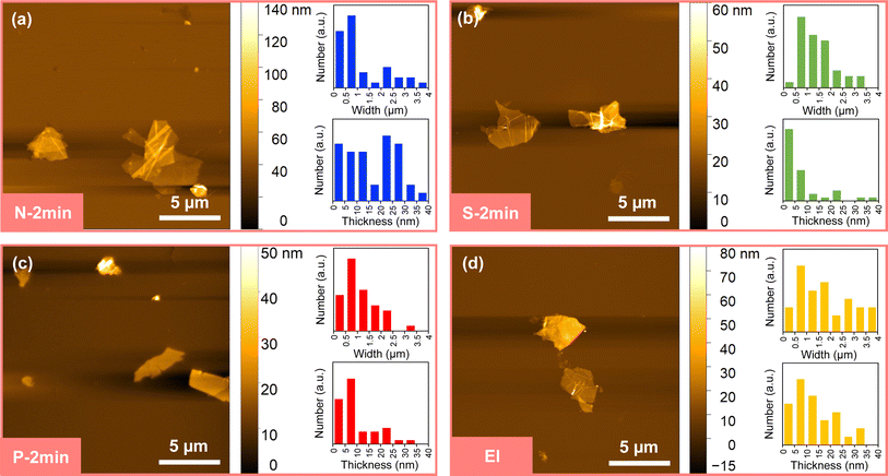 | ||
| Fig. 4 AFM images and morphological analyses of ECGs synthesized with two-step (a) nitric acid, (b) sulfuric acid, (c) phosphoric acid and one-step (d) electrochemical method. | ||
The morphological analyses confirm the potent oxidation strength of nitric and sulfuric acids, which led to a higher degree of oxidation and heavy graphite exfoliation. The use of these acids in the intercalation step weakened the interactions between the graphite layers, resulting in an increased exfoliation rate for the N-2 min and S-2 min samples. Additionally, the aggressive oxidation during intercalation enhanced the likelihood of separating and unzipping larger portions of HNO3-GIC and H2SO4-GIC. This yielded small graphene sizes and multilayer sheets for the N-2 min sample and large graphene sizes and single-layer sheets for the S-2 min sample. Conversely, dihydrogen phosphate ions (H2PO4−) diffused into the graphite layers, forming weaker interactions such as hydrogen bonds and van der Waals forces with the graphene sheets. This led to a more effective intercalation and a higher yield of single- and few-layer graphene sheets in the P-2 min sample. While nitrate ions intercalated between graphite layers, their planar geometry and charge distribution might have made them less effective in exfoliating graphene layers compared to phosphate ions and sulfur ions, which have a tetrahedral geometry.
The observations suggest that intercalation with phosphoric acid produced single- and few-layer graphene sheets. In contrast, nitric acid tends to produce small and multilayered graphene sheets, while sulfuric acid led to larger graphene sizes with single- and few-layer sheets. Moreover, the higher oxidation levels associated with nitric acid and sulfuric acid led to over-oxidation, resulting in structural damage and defects in the graphene sheets, as evidenced in the scanning electron microscope (SEM) images in Fig. S1.† Such over-oxidation can impede the ability to maintain separation between graphene sheets, increasing the likelihood of restacking or aggregation. In contrast, the milder acidity of phosphoric acid facilitated controlled exfoliation and reduction of graphite, minimizing damage to the graphene layers.
2.3. Rheology, printability and shape fidelity of inks
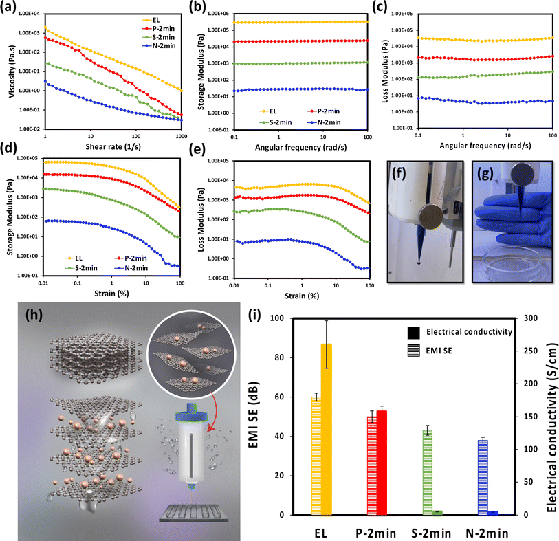
The preparation of ECG-based inks for 3D printing included the fabrication of pure ECGs' aqueous dispersions at three varying concentrations: 5%, 3%, and 2% by weight (wt%). To optimize printability and shape fidelity in DIW, it is essential to meticulously tailor the ink's rheological properties. This tailoring must strike a balance between facilitating flow during printing and ensuring shape retention afterwards.46–48 To assess and compare the rheological behavior of these inks, their rheological properties at 3 wt% concentration are presented here as a reference, while the properties of other concentrations can be found in the ESI.† To investigate the rheological behavior of the inks, a rotational shear test was initially conducted to evaluate their flow behavior (Fig. 5(a)). All inks exhibited a pronounced shear-thinning behavior; therefore, they are extrudable, as observed by any of the traditionally defined DIW inks.48 This behavior resulted from the disruption of interactions between the ECG sheets due to shear forces coupled with the alignment of these sheets along the direction of flow. This alignment significantly reduces the viscosity upon increasing the shear rate. Among them, P-2 min ink showed a more pronounced shear-thinning behavior. This significant shear-thinning behavior is highly desirable for DIW processes, as it ensures smooth flow through the nozzle during printing. As depicted in Fig. 5(a), El ink demonstrated the highest viscosity across the entire range of shear rates. Following this, P-2 min exhibited the highest value. The ink's high viscosity is beneficial for preserving its paste-like consistency.46 The viscosity of all ECG-based inks resides within the range of 1 to 1000 Pa s at a shear rate of 0.1 s−1, which is defined as the required viscosity for DIW, ensuring the printability of the inks.48Upon extruding from the printing head, the ink necessitates a rapid transformation from a shear-thinning fluid to a solid-like substance to preserve its geometric integrity. A key metric for assessing the ink's capability to uphold this geometric fidelity is the loss modulus (G′′) ratio to storage modulus (G′).21 To assess this pivotal metric, oscillatory measurements were performed. The linear viscoelastic region of the inks was determined by subjecting them to an increasing oscillating deformation (strain amplitude sweep test) from 0.01 to 100% at a constant frequency of 1 rad s−1. As Fig. 5(e and f) illustrate, G′ and G′′ exhibit a plateau in the graph at small strain amplitudes (approximately less than 0.1%). Consequently, the strain amplitude of 0.1% is an appropriate deformation magnitude for the frequency sweep test as it lies in the linear viscoelastic (LVE) region.
The frequency sweep test results are shown in Fig. 5(b and c), where G′ and G′′ are frequency invariant (plateau-like behavior), signifying the gel-like behavior of the inks. G′ is higher than G′′ in the entire probed frequency window, confirming the dominant solid-like behavior of the inks under small shear strains. After printing, this characteristic (G′ > G′′) can help the printed structure retain its shape until fully solidified and prevent the stacked filaments from sagging under their own weight and reduce the bending of spanning parts.48
Aligned with the flow curve results, a consistent trend is evident across all inks, with both G′ and G′′ following the order El > P-2 min > S-2 min > N-2 min. Evidently, there is a significant difference in the moduli of inks with identical concentrations. Naficy et al.49 research emphasized that the viscoelastic properties of GO-based ink are intricately tied to dispersion concentration. For the first time, it is shown here that, at a given concentration of GO-based ink, the functional groups play a crucial role in the rheological characteristics of the inks, consequently influencing their printability.
Several empirical formulae have been developed to evaluate the rheological properties of fluids containing 2D particles. One notable example is eqn (1) below, which is based on relatively rigid, disk-like particles, where h/d is the flake aspect ratio (thickness/diameter) and κ−1 is the Debye screening length (cf. Derjaguin–Landau–Verwey–Overbeek (DLVO) theory).50 This equation suggests that the critical volume fraction, which directly influences the rheology of a dispersion, is significantly affected by both the particles' surface charge and the surrounding matrix's electromagnetic permittivity.
 | (1) |
Empirically, this dependency can be quantified by measuring the zeta potential (ζ) of the particles (Table S2†). The results clearly indicate that the rheology of inks containing 2D nanomaterials, such as graphene, is highly influenced by the aspect ratio of the particles and their interactions with the matrix.46 Based on ζ results and characterization of synthesized ECGs, hydrogen bonding and electrostatic interaction coexist between ECG sheets and water molecules. ζ results show that N-2 min and S-2 min have higher negative charges resulting from, for example, their ionized carboxylic groups, leading to electrostatic repulsion between their layers.51 Inferior rheological properties of N-2 min and S-2 min imply that electrostatic interaction between N-2 min and S-2 min sheets is more effective than hydrogen bonding in N-2 min and S-2 min inks for their gelation and by extension, the rheology of their dispersion. This is mainly due to the fact that electrostatic interaction is a long-range force and it is usually stronger than that of a hydrogen bond.51 Since the synthesized ECGs have a large surface area, they are more sensitive to changes in electrostatic interactions.
The results from the amplitude sweep tests, as illustrated in Fig. 5(d and e), are consistent with frequency sweep test results, showing notable differences among the inks. For N-2 min and S-2 min, both G′ and G′′ exhibit lower values than those of El and P-2 min. A key observation across all the inks is that G′ consistently exceeds G′′, underscoring their predominantly elastic behavior. With increasing amplitude, a significant drop in viscoelastic moduli is observed, suggesting the disruption of the network structure by the break up of inter-sheet interactions and orientations of nanosheets parallel to the flow direction.
2.4. 3D printing
The high viscosity and viscoelastic moduli of El and P-2 min at a 5 wt% concentration pose challenges for ink extrusion (ESI, Fig. S2†), demanding high shear forces and compromising the energy efficiency of the printing process. Conversely, a 2 wt% concentration lacks the required moduli and viscosity for successful 3D printing across all inks (Fig. S3†). A concentration of 3 wt% was chosen for El and P-2 min to optimize energy usage and minimize material waste. For S-2 min and N-2 min, achieving similar rheological properties to the 3 wt% El and P-2 min inks, while also meeting the required rheological properties for 3D printing, necessitates a concentration of 5 wt%. Consequently, the formulated inks for 3D printing are at a 3 wt% concentration for El and P-2 min and 5 wt% for N-2 min and S-2 min. This strategic selection aims to balance ink rheology and energy-efficient printing processes and minimize any unnecessary waste of materials.The DIW process in 3D printing has encountered challenges when extruding El, N-2 min, and S-2 min inks, despite these inks meeting the key rheological criteria for 3D printing, such as shear-thinning behavior and sufficiently high storage modulus.52 This suggests that rheological measurements alone are inadequate for determining the printability of 2D materials in the DIW process. In this context, unlike granular particles, which interact through mutual compression (‘pushing’ against each other) under applied force, a mechanism that facilitates 3D printing, 2D materials tend to buckle during interactions with other neighboring sheets.46 This buckling is attributed to their inherent softness (e.g., high flexibility) along their longest dimension and the presence of OCGs on the nanomaterials, which are prone to form strong hydrogen bonds either among themselves or with water.
In the context of El, the low density of OCGs implies a reduced capacity for forming strong hydrogen bonds or electrostatic attractions with the matrix. Additionally, the van der Waals forces between the layers present an obstacle, hindering their effective dispersion in water.22 Furthermore, the weakness of repulsive forces tends to result in the formation of irregular aggregates. When pressure is applied, El nanosheets and water undergo separation, forming water droplets (see Fig. 5(f)). This phenomenon contributes to the clogging of injectors during 3D printing processes due to compromised ink integrity.11 During the 3D printing process, the El sheets and water cease to be homogeneously mixed. They instead segregate into distinct phases: one predominantly containing water and the other densely populated with El sheets. Thus, other than ink rheology, ink homogeneity plays a more important role in the printability/extrudability of the nanomaterials-based inks.
In the context of N-2 min and S-2 min inks, even though they demonstrated solid-like behavior at 5 wt% and fulfilled the required rheological criteria for DIW, they fell short in terms of yield stress, a crucial requirement for effective DIW printing. This shortfall is primarily ascribed to the solid-like behavior being predominantly driven by short-range hydrogen bonding in these two samples.46 Typically, to achieve the necessary yield stress for DIW, the particle concentration within the ink must be significantly increased beyond the critical concentration. This increase leads to the formation of a densely jammed structure, resulting from an increased number of interactions per nanosheet.46 However, in these two types of inks, high concentrations of nanosheets resulted in inhomogeneity, leading to inconsistencies in DIW. In addition, the low synthesis yield of N-2 min and S-2 min inks presented a significant challenge in achieving high particle concentrations necessary for effective DIW. Additionally, the high concentrations required greater energy input for extrusion, translating into higher energy demands during ink fabrication and DIW operations.
In addition, the conductivity of 50-micrometer-thick graphene films, as shown in Fig. 5(i), varies based on the acid used in Step I, following the order: El > P-2 min > S-2 min > N-2 min, in complete agreement by XRD, TGA, Raman, FTIR, XPS and AFM methods. The lower electrical conductivities observed in the S-2 min and N-2 min samples can be attributed to the presence of OCGs,53 while the high electrical conductivity of El can be attributed to the minimal presence of OCGs and the larger lateral size of its sheets. Specifically, the electrical conductivity of the P-2 min sample was 158 ± 14 S cm−1, aligning well with the magnitude previously reported in the literature for reduced graphene oxide (rGO) synthesized using the chemical method.54,55 Interestingly, the conductivity of the P-2 min ECGs is found to be one order of magnitude higher, as per ref. 56, and in some cases, even two orders of magnitude greater than the conductivity values reported for rGO reduced under extreme conditions, such as hydrazine reduction57 or microwave-assisted reduction.17 Remarkably, the high electrical conductivity value for the P-2 min sample has been achieved without any additional reduction steps. Fig. 5(i) also displays the ECG films' electromagnetic interference (EMI) shielding effectiveness (SE). The sample thickness plays a significant role in influencing the EMI SE. At a relatively low thickness of 50 μm, the total shielding effectiveness SET values are 60 dB for El, 50 dB for P-2 min, 38 dB for S-2 min, and 30 dB for N-2 min, respectively. These results indicate that El provides the highest EMI SE, followed by P-2 min, S-2 min, and N-2 min in descending order.
Conclusively, phosphoric acid, with its mild oxidizing power and ability to stabilize graphene sheets, emerges as an attractive electrolyte for the intercalation step in the two-step EC synthesis of ECGs. It is particularly effective when the goal is to produce high-quality, few-layered, or monolayer graphene. In contrast, the strong oxidizing power of nitric and sulfuric acids led to an increased number of superficial OCGs, but it also posed a higher risk of over-oxidation and structural damage to the graphene sheets. Among the ECGs, the P-2 min samples stood out, exhibiting the highest thermal stability, synthesis yield, electrical conductivity, and EMI SE, the least defective structure containing more monolayer graphene sheets alongside its decent printability. The ease of its synthesis complements the superior properties of P-2 min. The phosphoric acid electrolyte used in Step I can be reused until fully consumed, underscoring its potential for scalable graphene production. In contrast, with strong acids like nitric acid, the electrolyte must be disposed after each synthesis due to contamination produced during intercalation (Step I). Thus, the P-2 min method is more environmentally friendly and cost-effective for large-scale graphene ink production. Accordingly, our research now focuses exclusively on the development and optimizing P-2 min inks, which offers a more balanced approach between final material properties and energy efficiency.
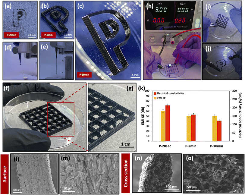
In this study, a multi-layer construct (shown as a ‘P’ letter), representative of phosphoric acid intercalated ECG was successfully 3D printed (Fig. 6(b)) due to adequate homogeneity and structural integrity of P-2 min ink, with no need for additives. By contrast, other work has relied on a binder or other rheological modifiers to achieve 3D printable ink. Jiang et al.,11 for instance, employed Ca2+ ions as cross-linkers to render homogeneous GO inks suitable for DIW.
Given the remarkable attributes of P-2 min and its commendable printability, we investigated the influence of intercalation duration on the ultimate properties and printability of ECGs intercalated with phosphoric acid. The duration of intercalation (Step I) in phosphoric acid was altered systematically, ranging from a brief span of 20 s, extending to 2 min and further to 10 min. To distinguish the resultant synthesized graphene samples based on their intercalation periods, they were labelled as P-20 s, P-2 min, and P-10 min, respectively.
In our study, as depicted in Fig. 6(a and d), the P-20 s ink was found to be non-extrudable and, consequently, unsuitable for printing. By contrast, the P-10 min ink demonstrated exceptional printing quality, exemplified by its ability to create a multi-layered structure (Fig. 6(c)). This structure effectively showcased the ink's shape stability and fidelity in all directions, a critical factor for the performance of 3D-printed electronics, which heavily relies on the quality of the printed lines and structures. Hence, P-10 min ink emerges as a promising candidate for printed electronics applications.
The post-printing stability of the multi-layered structures, as depicted in Fig. 6(f and g), distinctly demonstrates the ink's exceptional shape retention. Crucially, filament spreading was absent, which often results from gravitational forces or the load of successive layers. This observation suggests the formation of a robust network within the printed structure, sufficiently resilient to support its own weight. Such a characteristic is indicative of the ink's potential for intricate and reliable 3D printing applications.
Fig. 6(h–j) showcase the high electrical conductivity of the printed structures post-drying, which were sufficient to power an LED without necessitating any post-printing reduction processes. Fig. 6(k) shows a comparison between the electrical conductivity and EMI SE of samples intercalated with phosphoric acid for different durations. The sample P-10 min exhibited the lowest conductivity and EMI SE, whereas P-20 s demonstrated the highest values in both categories.
Fig. 6(l–o) present the SEM micrographs of the aerogel derived from filaments based on P-10 min ink, which was freeze-dried after extrusion. These images reveal the aerogel filament's porous structure at cross-section and laminates on the surface.11Fig. 6(o) shows that the 3D-printed P-10 min ink illustrates that the nanosheets were not merely stacked; they formed a highly interconnected porous structure, making them suitable for applications where large surface area is a prerequisite. This structure is conducive to electron transport and offers a large surface area, making it suitable for various applications. As aforementioned, on the walls or surfaces of the filaments, a distinctly different arrangement of sheets was evident. This is likely due to the shear forces exerted as the inks flow through the nozzle tip, inducing a maximal shear rate at the walls, leading to the alignment of nanosheets along the direction of shear.
The distinction in the electrical properties among the P-samples was further elucidated by XPS, Fig. S4,† and the oxygen content data is summarized in the accompanying table (Table S3†). Specifically, the P-10 min sample, with extended intercalation time, showed a higher oxygen content compared to P-20 s, which correlates with its reduced electrical conductivity and EMI SE. In this regard, as the intercalation time increased from 20 s to 2 and 10 min, the oxygen content in the samples rose from 14.5% to 15.3%. This observation indicates a trade-off between electrical conductivity and printability among inks intercalated in phosphoric acid for varying durations. As it is obvious from 3D printing, P-10 min ink was more homogenous due to longer intercalation time, which allowed phosphorous ions more time to diffuse into the graphite layers and affect the graphite layers more evenly and, as a result, have a more homogenous ink. Therefore, a 10 minutes intercalation period in phosphoric acid led to an ink that could be extruded uniformly.
All in all, this study elucidated the critical role of the type of intercalating agent and intercalation duration on the printability and performance of EGC inks in 3D printing. While the El, N-2 min, and S-2 min inks failed in extrusion due to lack of integrity and inhomogeneity, phosphoric acid intercalated ECG (P-2 min) demonstrated successful 3D printing. By systematically varying intercalation duration, it was found that P-10 min ink exhibited the highest print quality, indicating an optimal intercalation period for phosphorus ions. A balance between printability and electrical performance was observed, highlighting the potential of optimized graphene inks in 3D-printed electronics. Overall, this work paves the way for further exploration of the fine-tuning of non-covalent interactions and intercalation processes to develop advanced 2D materials-based inks for 3D printing.
3. Conclusion
In this study, we employed a two-step electrochemical synthesis to develop graphene inks that were not only highly printable and conductive but water-based and additive-free. These inks were designed for applications in 3D-printed electronics and EMI shields. A key aspect was fine-tuning intercalation conditions to optimize the rheological properties and the dispersion of the inks. A careful modulation was crucial in achieving a delicate balance between printability and the desired final properties, with a special focus on conductivity. We found that the type of the acid used for intercalation and the duration of this process significantly influenced the printing fidelity. Notably, intercalation in phosphoric acid markedly improved the ink printability, allowing the inks to be shaped into pre-designed structures with superior conductivity (158 S cm−1) and high EMI SE (50 dB at 50 μm thickness). This process effectively eliminated the need for any post-printing reduction procedures. Among the inks intercalated in phosphoric acid, those treated for 10 min exhibited the highest printing fidelity. Our two-step synthesis method marks a significant advancement in the field, facilitating rapid, continuous, and large-scale production of conductive, and environmentally friendly inks. These inks are additive-free and do not require harmful reductant chemistries or high-temperature post-processing. This innovation opens new avenues in the realm of advanced printed electronics, offering substantial benefits in terms of environmental impact and production efficiency.4. Experimental section
4.1. Graphene ink synthesis
The electrochemical exfoliation of graphite has been used to synthesize ECGs. Except for the El sample, which was directly exfoliated and oxidized in ammonium sulphate [(NH4)2SO4] other samples were intercalated with concentrated acids and then subjected to the same exfoliation/oxidation process. Schematic of the synthesis process is illustrated in Fig. 1. In the synthesis process, the graphite foil (graphite store, 0.5 mm thick) anode and platinum mesh (Thermo Scientific Chemicals, gauze woven from wire 45 mesh, Ø 0.198 mm (0.0078 in)) cathode were cut into 1 × 5 cm2 slices and placed parallel to each other at a constant distance of 2 cm. In the intercalation process, the cathode and anode were immersed in three different concentrated acids, including nitric acid (Certified ACS Plus, Fisher Chemical), sulfuric acid (95–98% ACS, FCC, VWR Chemicals) and phosphoric acid (ortho-phosphoric acid 85% ACS reagent, Sigma-Aldrich) as electrolyte and a constant voltage of 2 V powered by a DC power supply (Siglent Technologies) for 2 min to make graphite intercalated compound (GIC). The obtained GICs were named based on the intercalant agent as follows: H3PO4-GIC, H2SO4-GIC, and HNO3-GIC. After intercalation, the exfoliation/oxidation of the GICs was carried out in a 0.1 M ammonium sulphate [(NH4)2SO4] electrolyte at a constant voltage of 10 V until the current dropped to zero (graphite completely exfoliated). Exfoliated powders were separated and washed with deionized water (DI-water) to remove all surface impurities using a vacuum filtration process with a nylon membrane (Whatman, pore size 0.45 μm). This ensured that no residual ions remained in the synthesized powder, and a neutral pH was recorded. The washed product was re-dispersed in deionized water, bath sonicated for 15 minutes, and allowed to settle overnight. The residues obtained from the settling step were centrifuged at 3000 rpm for 10 minutes to separate the graphene from the unexfoliated graphite. Part of the suspension after centrifugation was freeze-dried and stored for further characterizations, including XRD, TGA, Raman, and other analyses, while the other part was used for preparing the inks. For preparing the inks, the washed product was re-dispersed in N,N-dimethylformamide (DMF) and bath-sonicated. An aqueous ECG dispersion was obtained through a solvent exchange method from the DMF-based GO dispersion, followed by overnight settling. Further rheological, EMI shielding, electrical, and printability tests were conducted on the prepared inks.After characterizing and evaluating the performance of each acid as an intercalating precursor based on desired properties and printability, phosphoric acid was chosen as a suitable electrolyte for the first step. The intercalation duration for this system varied from 20 seconds to 2 minutes and up to 10 minutes. The sample codes and conditions for each synthesis are shown in Table 1.
4.2. Characterization
4.3. 3D printing
The 3D printing of graphene suspensions was carried out using a BIO X Bioprinter (CELLINK, Gothenburg, Sweden). The bioprinter uses piston-driven syringe heads and the material flow is regulated by the print pressure. The ink loaded into a 3 mL syringe (NormJect, Air-Tite Products Co., Inc., U.S.A.) and centrifuged for 2 min at 2500 rpm. After removing trapped air, the syringe was attached by a luer lock to a pressure sensor to evaluate the pressure applied to the ink during the printing process. The inks were printed at room temperature on plastic and glass Petri dishes using 22 gauge (410 μm diameter) tapered tips (CELLINK) at a print pressure of 70–90 kPa at a speed of 15 mm s−1. A rectangular grid (3 cm × 3 cm × 0.5 cm, length, width and height) with a rectilinear infill pattern and 15% infill density were printed, as well as specially designed shapes. The 3D-printed structures were placed in a freezer overnight after the printing. Then, frozen meshes were freeze-dried with a FreeZone 2.5 L Benchtop FreezeDryer for 48 h at −49 °C and 0.05 mbar.Data availability
The data that support the findings of this study, “Additive-Free Aqueous-Based Graphene Inks for 3D Printing Functional Conductive Aerogels,” are available upon reasonable request. The data are stored in the corresponding author's institution's repository and can be accessed by contacting Dr Milad Kamkar at E-mail: milad.kamkar@uwaterloo.ca; or Dr Uttandaraman Sundararaj at u.sundararaj@ucalgary.ca. Due to ethical and privacy concerns, access to the data is restricted and will require approval from the relevant institutional review board.Conflicts of interest
Elnaz Erfanian, Farbod Sharif, Uttandaraman Sundararaj, and Milad Kamkar are inventors on a U.S. Provisional patent application related to this work with application serial number US 63/693,930, filed on September 12, 2024. The authors declare that there are no other competing interests.Acknowledgements
The authors gratefully acknowledge the financial support of the Natural Sciences and Engineering Research Council of Canada (NSERC) Discovery Grant 05503/2020, the Canada Excellence Research Chair Program (CERC-2018-00006) and Canada Foundation for Innovation (Project number 38623). We acknowledge Ms Faezeh Bakhshi for her help with drawing the schematics.References
- Z. Sun, S. Fang and Y. H. Hu, 3D graphene materials: from understanding to design and synthesis control, Chem. Rev., 2020, 120(18), 10336–10453 CrossRef CAS PubMed.
- A. G. Olabi, et al., Application of graphene in energy storage device–A review, Renewable Sustainable Energy Rev., 2021, 135, 110026 CrossRef CAS.
- J. Roman, et al., Lignin-graphene oxide inks for 3D printing of graphitic materials with tunable density, Nano Today, 2020, 33, 100881 CrossRef CAS.
- J. Zhao, et al., Printable ink design towards customizable miniaturized energy storage devices, ACS Mater. Lett., 2020, 2(9), 1041–1056 CrossRef CAS.
- K. Fu, et al., Progress in 3D printing of carbon materials for energy-related applications, Adv. Mater., 2017, 29(9), 1603486 CrossRef PubMed.
- C. H. Lui, et al., Ultraflat graphene, Nature, 2009, 462(7271), 339–341 CrossRef CAS PubMed.
- A. Kamyshny and S. Magdassi, Conductive nanomaterials for 2D and 3D printed flexible electronics, Chem. Soc. Rev., 2019, 48(6), 1712–1740 RSC.
- J. Qin, et al., Two-dimensional mesoporous materials for energy storage and conversion: Current status, chemical synthesis and challenging perspectives, Electrochem. Energy Rev., 2023, 6(1), 9 CrossRef CAS.
- R. Raj, S. C. Maroo and E. N. Wang, Wettability of graphene, Nano Lett., 2013, 13(4), 1509–1515 CrossRef CAS PubMed.
- A. Kamyshny and S. Magdassi, Conductive nanomaterials for printed electronics, Small, 2014, 10(17), 3515–3535 CrossRef CAS PubMed.
- Y. Jiang, et al., Direct 3D printing of ultralight graphene oxide aerogel microlattices, Adv. Funct. Mater., 2018, 28(16), 1707024 CrossRef.
- B. Yao, et al., 3D-printed structure boosts the kinetics and intrinsic capacitance of pseudocapacitive graphene aerogels, Adv. Mater., 2020, 32(8), 1906652 CrossRef CAS PubMed.
- F. Liu, et al., Synthesis of graphene materials by electrochemical exfoliation: Recent progress and future potential, Carbon Energy, 2019, 1(2), 173–199 CrossRef.
- N. Zaaba, et al., Synthesis of graphene oxide using modified hummers method: solvent influence, Procedia Eng., 2017, 184, 469–477 CrossRef CAS.
- J. Ma, et al., Highly conductive, mechanically strong graphene monolith assembled by three-dimensional printing of large graphene oxide, J. Colloid Interface Sci., 2019, 534, 12–19 CrossRef CAS PubMed.
- J. J. Moyano, et al., Filament printing of graphene-based inks into self-supported 3D architectures, Carbon, 2019, 151, 94–102 CrossRef CAS.
- Y. Zhu, et al., Microwave assisted exfoliation and reduction of graphite oxide for ultracapacitors, Carbon, 2010, 48(7), 2118–2122 CrossRef CAS.
- S. D. Lacey, et al., Extrusion-based 3D printing of hierarchically porous advanced battery electrodes, Adv. Mater., 2018, 30(12), 1705651 CrossRef PubMed.
- S. Abdolhosseinzadeh, et al., A Universal Approach for Room-Temperature Printing and Coating of 2D Materials, Adv. Mater., 2022, 34(4), 2103660 CrossRef CAS PubMed.
- B. Guo, et al., 3D printing of reduced graphene oxide aerogels for energy storage devices: a paradigm from materials and technologies to applications, Energy Storage Mater., 2021, 39, 146–165 CrossRef.
- A. Schwab, et al., Printability and shape fidelity of bioinks in 3D bioprinting, Chem. Rev., 2020, 120(19), 11028–11055 CrossRef CAS PubMed.
- E. Erfanian, et al., Electrochemically synthesized graphene/TEMPO-oxidized cellulose nanofibrils hydrogels: Highly conductive green inks for 3D printing of robust structured EMI shielding aerogels, Carbon, 2023, 210, 118037 CrossRef CAS.
- C. Zhu, et al., Supercapacitors based on three-dimensional hierarchical graphene aerogels with periodic macropores, Nano Lett., 2016, 16(6), 3448–3456 CrossRef CAS PubMed.
- T. S. Tran, et al., 3D printed graphene aerogels using conductive nanofibrillar network formulation, NanoTrends, 2023, 2, 100011 Search PubMed.
- S. Yang, et al., Emerging 2D materials produced via electrochemistry, Adv. Mater., 2020, 32(10), 1907857 CrossRef CAS PubMed.
- S. Pei, et al., Green synthesis of graphene oxide by seconds timescale water electrolytic oxidation, Nat. Commun., 2018, 9(1), 145 CrossRef PubMed.
- J. Cao, et al., Two-step electrochemical intercalation and oxidation of graphite for the mass production of graphene oxide, J. Am. Chem. Soc., 2017, 139(48), 17446–17456 CrossRef CAS PubMed.
- K. Parvez, et al., Electrochemically exfoliated graphene as solution-processable, highly conductive electrodes for organic electronics, ACS Nano, 2013, 7(4), 3598–3606 CrossRef CAS PubMed.
- W. Forsman, et al., Chemistry of graphite intercalation by nitric acid, Carbon, 1978, 16(4), 269–271 CrossRef CAS.
- K. Elmore, et al., Dissociation of phosphoric acid solutions at 25, J. Phys. Chem., 1965, 69(10), 3520–3525 CrossRef CAS.
- M. Sadeghi, et al., Dual-mode sorption of inorganic acids in polybenzimidazole (PBI) membrane, J. Macromol. Sci., Part B: Phys., 2010, 49(6), 1128–1135 CrossRef CAS.
- B. Gurzęda and P. Krawczyk, Electrochemical formation of graphite oxide from the mixture composed of sulfuric and nitric acids, Electrochim. Acta, 2019, 310, 96–103 CrossRef.
- C.-H. Chen, et al., Towards the continuous production of high crystallinity graphene via electrochemical exfoliation with molecular in situ encapsulation, Nanoscale, 2015, 7(37), 15362–15373 RSC.
- K. Parvez, Z.-S. Wu, R. Li, X. Liu, R. Graf, X. Feng and K. Müllen, Exfoliation of Graphite into Graphene in Aqueous Solutions of Inorganic Salts, J. Am. Chem. Soc., 2014, 136(16), 6083–6091 CrossRef CAS PubMed.
- T. Qiu, et al., The preparation of synthetic graphite materials with hierarchical pores from lignite by one-step impregnation and their characterization as dye absorbents, RSC Adv., 2019, 9(22), 12737–12746 RSC.
- M. Goodarzi, G. Pircheraghi and H. A. Khonakdar, Tailoring the graphene polarity through the facile and one-step electrochemical exfoliation in low concentration of exfoliation agents, FlatChem, 2020, 22, 100181 CrossRef CAS.
- W. Liu and G. Speranza, Tuning the oxygen content of reduced graphene oxide and effects on its properties, ACS Omega, 2021, 6(9), 6195–6205 CrossRef CAS PubMed.
- K. Dai, et al., Covalently-functionalized graphene oxide via introduction of bifunctional phosphorus-containing molecules as an effective flame retardant for polystyrene, RSC Adv., 2018, 8(44), 24993–25000 RSC.
- R. Zhong, et al., Improving thermo-oxidative stability of nitrile rubber composites by functional graphene oxide, Materials, 2018, 11(6), 921 CrossRef PubMed.
- A. Eckmann, et al., Probing the nature of defects in graphene by Raman spectroscopy, Nano Lett., 2012, 12(8), 3925–3930 CrossRef CAS PubMed.
- K. N. Kudin, et al., Raman spectra of graphite oxide and functionalized graphene sheets, Nano Lett., 2008, 8(1), 36–41 CrossRef CAS PubMed.
- N. Díez, et al., Enhanced reduction of graphene oxide by high-pressure hydrothermal treatment, RSC Adv., 2015, 5(100), 81831–81837 RSC.
- J. Y. Lee, et al., Nitrogen-doped graphene-wrapped iron nanofragments for high-performance oxygen reduction electrocatalysts, J. Nanopart. Res., 2017, 19(3), 98 CrossRef.
- B. Huang, et al., Effect of nitric acid modification on characteristics and adsorption properties of lignite, Processes, 2019, 7(3), 167 CrossRef CAS.
- J. Chen, et al., Synthesis of phosphonated graphene oxide by electrochemical exfoliation to enhance the performance and durability of high-temperature proton exchange membrane fuel cells, J. Energy Chem., 2023, 76, 448–458 CrossRef CAS.
- P. Wei, et al., Go with the flow: Rheological requirements for direct ink write printability, J. Appl. Phys., 2023, 134(10), 100701 CrossRef CAS.
- C. L. C. Chan, J. M. Taylor and E. C. Davidson, Design of soft matter for additive processing, Nat. Synth., 2022, 1(8), 592–600 CrossRef.
- M. Saadi, et al., Direct ink writing: a 3D printing technology for diverse materials, Adv. Mater., 2022, 34(28), 2108855 CrossRef CAS PubMed.
- S. Naficy, et al., Graphene oxide dispersions: tuning rheology to enable fabrication, Mater. Horiz., 2014, 1(3), 326–331 RSC.
- S. Jogun and C. Zukoski, Rheology and microstructure of dense suspensions of plate-shaped colloidal particles, J. Rheol., 1999, 43(4), 847–871 CrossRef CAS.
- H. Bai, et al., On the gelation of graphene oxide, J. Phys. Chem. C, 2011, 115(13), 5545–5551 CrossRef CAS.
- D. A. Rau, C. B. Williams and M. J. Bortner, Rheology and Printability: A Survey of Critical Relationships for Direct Ink Write Materials Design, Prog. Mater Sci., 2023, 101188 CrossRef.
- G. Eda and M. Chhowalla, Chemically derived graphene oxide: towards large-area thin-film electronics and optoelectronics, Adv. Mater., 2010, 22(22), 2392–2415 CrossRef CAS PubMed.
- S. Park, et al., Colloidal suspensions of highly reduced graphene oxide in a wide variety of organic solvents, Nano Lett., 2009, 9(4), 1593–1597 CrossRef CAS PubMed.
- V. B. Mohan, et al., Characterisation of reduced graphene oxide: Effects of reduction variables on electrical conductivity, Mater. Sci. Eng., B, 2015, 193, 49–60 CrossRef CAS.
- L. G. Guex, et al., Experimental review: chemical reduction of graphene oxide (GO) to reduced graphene oxide (rGO) by aqueous chemistry, Nanoscale, 2017, 9(27), 9562–9571 RSC.
- S. Stankovich, et al., Synthesis of graphene-based nanosheets via chemical reduction of exfoliated graphite oxide, Carbon, 2007, 45(7), 1558–1565 CrossRef CAS.
- J.-B. Wu, et al., Raman spectroscopy of graphene-based materials and its applications in related devices, Chem. Soc. Rev., 2018, 47(5), 1822–1873 RSC.
- S. S. Nanda, D. K. Yi and K. Kim, Study of antibacterial mechanism of graphene oxide using Raman spectroscopy, Sci. Rep., 2016, 6(1), 28443 CrossRef CAS PubMed.
- A. C. Ferrari and D. M. Basko, Raman spectroscopy as a versatile tool for studying the properties of graphene, Nat. Nanotechnol., 2013, 8(4), 235–246 CrossRef CAS PubMed.
Footnote |
| † Electronic supplementary information (ESI) available. See DOI: https://doi.org/10.1039/d4ta03082f |
| This journal is © The Royal Society of Chemistry 2024 |


