
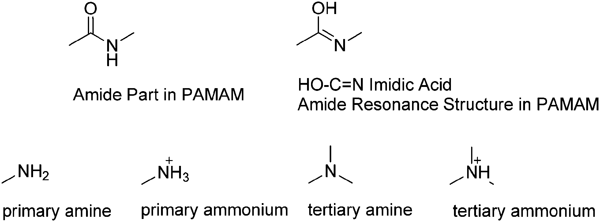
Poly-amidoamine structure characterization: amide resonance structure of imidic acid (HO–C![[double bond, length as m-dash]](https://www.rsc.org/images/entities/h2_char_e001.gif) N) and tertiary ammonium
N) and tertiary ammonium
Yan Jia,
XiaoLiang Yangb and
Ying Qian*a
aSchool of Chemistry and Chemical Engineering, Southeast University, Nanjing, 211189, China. E-mail: yingqian@seu.edu.cn; jiyan98@163.com
bSchool of Chemistry and Chemical Engineering, Nanjing University, Nanjing, 210093, China
First published on 16th September 2014
Abstract
The fluorescence emission phenomena of polyamidoamine (PAMAM) have been discovered and characterized in recent decades. The amide, primary amine, and tertiary amine groups are present in PAMAM, which are not the traditional or typical fluorescence emission groups. The fluorescence emission groups or mechanism of PAMAM were yet not clear but cause cares. In this study, PAMAM was characterized using NMR (15N NMR, 13C NMR, 1H NMR, and N–H 2D NMR), IR, and MS. The results proved that amide resonance structures, corresponding to imidic acid and tertiary ammonium groups, existed in PAMAM. The new imidic acid and tertiary ammonium groups found in PAMAM might help explain the intrinsic-fluorescence phenomena.
Introduction
Polyamidoamine (PAMAM) dendrimers were first synthesized by Tomalia in 1985.1 PAMAM dendrimers have water soluble interval space or internal holes2 and several modifiable chemical groups;3 moreover, they are highly biocompatible,4 and they can package nanoparticles,5 and can also deliver small molecules or drugs.6 In recent years, the observed fluorescence emission phenomena of PAMAM have generated considerable interest.7 The PAMAM dendrimers contain amides, primary amines, and tertiary amines; however, there are no traditional or typical fluorescence emission groups in PAMAM. Some other dendrimers, such as poly(amino ester)s,8b poly(propyl ether imine),8e poly(propyleneimine) (PPI) dendrimer, and poly(ethyleneimine) (PEI) dendrimer9 show fluorescence emission, which is commonly known as intrinsic fluorescence.10 Therefore, the fluorescence emission phenomena and mechanism of PAMAM have been observed and studied by many research groups.8In this study, we have discussed the amide resonance structure of imidic acid (HO–C![[double bond, length as m-dash]](https://www.rsc.org/images/entities/char_e001.gif) N) in PAMAM. This structure, under some situations, seems to be connected with the fluorescence emission of PAMAM. We used 15N NMR, 1H NMR, 13C NMR, N–H 2D NMR, and IR spectra to determine the amide resonance structure of the newly found imidic acid and the tertiary ammonium groups in PAMAM.
N) in PAMAM. This structure, under some situations, seems to be connected with the fluorescence emission of PAMAM. We used 15N NMR, 1H NMR, 13C NMR, N–H 2D NMR, and IR spectra to determine the amide resonance structure of the newly found imidic acid and the tertiary ammonium groups in PAMAM.
The amide resonance structure of imidic acid (HO–CN) has been studied during previous experiments9 and quantum chemical studies.10 Note that amide and imidic acid are tautomers of each other, and the tautomeric ratios11 depend on conditions such as the temperature, solvent, and pH. The amide to imidic acid transition in PAMAM could be related to the above conditions. The amide/imidic acid mechanism can be used to explain some of the influences (such as pH) on the amide to imidic acid transition. The amide resonance structure of imidic acid in PAMAM derivatives that are responsible for the fluorescence emission have been described in a related report.12 The novel elucidated structures of the imidic acid and the tertiary ammonium groups found in PAMAM could help explain the phenomenon of intrinsic fluorescence.
Results and discussions
PAMAM (Fig. 1) exhibited weak fluorescence when synthesized initially. However, the PAMAM dendrimers staled in air or added acid, owing to which PAMAM produced fluorescence emission. The fluorescence emission phenomena of PAMAM have been proved by many research groups.7–10 | ||
| Fig. 1 The PAMAM-G1 amide structure and PAMAM-G1 amide resonance structure of imidic acid. | ||
In order to determine the chemical structure of the PAMAM fluorescence emission centers, the NMR (15N NMR; 1H NMR; 13C NMR; N–H 2D NMR), MS, and IR spectra were analyzed to characterize the structures of PAMAM-G1.
The sticky PAMAM dendrimers were in the liquid state at room temperature. The pure PAMAM-G1 exhibited strong fluorescence emission, but the PAMAM solution exhibited weak fluorescence. The traditional NMR was performed in solution, which might influence the fluorescence emission center of PAMAM. Therefore, the pure PAMAM-G1 was used to perform NMR experiments and to obtain structural information on the pure state of PAMAM-G1. Accordingly, the 15N NMR, 1H NMR, 13C NMR, N–H 2D NMR, and IR spectra of pure PAMAM-G1 were analyzed.
The 15N NMR was carried out using a 600 MHz NMR liquid spectrometer. The pure PAMAM-G1 was a sticky liquid when the sample temperature was set to 60 °C during the 15N NMR testing. The abundance of 15N in nature is 0.36%;13 therefore, 15N NMR was recorded using a pure liquid PAMAM-G1 sample without adding any solvents. Note that the 15N NMR testing time was about 30 hours, and there were three kinds of N atoms in the PAMAM-G1, i.e., amides, primary amines, and tertiary amines.
Fig. 2 shows the 15N NMR spectra of pure PAMAM-G1. The 15N NMR was tested in the range from −100 to 1000 ppm. The peaks at about 82.60 were attributed to the N atom of the tertiary amine. The peaks ranging from 26.47 to 37.49 were attributed to the primary amine and the tertiary amine. The peaks ranging from 34.39 to 37.49 were attributed to the ammonium (primary ammonium and tertiary ammonium) group, thus proving the existence of different levels of protonation of N atoms in the primary and tertiary amines.
 | ||
| Fig. 2 The 15N NMR spectra of PAMAM-G1 at pure liquid state. δ (600 MHz 15N NMR): 119.58, 118.21, 116.81 (amide); 82.60 (tertiary amine); 26.47 to 37.49 (amine and ammoniums). | ||
The three peaks corresponding to 119.58, 118.21, and 116.81 were attributed to the N atoms of the amide. The integrated area ratios were 1.00/0.91/1.12. The NMR peaks of the amide N atom were split into three peaks, thus showing that there were three states of the amide N atoms. The three peaks corresponded to the resonance structures of the amide group in PAMAM-G1, and one of these states corresponded to imidic acid. In the N–O bond NMR signal range (200–1000 ppm), there were 15N NMR 499.73 small peaks, and these peaks were attributed to the N–O bond. This bond was very weak, probably because the imidic acid hydroxyl was connected with the amine ion to form salts. The N–O bond peak was so weak that it excluded the possibility of PAMAM producing a lot of oxime. Thus, the reverse reaction corresponding to the oxime/amide Beckmann rearrangement was excluded.
The N–H 2D NMR (Fig. 3) shows the (1H/15N NMR) 7.59, 119.72 peak. This peak corresponds to the N–H 2D-NMR of the amide group. There were three peaks corresponding to 116.81, 118.21, and 119.58 in the 15N NMR spectrum. This shows that the peak corresponding to 119.72 (in the N–H 2D NMR) or the peak corresponding to 119.58 (in the 1D N NMR) were related to the single H atom. Moreover, the other nearby amide peaks corresponding to 116.81 and 118.21 (in 1D N NMR) were not related to the H atom, thus proving that the amide exists in a state in which the N atom does not form a bond with the H atom. The amide resonance structure of the imidic acid shows that the N atom is not bonded with the H atom. Furthermore, the peaks at 116.81 and 118.21 were attributed to the imidic acid state and to the middle transitional state of the amide.
 | ||
| Fig. 3 The 15N–1H 2D (two dimensional) NMR of PAMAM-G1 in pure liquid state. δ (600 MHz 2D 1H/15N NMR): 7.59/119.72 (amide). | ||
The 1H NMR (Fig. 4) has a strong peak at 4.60, which was attributed to the hydroxyl NMR peaks. The pure PAMAM-G1 was tested in the absence of a solvent, thereby excluding the detection of H2O or other hydroxyl groups. The H NMR showed that there was OH hydroxyl structure formed in PAMAM. This shows that the oxygen atoms are connected to the H atoms. The hydroxyl corresponded to the OH group of the amide resonance structure in imidic acid (HO–CN). The peaks at 7.17, 6.75, and 6.25 were attributed to the ammonium H atom belonging to the primary and tertiary ammonium groups, whereas the peak at 7.57 was attributed to the amide group.
 | ||
| Fig. 4 The 1H NMR spectrum of PAMAM-G1 in pure liquid state. δ (600 MHz 1H NMR): 7.76–7.57 (amide); 7.17–6.25 (ammoniums); 4.60 (hydroxyl of HO–CN); 1.67–2.66 (amine 2H–N). | ||
The 13C NMR (Fig. 5) shows peaks in the range of 163.39–163.40 and 172.54–173.79, thus proving the existence of two states of the amide C atoms, and these two states correspond to the amide and the imidic acid, respectively.
 | ||
| Fig. 5 The 13C NMR spectrum of PAMAM-G1 at pure liquid state. δ (600 MHz 13C NMR): 172.54–173.79 (amide); 163.39–163.40 (imidic acid HO–CN); 31.61–50.39 (methylene). | ||
The PAMAM-0.5G, PAMAM-1G, PAMAM-1.5G, and PAMSM-2.0G (Fig. 6) IR spectra provided information regarding the carbonyl groups and double bonds (Fig. 7). The peak at 1733 cm−1 in the IR spectrum of PAMAM-0.5G was attributed to the carbonyl group of the ester. The peaks at 1733 and 1659 cm−1 in the IR spectrum of PAMAM-1.5G were attributed to the carbonyl group of the ester and to the double bonds, respectively. The peaks at 1647 and 1644 cm−1 in the IR spectra of PAMAM-1.0G and PAMAM-2.0G were attributed to double bonds. However, there were no peaks at 1733 cm−1 in the IR spectrum of PAMAM-1.0G and PAMAM-2.0G, thus proving that there were no carbonyl groups in PAMAM-1.0G and PAMAM-2.0G. The 1644–1659 double bonds IR absorption signal peaks, which should be attributed to the CN of imidic acid of PAMAM.
 | ||
| Fig. 6 The structure (amide resonance structure; imidic acid formula) of PAMAM-0.5G, PAMAM-1.5G, PAMAM-1.0G, and PAMAM-2.0G. | ||
 | ||
| Fig. 7 The IR spectra of PAMAM-0.5G, PAMAM-1.5G, PAMAM-1.0G, and PAMAM-2.0G. | ||
The MS spectra (Fig. 8) of PAMAM-G1 show the [M + 2H]2+ peaks. Amide resonance structures were detected and the amine protonation also changed in PAMAM-G1; however, no other chemical groups were present in PAMAM-G1. The amide resonance structure of the imidic acid did not change the molecular weight of PAMAM. The MS spectra cannot exclude the possibility of an amide resonance structure of imidic acid. The molecular peaks from 517.4 to 520.3 show protonation, thereby proving the existence of ammonium groups in the PAMAM structure.
 | ||
| Fig. 8 The MS spectra (two samples) of PAMAM-G1 (Fig. 1) (calculated: 516.4 [M], 517.4 [M + H]+, 518.4 [M + 2H]2+; found: 518.3 [M + 2H]2+, 519.3 [M + 3H]3+, and 520.3 [M + 4H]4+). | ||
The spectral data in Table 1 (15N NMR; 1H NMR; 13C NMR; N–H 2D NMR, IR, and MS) show that amide resonance structures of imidic acid exist in PAMAM. This imidic acid structure (Fig. 9) could correspond to the fluorescence emission group; moreover, this group shows a rigid co-plane structure and comprises CN double bonds, p–π conjugated structures, and electron-donating hydroxyl groups. The imine CN double bonds exhibit fluorescence emission under some conditions.14 The CN in imidic acid was similar to the CN in imine, and both of these groups exhibited fluorescence emission. The amide, imidic acid, primary amine, primary ammonium, tertiary amine,15 and tertiary ammonium groups in PAMAM are shown in Fig. 9. The characterized structures were associated with the fluorescence emission properties, thereby accounting for the intrinsic fluorescence phenomena.
| Spectraa | Fig. | Primary amine or ammonium | Tertiary amine or ammonium | Amide | Imidic acid (HO–CN) |
Ester (CO) |
|---|---|---|---|---|---|---|
| a Footnote: unit of NMR (ppm), IR (cm−1). | ||||||
| 15N NMR | 2 | 26.47–37.49 | 82.60 | 119.58 | 116.81; 118.21 | |
| 1H NMR | 4 | (1.67–2.66 amine) (7.17; 6.75; 6.25 ammonium) | 7.57, 7.76 | 4.60 | ||
| 1H/15N 2D NMR | 3 | 7.59/119.72 | ||||
| 13C NMR | 5 | 31.61–50.39 | 172.54–173.79 | 163.39–163.40 | ||
| IR PAMAM-0.5G | 7 | — | 1733 | |||
| IR PAMAM-1.5G | 7 | 3387 | 1659 | 1733 | ||
| IR PAMAM-1.0G | 7 | 3325 | 1647 | |||
| IR PAMAM-2.0G | 7 | 3385 | 1644 | |||
 | ||
| Fig. 9 The parts of amide (P-01); imidic acid parts (P-02); primary amine (P-03); primary ammonium (P-04); tertiary amine (P-05); and tertiary ammonium (P-06); in PAMAM. | ||
Experimental methods
1. Synthesis
PAMAM 0.5G, 1.0G, 1.5G, 2.0G.1 Methyl acrylate (MA) and ethylenediamine (EDA) were used as substrates.(1) The Michael addition of the amine groups in EDA to MA below 50 °C in a methanol solution affords the dendritic product of 0.5 generation (G) with the terminated ester groups.
(2) The amidation of the terminal ester groups of the 0.5G dendrimer after dissolving in a methanol solution by excessive EDA below 50 °C affords the 1G dendrimer with the terminal amine groups.
(3) Distillation of the exceeded EDA under reduced pressure and washed by ethyl ether gives the purified 1G dendrimer. The PAMAM dendrimers presented as a yellow sticky liquid.
(4) Repeated step (1), (2), and (3) to obtain 1.5G, 2.0G.
2. Instruments
NMR by BRUKER 600 MHz NMR spectrometer. Pure PAMAM-G1 sample without adding solvents, was maintained at a temperature of 60 °C, external standard method, testing time 30 h; IR by Nicolet 750 infrared spectrometer, pure PAMAM KBr smear; MS by LC-Q TOF MS.Conclusions
The amide of PAMAM exists as a resonance structure of imidic acid. The tertiary amine transfers to the ammonium group under some conditions. The 15N NMR, 13C NMR, 1H NMR, N–H 2D NMR, IR of the pure liquid PAMAM-G1, and the MS spectra proved the existence of imidic acid and tertiary ammonium structures in PAMAM. In conclusion, the newly discovered imidic acid and tertiary ammonium structures in PAMAM could be connected with its fluorescence emission properties. These results can help explain and understand the intrinsic fluorescence phenomena exhibited by dendrimers.Acknowledgements
The National Natural Science Foundation of China (no. 61178057) and the Scientific Research Foundation of Graduate School of Southeast University (no. YBPY1209) were greatly appreciated for financial support.Notes and references
- D. A. Tomalia, H. Baker, J. Dewald, M. Hall, G. Kallos, S. Martin, J. Roeck, J. Ryder and P. Smith, Polym. J., 1985, 17, 117–132 CrossRef CAS.
- J. F. Jansen, E. M. de Brabander-van den Berg and E. W. Meijer, Science, 1994, 266, 1226–1229 CAS.
- (a) A. I. Cooper, J. D. Londono, G. Wignall, J. B. McClain, E. T. Samulski, J. S. Lin, A. Dobrynin, M. Rubinstein, A. L. C. Burke, J. M. J. Frechet and J. M. DeSimone, Nature, 1997, 389, 368–371 CrossRef CAS; (b) R. M. Crooks, M. Zhao, L. Sun, V. Chechik and L. K. Yeung, Acc. Chem. Res., 2001, 34, 181–190 CrossRef CAS PubMed; (c) Y. Ji and Y. Qian, RSC Adv., 2014, 4, 25510–25519 RSC.
- (a) C. S. Braun, M. T. Fisher, D. A. Tomalia, G. S. Koe, J. G. Koe and C. R. Middaugh, Biophys. J., 2005, 88(6), 4146–4158 CrossRef CAS PubMed; (b) X. Y. Shi, I. J. Majoros, A. K. Patri, X. D. Bi, M. T. Islam, A. Desai, T. R. Ganser and J. R. Baker Jr, Analyst, 2006, 131(3), 374–381 RSC; (c) L. Fernandez, M. Gonzalez, H. Cerecetto, M. Santoc and J. Silberet, J. Supramol. Chem., 2006, 18(8), 633–643 CrossRef CAS; (d) K. J. Landmark, S. DiMaggio, J. Ward, C. Kelly, S. Vogt, S. Hong, A. Kotlyar, A. Myc, T. P. Thomas, J. E. Penner-Hahn, J. R. Baker Jr, M. M. B. Holl and B. G. Orr, ACS Nano, 2008, 2, 773–783 CrossRef CAS PubMed; (e) R. K. Tekade, P. V. Kumar and N. K. Jain, Chem. Rev., 2009, 109, 49–87 CrossRef CAS PubMed.
- (a) S. P. Mukherjee and H. J. Byrne, Nanomedicine, 2013, 9(2), 202–211 CrossRef CAS PubMed; (b) C. M. Lamy, O. Sallin, C. Loussert and J. Y. Chatton, ACS Nano, 2012, 6(2), 1176–1187 CrossRef CAS PubMed; (c) Y. J. Tsai, C. C. Hu, C. C. Chu and T. Imae, Biomacromolecules, 2011, 12(12), 4283–4290 CrossRef CAS PubMed; (d) M. F. Ottaviani, F. Montalti, N. J. Turro and D. A. Tomalia, J. Phys. Chem. B, 1997, 101, 158–166 CrossRef CAS.
- (a) S. Sadekar and H. Ghandehari, Adv. Drug Delivery Rev., 2012, 64(6), 571–588 CrossRef CAS PubMed; (b) L. Albertazzi, B. Storti, L. Marchetti and F. Beltram, J. Am. Chem. Soc., 2010, 132(51), 18158–18167 CrossRef CAS PubMed; (c) B. K. Biswal, M. Kavitha, R. S. Verma and E. Prasad, Cytotechnology, 2009, 61(1–2), 17–24 CrossRef PubMed; (d) H. Wang, H. B. Shi and S. K. Yin, Exp. Ther. Med., 2011, 2, 777–781 CAS; (e) F. Zeng and S. C. Zimmerman, Chem. Rev., 1997, 97, 1681–1712 CrossRef CAS PubMed; (f) A. W. Bosman, H. M. Janssen and E. W. Meijer, Chem. Rev., 1999, 99, 1665–1966 CrossRef CAS PubMed; (g) L. J. Twyman, A. S. H. King and I. K. Martin, Chem. Soc. Rev., 2002, 31, 69–82 RSC.
- (a) K. E. Sapsford, L. Berti and I. L. Medintz, Angew. Chem., Int. Ed., 2006, 45(28), 4562–4588 CrossRef CAS PubMed; (b) A. M. Caminade, A. Hameau and J. P. Majoral, Chem.–Eur. J., 2009, 15(37), 9270–9285 CrossRef CAS PubMed; (c) G. R. Newkome and C. D. Shreiner, Polymer, 2008, 49(1), 1–173 CrossRef CAS; (d) U. Boas, J. B. Christensen and P. M. H. Heegaard, J. Mater. Chem., 2006, 16(38), 3786–3798 RSC; (e) C. L. Larson and S. A. Tucker, Appl. Spectrosc., 2001, 55, 679–683 CrossRef CAS.
- (a) D. J. Wang and T. Imae, J. Am. Chem. Soc., 2004, 126, 13204–13205 CrossRef CAS PubMed; (b) D. C. Wu, Y. Liu, C. B. He and S. H. Goh, Macromolecules, 2005, 38(24), 9906–9909 CrossRef CAS; (c) Y. F. Fan, Y. G. Fan and Y. N. Wang, J. Appl. Polym. Sci., 2007, 106(3), 1640–1647 CrossRef CAS; (d) P. L. Wang, X. Wang, K. Meng, S. Hong, X. Liu, H. Cheng and C. C. Han, J. Polym. Sci., Part A: Polym. Chem., 2008, 46(10), 3424–3428 CrossRef CAS; (e) G. Jayamurugan, C. P. Umesh and N. Jayaraman, Org. Lett., 2008, 10, 9–12 CrossRef CAS PubMed.
- (a) L. Antonov, Tautomerism: Methods and Theories, Wiley-VCH, Weinheim, 1st edn, 2013 Search PubMed; (b) J. Oszczapowicz, Basicity, H-bonding, tautomerism and complex formation of imidic acid derivatives, in Amidines and Imidates: Volume 2 (1991), ed. S. Patai and Z. Rappoport, John Wiley & Sons, Ltd., Chichester, UK, 2010 Search PubMed; (c) I. Buśko-Oszczapowicz and J. Oszczapowicz, Detection and determination of imidic acid derivatives, in Amidines and Imidates: Volume 2 (1991), ed. S. Patai and Z. Rappoport, John Wiley & Sons, Ltd., Chichester, UK, 2010 Search PubMed.
- C. R. Kemnitz and M. J. Loewen, J. Am. Chem. Soc., 2007, 129(9), 2521–2528 CrossRef CAS PubMed.
- R. M. Balabin, J. Chem. Phys., 2009, 131(15), 154307 CrossRef PubMed.
- D. Staneva, P. Bosch, A. M. Asiri, L. A. Taib and I. Grabchev, Dyes Pigm., 2014, 105, 114–120 CrossRef CAS.
- A. O. Nier, Phys. Rev., 1950, 77, 789 CrossRef CAS.
- (a) J. S. Wu, W. M. Liu, J. C. Ge, H. Y. Zhang and P. F. Wang, Chem. Soc. Rev., 2011, 40, 3483–3495 RSC; (b) J. S. Wu, W. M. Liu, X. Q. Zhuang, F. Wang, P. F. Wang, S. L. Tao, X. H. Zhang, S. K. Wu and S. T. Lee, Org. Lett., 2007, 9, 33–36 CrossRef CAS PubMed; (c) W. Liu, L. Xu, R. Sheng, P. Wang, H. Li and S. Wu, Org. Lett., 2007, 9, 3829–3832 CrossRef CAS PubMed; (d) D. Ray and P. K. Bharadwaj, Inorg. Chem., 2008, 47, 2252–2254 CrossRef CAS PubMed; (e) V. Chandrasekhar, P. Bag and M. D. Pandey, Tetrahedron, 2009, 65, 9876–9883 CrossRef CAS; (f) H. S. Jung, K. C. Ko, J. H. Lee, S. H. Kim, S. Bhuniya, J. Y. Lee, Y. Kim, S. J. Kim and J. S. Kim, Inorg. Chem., 2010, 49, 8552–8557 CrossRef CAS PubMed; (g) M. Suresh, A. K. Mandal, S. Saha, E. Suresh, A. Mandoli, R. D. Liddo, P. P. Parnigotto and A. Das, Org. Lett., 2010, 12, 5406–5409 CrossRef CAS PubMed; (h) Z. Li, M. Yu, L. Zhang, M. Yu, J. Liu, L. Wei and H. Zhang, Chem. Commun., 2010, 46, 7169–7171 RSC.
- (a) C. C. Chu and T. Imae, Macromol. Rapid Commun., 2009, 30(2), 89–93 CrossRef CAS PubMed; (b) L. Cao, D. D. Jia, S. F. Wang, Y. L. Rong, C. Liu and D. J. Wang, Chem. Lett., 2014, 43(2), 246–248 CrossRef CAS; (c) M. Sun, C. Y. Hong and C. Y. Pan, J. Am. Chem. Soc., 2012, 134, 20581–20584 CrossRef CAS PubMed.
| This journal is © The Royal Society of Chemistry 2014 |