 Open Access Article
Advances and prospects of dynamic DNA nanostructures in biomedical applications
Yiling Chen and
Sirong Shi*
State Key Laboratory of Oral Diseases, National Clinical Research Center for Oral Diseases, West China Hospital of Stomatology, Sichuan University, Chengdu 610041, P. R. China. E-mail: sirongshi@scu.edu.cn
First published on 24th October 2022
Abstract
With the rapid development of DNA nanotechnology, the emergence of stimulus-responsive dynamic DNA nanostructures (DDNs) has broken many limitations of static DNA nanostructures, making precise, remote, and reversible control possible. DDNs are intelligent nanostructures with certain dynamic behaviors that are capable of responding to specific stimuli. The responsible stimuli of DDNs include exogenous metal ions, light, pH, etc., as well as endogenous small molecules such as GSH, ATP, etc. Due to the excellent stimulus responsiveness and other superior physiological characteristics of DDNs, they are now widely used in biomedical fields. For example, they can be applied in the fields of biosensing and bioimaging, which are able to detect biomarkers with greater spatial and temporal precision to help disease diagnosis and live cell physiological function studies. Moreover, they are excellent intelligent carriers for drug delivery in treating cancer and other diseases, achieving controlled release of drugs. And they can promote tissue regeneration and regulate cellular behaviors. Although some challenges need further study, such as the practical value in clinical applications, DDNs have shown great potential applications in the biomedical field.
1 Introduction
Since Nadrian Seeman first proposed the notion of constructing various DNA nanostructures in 1982,1 DNA nanotechnology has been evolving quickly and continuously. DNA nanostructures have been designed in various sizes and shapes, which can regulate the biological behavior of cells and organisms.2 DNA nanostructures are synthesized via self-assembly from single-stranded DNAs (ssDNAs) based on the rule of complementary base-pairing. However, the traditional methods to design DNA nanostructures are quite complicated and troublesome. Therefore, computer-aided design software has been invented by scientists to reduce the workload, and the main design software include CaDNAno, CanDo, Daedalus, vHelix, etc.3At present, through the continuous attempts by researchers, DNA nanotechnology has been combined with various stimulus-responsive functional units, realizing the transformation of DNA nanotechnology from structural DNA nanotechnology to dynamic DNA nanotechnology.4 DDNs are DNA structures constructed using programmable DNA self-assemblies that reconfigure their conformations in response to exogenous stimuli, such as metal ions, pH, and endogenous stimuli, such as adenosine triphosphate (ATP), glutathione (GSH), microRNAs (miRNAs), etc.5 External stimuli allow for remote control, while internal stimuli allow DNA nanostructure to be better coordinated with complex biological systems in vivo.6 Due to the highly specific programmable ability of DDNs to recognize environmental stimuli, they can serve as targeted delivery carriers to target sites and reduce non-specific toxicity to adjacent healthy tissues. Besides, morphological changes in DDNs can be detected by imaging techniques or other detectable readout signals,5 which have broad potential applications in the biosensing and bioimaging field.
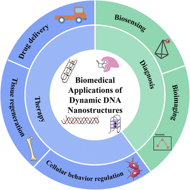
In this review, we reviewed the latest research findings in the field of DDNs. We discussed the synthesis mechanisms of DDNs, and their biomedical applications, including biosensing, bioimaging, drug delivery, tissue regeneration, and cellular behavior regulation (Fig. 1). At last, we have pointed out the current challenges and future research directions of DDNs.
2 Classifications
2.1 Dynamic tetrahedral DNA nanostructures
Four specifically designed isometric ssDNAs were constructed into tetrahedral three-dimensional structures, named tetrahedral DNA nanostructures (TDNs). Compared with other types of DNA nanostructures, TDNs have the following advantages, including high mechanical strength, good structural stability, excellent editability, etc.7 The addition of stimulus-responsive units to TDNs allows them to undergo conformational changes in response to the corresponding stimuli,8 protecting drugs for targeted delivery and releasing them at the appropriate site. In addition to delivering drugs, they are also promising tools in bioimaging, biosensing,9 and diagnosis,10 etc.2.2 Switchable DNA origami nanostructures
Since Paul Rothemund developed DNA origami technology in 2006,11 it has become an important milestone in DNA nanotechnology. Several outstanding advantages make it an indispensable member of DNA nanostructures.12 Firstly, the shape of DNA origami is well defined. Moreover, it can be easily designed with the help of various software. With the continuous development of DNA origami technology, a variety of DDNs have been constructed, such as DNA nanocapsules,13 DNA nanochannels,14 DNA nanorobots,15 DNA circuits,16 etc. This top-down origami approach is a simple yet powerful technique in the field of DNA nanotechnology.2.3 Other DNA structures with dynamic behavior
In addition to the DDNs mentioned above, others such as DNA hydrogel, DNA walkers, etc. also can be designed to respond to a variety of stimuli.DNA hydrogels are prepared by physical entanglement or chemical ligation of DNA strands. At present, various DNA hydrogels with dynamic behavior have been developed. For example, they can respond to NIR light,17 pH,18 miRNAs,19 etc. DNA hydrogels show strong advantages in the preparation of injectable hydrogels due to their good thixotropic properties,20 which make them important prospects for biomedical applications.
DNA walkers are DNA devices that can travel on a variety of tracks, divided into inchworm walker, Bipedal walker and spider walker.3 In recent years, DNA walkers that can respond to antibody,21 miRNAs,22 light,23 etc. have been constructed. And DNA walker's strong migration capabilities and excellent flexibility make it a versatile tool for the quantitative detection of biomolecules.
3 Major stimuli-responsive DNA systems in DDNs
3.1 Metal ions-responsive systems
DNA nanostructures respond to metal ion stimuli in the following two forms. Firstly, they respond to the electrostatic repulsion caused by the negatively charged phosphate main chain under cationic conditions. Meanwhile, Bivalent cations can help DNA strands overcome the electrostatic interaction between them, which is necessary for most self-assembly of DNA nanostructures. Secondly, they rely on ion-stimulated molecules, which mainly refer to G-quadruplex. In the absence of particular metal ions such as K+,24 Mg2+, Pb2+,25 Hg2+, Ag+, Cu2+, Ca2+, Sr2+, etc., G-rich ssDNA undergoes conformational change to form stabilized G-quadruplex.26Generally, those DNA nanostructures mediated by metal ions can be used to detect the presence of specific metal ions, which become potential intelligent materials in the applications in the field of environmental and food safety monitoring.27 In addition, scientists have expanded their reach to biomedical applications in recent years. For example, Mengyuan Li et al. developed a spherical metal–DNA nanostructure by mixing DNA molecules and ferrous ions. The size and structure of metal–DNA nanostructures can be precisely controlled by adjusting the ratio and concentration of DNA molecules and ferrous ions. This metal–DNA nanostructure can effectively deliver nucleic acid drugs to different cells, which shows efficient biological recognition in vitro and in vivo.28 Considering the elevated extracellular ion concentration in tumor microenvironments,29 ion-stimuli DNA nanostructure holds great application prospects in the future.
3.2 Photo-responsive systems
As a non-invasive external stimulus, light can activate the conformational change of photo-responsive DNA structure and release the drugs carried therein. Photo-cleavable linker or chromophore is introduced as a photo-responsive structure into the polymer backbone or matrix of DNA carrier.30 The responsive light sources include ultraviolet light (UV), near-infrared light (NIR) and visible light. The mechanisms of UV-mediated DNA structure change mainly include photocleavage, photoisomerization and photocrosslinking.31 NIR is considered more suitable for clinical application because of its high maximum permissible exposure and tissue penetration depth, leaving less damage to healthy cells.32 Therefore, NIR-induced photothermal therapy is a hot research field at present.Because instantaneous manipulation can be achieved in light stimulation with high precision in time and space, and switching profiles can be regulated by adjusting the wavelength, intensity and exposure duration of the light,33 light stimulation has been broadly employed as an external stimulus to provoke structural changes. However, there are still some challenges to be solved. Firstly, high doses of UV radiation may lead to the destruction of DNA main chain structure and cause tissue damage,34 while low doses of UV radiation may not be able to trigger photosensitive molecules. The dose control of UV light still needs further discussion in practical application. Secondly, it is necessary to minimize toxicity to normal cells or tissues and increase tissue specificity to light stimuli. Currently, imaging-guided photo-responsive drug delivery may become a promising solution.32 In addition, although NIR has better tissue penetrability than UV, it is still difficult to reach the target position of deep tissue. Hence, there is still an urgent need to develop a corresponding optical delivery system with stronger tissue penetrability.
3.3 pH-Responsive systems
The mechanisms of pH-responsive DNA nanostructures in biomedical applications mainly include the following points. The first is based on the different pH of specific organelles in cells. The pH in lysosomes is about 4.5–5.5, the acidic environment and its degradation mechanisms can trigger the release of drugs embedded in DNA nanostructures.35 The second mechanism is based on the different pH between some diseased target cells and other normal cells. The pH inside the tumor cells is higher than normal cells, which is about 7.3–7.6. Elevated pH is necessary for the proliferation and metastasis of tumor cells and can reduce their apoptosis. The extracellular pH of tumor cells is about 6.8–7.0, which is lower than that of normal extracellular environment. This may be associated with the accumulation of acid metabolites such as lactic acid.36 The reverse pH in tumor cells provides a potential method for targeted delivery of drugs in DDNs. The third mechanism is based on the abnormal pH in organelles in some diseases. For example, in neuronal ceroid lipofuscinosis, the pH in lysosomes increases abnormally.37In pH-responsive DNA nanostructures, the most commonly used functional units are i-motif and DNA triplex structures. I-motif, a cytosine-rich oligonucleotide, shows pH-responsive structure change in acidic conditions.38 And DNA triplex, mainly constructed by Watson–Crick and Hoogsteen interactions, forms typical base pairing, T–A·T and C–G·C+.39 And its stability can be greatly improved in low pH environment.40 Moreover, DNA triplex structure can be modified by varying the relative content of CGC/TAT triplets, which offers a higher degree of programmability.41
3.4 Endogenous small molecules responsive systems
Small molecules also act as endogenous stimuli to induce conformational changes in DNA nanostructures, such as ATP, GSH, etc. These small molecules-responsive DNA nanocarriers provide a new perspective for the design of tumor diagnosis and therapy strategies.42–44ATP concentrations are different at the intracellular level, extracellular level, and different organelles. In addition, in tumor cells, due to the rapid proliferation of tumor cells, the intracellular ATP level is much higher than that of normal cells. Moreover, the ATP level in the extracellular microenvironment is also higher than that of normal tissues.45 Therefore, these mechanisms can be used to design DNA nanostructures that respond to ATP stimulation. ATP aptamers are mostly used in ATP-responsive delivery systems.46
GSH, as an important intracellular regulatory metabolite, can participate in a variety of important biochemical reactions and plays an antioxidation role in vivo. The intracellular GSH concentration is higher than that in the extracellular fluid, which can be used for GSH-responsive DNA nanocarriers.47
4 Biomedical applications of dynamic DNA nanostructures
4.1 Biosensing
DDNs are attractive tools in biosensing. They show high sensitivity in the detection of small molecules in cells, and show potential application value in the early diagnosis of diseases.Cheng Jing et al. embedded aptamer configuration in TDNs and developed a novel ATP electrochemical aptamer sensor. In the presence of ATP, aptamer binds to ATP, making TDNs in a tense state and forming a G-quadruplex configuration at the edge. While without ATP, TDNs are in a relaxed state and G-quadruplex cannot be formed. The detection limit of this biosensor is 50 pm, which has good sensitivity compared with the traditional ATP detection kit.48 Peng Yang and coworkers designed an antibody-responsive DNA walker, which could continuously move on the three-dimensional orbit composed of DNA-functionalized gold nanoparticles. Delightfully, this DNA walker was proved to have the function of detecting antibodies and small molecules in buffer and human serum samples.21
miRNAs have been reported as a reliable biomarker for the early diagnosis and prognosis of numerous diseases, such as cancer, rheumatoid arthritis, multiple sclerosis, etc. Accurate and sensitive detection of miRNAs is particularly necessary for early diagnosis and early therapeutic intervention. For example, miRNA-141 is an important biomarker in prostate cancer.49 Zhangcheng Fu et al. constructed an electrochemical biosensor to detect miRNA-141 using in situ catalytic hairpin assembly (CHA) actuated TDN interfacial probes. Researchers confirmed that this biosensor has good sensitivity and specificity for the detection of miRNA-141 in serum samples.50 Wen-Hsin Chang et al. designed a DNA-based acrylamide hydrogel microcapsule based on the mechanism of competitive sequence replacement between target miRNA and bridging DNA in the microcapsule shell. Furthermore, their abilities to detect miRNA-141 have been proven to be highly effective.19
In addition, tumor-derived exosomes, which contain a variety of proteins from cancer cells, are also one of the biomarkers for cancer diagnosis. Yanyan Yu et al. designed an exosome detection platform based on the three-dimensional DNA motor. The DNA motor is activated by binding to the target protein on the exosome with its aptamer, and autonomously walks on the gold nanoparticle track driven by the restriction endonuclease. The difference in exosome signal protein intensity from different tumor cell sources can be identified by these DNA motors. Pleasantly, they show a good detection effect in serum samples from breast cancer patients, and have potential application value in clinical diagnosis.51
In the diagnosis of most diseases, it is often necessary to detect more than one biomarker at the same time. Meanwhile, the biosensor for single biomarker detection will be limited in clinical application. To overcome the limitation of single biomarker analysis, molecular logic gates have been introduced into the diagnosis of multi-factor diseases.52 Ali Ebrahimi et al. designed DNA logic gate cascaded logical operator, which can monitor four miRNAs (HAS-miR-143-3P, HAS-miR-18b-5P, HAS-miR-424-5P and HAS-miR-93-5P) associated with Alzheimer's disease.53 They revealed a novel method for analyzing molecular circuits, which provides a new idea for the detection of disease biomarkers.
We believe that with more in-depth research in DDNs, wider applications of DNA machinery for sensing and diagnostic uses will be found and greater prospects in clinical use could be anticipated.
4.2 Bioimaging
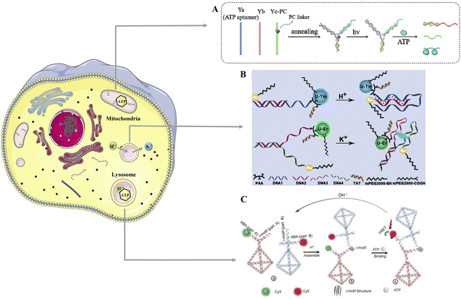
Bioimaging plays an important role in understanding the tissue structure and illustrating the various physiological functions of living organisms. Based on the good addressability of DDNs, they can be used to detect target biomolecules with high spatial and temporal accuracy by carrying fluorescent molecules,54 which has become a promising tool for live cell analysis (Fig. 2). | ||
| Fig. 2 Applications of DDNs bioimaging tools in revealing the physiological activities of living organisms. (A) Design of spatiotemporally controlled nanodevice and its mechanism in the visualization of ATP in mitochondria under hypoxic conditions. Reproduced with permission.55 Copyright 2021, American Chemical Society. (B) The schematic diagram of the assembly of DNA nanosensor to monitor K+ and pH in the lysosome. Reproduced with permission.56 Copyright 2021, John Wiley and Sons. (C) Design of framework nucleic acid detection platform to image ATP in acidic environment in lysosomes. Reproduced with permission.57 Copyright 2021, John Wiley and Sons. | ||
Jin Liu et al. constructed a reductase and light-responsive nanodevice for the visualization of ATP in mitochondria under hypoxic conditions by combining light-responsive DNA probes into a hypoxia-responsive metal–organic framework (Fig. 2A).55 This study bridges the gap between spatial and temporal controlled imaging studies of biomolecules in organelles under hypoxic conditions. By detecting histological changes associated with hypoxia at the organelle level, researchers can increase their understanding of hypoxia-related metabolic pathways. Feng Chen and coworkers developed a DNA nanosensor that can simultaneously image K+ and H+ in the lysosomes. This sensor uses a pH-responsive DNA triplex to detect H+ and generates blue fluorescence, a K+-responsive G-quadruplex to detect K+, and generates green fluorescence (Fig. 2B).56 Using this sensor, researchers demonstrated that H+ influx is accompanied by K+ efflux during lysosomal acidification. Moreover, Pai Peng et al. constructed a framework nucleic acid detection platform containing an ATP aptamer and an i-motif sequence that can respond to the acidic environment in lysosomes. This nanodevice can image ATP in lysosomes, helping scientists better understand the biological function of lysosomes (Fig. 2C).57
The development of non-invasive in situ imaging techniques has promising applications in disease diagnosis. Effective and sensitive detection of cancer-associated miRNAs is strongly needed in cancer diagnosis and related biological studies. Compared to other imaging methods which need pre-processing to extract miRNA from cell lysates,58 DDNs allow in situ bioimaging of miRNA expression in living cells with better cell specificity and temporal accuracy.
Yu-Heng Liu et al. established a photo-responsive DNA biosensor that uses UV photocleavage to activate walking and MnO2 nanosheets to provide intrinsic power. Not only was it able to measure survivin mRNA, which is highly associated with malignant tumors, in live cancer cells with accuracy and sensitivity, but also can track survivin mRNA dynamically.59 Juan Zhang et al. built a Cu2+-dependent DNAzyme logic circuit to analyze and image multiple miRNAs in living cells. The Cu2+-dependent DNAzyme logic gate was activated to output a fluorescent signal only when both miRNA-155 and miRNA-21 were present.60 This logic circuit can rapidly identify different miRNAs within cells in a complex cellular environment, providing a promising application of DNA biocomputing circuits in living cells.
Moreover, overexpression of telomerase activity and shortening of telomere length are closely associated with the proliferation of cancer cells.61 Ruixue Zhang et al. designed a pH-responsive dynamic DNA tetrahedron docking assembly, which can only maintain structural integrity in the acidic environment and release strands containing telomerase substrates in the alkaline environment. Therefore, this nanodevice can specifically label cancer cells based on the extracellular acidic environment, intracellular alkaline environment, and increased intracellular telomerase activity of cancer cells.62 Yuhong Lin et al. devised DNA nanohydrogels that can image intracellular telomerase activity in vitro and release therapeutic siRNAs for tumor cell gene therapy upon stimulation of telomerase in experimental animals.63 This nanohydrogel creatively combines the detection of biomarkers and stimulus-induced therapy in an effective way.
In conclusion, on the one hand, dynamic DNA-based biosensing and bioimaging nanodevice can reveal the physiological activities occurring at the organelle, cell, tissue, or organism level. On the other hand, they can analyze the role of certain biomarkers in diseases, deepening scientists' understanding of pathogenesis, and therefore opening up new ways for disease diagnosis (Table 1).
| Classification | Disease | Biomarker | DNA structure | Stimulus responsive unit | Output signal | Ref. |
|---|---|---|---|---|---|---|
| Biosensing | Prostate cancer | miRNA-141 | TDN probes | CHA | Electrochemical signal | 50 |
| DNA-based acrylamide hydrogel microcapsule | Bridged DNA | Fluorescent quantum dots | 19 | |||
| Alzheimer's disease | HAS-miR-143-3P, HAS-miR-18b-5P, HAS-miR-424-5P, HAS-miR-93-5P | DNA logic gate | CHA | UV-vis absor1bance | 53 | |
| Breast cancer | Exosome | DNA motor | CD63 aptamer | Fluorescence intensity | 51 | |
| Bioimaging | Cancer | Survivin mRNA | Photo-gated DNA walker | MnO2 nanosheets | Fluorescent signal | 59 |
| miRNA-155 and miRNA-21 | DNAzyme logic circuit | Cu2+ | Fluorescent signal | 60 | ||
| Telomerase | pH-responsive dynamic DNA tetrahedron docking assembly | DNA triplex | 62 | |||
| DNA nanohydrogels | Telomeric primer sequence | 63 |
4.3 Drug delivery
DOX is one of the most commonly used drugs in cancer treatment. It can inhibit DNA and RNA synthesis and can also generate free radicals causing DNA and cell membrane damage, thus killing cancer cells.65 Taking the advantage of the fact that the concentration of ATP in the extracellular microenvironment of tumor cells is much lower than that in the intracellular microenvironment and the level of ATP in tumor cells is much higher than that in normal cells, Yao Jiang et al. designed a 3D DNA nanostructure that responds to the high concentration of ATP in tumor cells and can release DOX to carry out anti-tumor effects.66 In addition to responding to ATP stimulation, Jiayu Yu et al. synthesized a DNA–BSA nanocarrier by combining bovine serum albumin (BSA) with DNA, which was loaded with DOX. Because it contains a pH-sensitive i-motif sequence and can dynamically release DOX in the presence of acidic pH and DNaseI, it exerts targeted anti-tumor effects.67 Under the irradiation of NIR, the DNA–azobenzene nanopump created by Yue Zhang et al. induces DNA hybridization and de-hybridization by photoisomerization of azo molecules to complete controlled release of DOX and achieve localizable and efficient drug release.68 Ping-Ping He et al. also used NIR as a stimulus trigger and formed an MXene–DNA hydrogel delivering DOX in a targeted manner, performing as localized synergistic photothermal-chemo therapy in cancer treatment.17 Furthermore, to tackle the problems of poor tissue penetration, insufficient local concentration, and poor efficacy of drugs in multidrug resistant (MDR) cancer, Jianqin Yan et al. fabricated a DOX-equipped PSP/TDNs nanocarrier with membrane-breaking and size-shrinkage properties. This nanocomplex can be cleaved by the high concentration of GSH in tumor cells, triggering the volume shrinkage of the nanocomplex and releasing DOX. Thus, DOX shows uniform distribution in tumor tissues with enhanced penetration ability, improving the therapeutic effect in MDR tumors.69 Moreover, Yifan Jiang et al. designed DNA nanospheres that can respond to tumor-associated TK1 mRNA in cancer cells. These DNA nanospheres can make specific recognition of TK1 mRNA through IsDNA structure, so it can automatically regulate DOX release through the changing level of mRNA, which also has a better therapeutic effect for drug-resistant tumor cells.70
Other than DOX, a series of chemotherapeutic drugs can be successfully encapsulated in DNA nanocarriers and exert stimulus-responsive antitumor effects. Jiao Zhang et al. constructed a DNA tetrahedron equipped with carbon ethyl bromide-modified camptothecin (CPT), which improved the hydrophilicity of the drug and could responsively release CPT in response to high levels of GSH in cancer cells.71 Moreover, an injectable CPT-conjugated DNA hydrogel synthesized by scientists can penetrate into tumor tissues by breaking them down into nanoscale particles under the degradation of enzymes. This hydrogel is also GSH-responsive and exhibits sensitive and sustained targeted drug delivery to tumor cells, effectively preventing cancer recurrence.72
PTX can also be successfully incorporated into spherical nucleic acid nanostructures (SNAs), and the multifunctional PTX–SNAs exert GSH-induced tumor targeting, thus reversing tumor tissue resistance in vitro and in vivo.73 Yi Ma et al. designed a DNA icosahedral structured carrier with telomerase responsiveness that can dissociate with the aid of telomerase and release caged platinum in the plasma surrounding cancer cells. Better therapeutic efficacy, especially for drug-resistant cancer cells, and reduced toxicity to normal tissues can be achieved through precise drug release.74
Recently, Wenjuan Ma and coworkers combines HER2-targeted DNA-aptamer with TDNs to deliver maytansine (DM1). In order to improve the biosafety and the accuracy of drug delivery, they established a camouflage by combining the erythrocyte membrane with pH-responsive liposomes, which exhibit excellent tumor-stimulus drug delivery behavior.75 Moreover, there are also studies combining chemotherapy and immunotherapy together. Scientists built the pH-responsive smart nanocubes to deliver DOX and immune stimulating agents targeting tumor cells and tumor-associated immune cells.76
Based on the series of precise drug-loaded DNA nanostructure platforms created in the above study, many other chemotherapeutic drugs might be considered to be similarly attached to DNA nanostructure platforms, and more drug-containing nanostructures with precise contents and structures could be designed.
MiRNA is an endogenous molecule with differential expression in tumor cells and normal cells, making it a potential tumor biomarker. Moreover, a series of miRNA-responsive DNA nanostructures have been designed for clinical diagnosis of cancer.78 Fan Zhang et al. innovatively combined endogenous miRNAs that can be used for clinical cancer diagnosis with exogenous therapeutical miRNAs and designed a miRNA-responsive DNA nano-drug delivery system for intelligent responsive gene regulation. They combined DNA–miRNA hybrids of let-7a and the complementary DNA of miR-155, and encapsulated them in exosomes. Induced by miR-155 overexpressed in breast cancer cells, it cleaves and releases let-7a and miR-155 with complementary DNA, inhibiting HMGA1 expression and promoting SOX1 expression.79
RNAi-based therapies are also a powerful strategy in cancer treatment. Small interfering RNA (siRNA), a functional factor of RNAi, is an important nucleic acid therapeutic agent that can inhibit the expression of target genes intracellularly and can regulate the expression of target genes in vitro and in vivo. Chang Xue et al. formed a gold nanoparticle/multi-functional 3D DNA self-assembled multilayer core/shell nanostructure (siRNA/Ap-CS), which is capable of releasing siRNA in response to endogenous miRNA and directly into the microenvironment of tumor cells, leading to the silence of target genes and inhibition of tumor-associated protein expression significantly, therefore inducing apoptosis and inhibiting tumor growth.80 Yuanyuan Guo and coworkers successfully attached pheophorbide A (PPA) photosensitizer to the DNA structure and further bound it to programmed death ligand-1 (PD-L1) siRNA linker, forming siRNA and PPA encapsulated nucleic acid nanogels. This nanogel not only kills tumor cells photo dynamically, but also releases siRNA to downregulate PD-L1 expression in tumor cells, synergistically promoting cytotoxic T lymphocytes (CTL)-mediated tumor cell killing.81 Yang Gao et al. designed a TDNs based nanobox, which allows anti-inflammatory tumor necrosis factor (TNF)-α targeted siRNA release in a pH-responsive manner when entering into lysosomes.82
Some studies synergize chemotherapeutic drugs with therapeutic nucleic acid drugs to enhance tumor suppression, offering a promising prospect for the development of targeted DNA nanoplatforms of drug delivery for synergistic therapy. Yuwei Li et al. co-loaded DOX and anaplastic lymphoma kinase (ALK)-specific siRNA on DNA nanomicelles, which released therapeutic drugs when the DNA nanomicelles underwent conformational changes in acidic pH in endosomes and lysosomes in cancer cells, offering synergistic treatment of anaplastic large cell lymphoma.83 Similarly, Zhaoran Wang and coworkers co-loaded siRNAs targeting Bcl2 and P-glycoprotein (P-gp) as well as DOX into tubular DNA nanodevice based on DNA origami technique. Under the stimulation of GSH in cells, the disulfide bonds break and the nanodevice opens and releases the drugs therein to exert anti-tumor effects.84 In addition, studies have also constructed a DNA hydrogel containing DOX and CpG, which breaks down under acidic conditions and releases DOX and CpG.85 CpGs can be recognized by Toll-like receptor 9 (TLR9) located in host cell endosomes, leading to the secretion of inflammatory cytokines including TNF-α, interleukin (IL)-6 and triggering a strong immune response against tumor tissue.86
Due to the good compatibility, homogeneity, programmability and biodegradability of DNA nanostructures, they will become promising and excellent delivery vehicles for therapeutic oligonucleotides in the future. And with the continuous research in this area, we believe that this new drug carrier will have a great impact on the biomedical field in the near future.
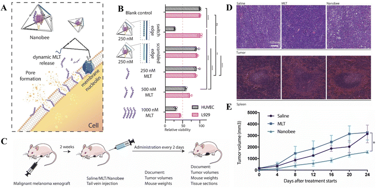
 | ||
| Fig. 3 Dynamic framework nucleic acid loaded with melittin as nanobee in treating malignant melanoma. (A) Schematic diagram of the working mechanism of nanobee. (B) In vitro evaluation of toxicity of nanobee. The nanobee can selectively target the membrane of nucleolin-positive HUVECs. (C) The construction of malignant melanoma (A375) xenografted tumor-bearing mice model to access the therapeutic value of nanobee in vivo. (D) HE staining showed the intense liquefaction necrosis of the tumor after nanobee treatment and spleen damage after the administration of melittin. (E) The injection of nanobee significantly inhibited the growth of tumor in the mice model. Reproduced with permission.87 Copyright 2020, John Wiley and Sons. | ||
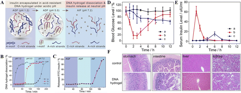
In addition to applications in cancer treatment, DDNs can also be used in the treatment of other diseases. To avoid the low bioavailability of antibiotics during Staphylococcus aureus biofilm infections, zwitterionic nanoparticles containing nucleic acid nanostructures have been fabricated for the delivery of vancomycin. Under the corresponding stimulation of the biofilm pH, the nanoparticles were able to achieve the desired charge reversal, improving bacterial binding and permeation of biofilms, which is highly effective to treat biofilm infections.88 Moreover, a DNA hydrogel resistant to acid while responding to physiological pH was also designed as an oral delivery vehicle for insulin. In the mimicking gastric environment and the mimicking duodenal environment, the loaded insulin is protected by the DNA hydrogel. While in the mimicking small intestine environment, the DNA hydrogel dissociates and releases insulin, forming an ideal oral insulin delivery system (Fig. 4).89
 | ||
| Fig. 4 Acid-resistant and physiological pH-responsive DNA hydrogel in oral insulin delivery. (A) Schematic diagram of the construction of DNA hydrogel and its working mechanism. (B) Sequential dissociation profiles of DNA hydrogels under pH 1.2, 5.0, and 7.2. Curve (a): DNA hydrogels cross-linked by A-motif and i-motif. Curve (b): only A-motif. Curve (c): only i-motif. (C) Release curve of fluorescein isothiocyanate-labeled insulin in artificial gastric fluid (pH 1.2), artificial duodenal fluid (pH 5.0) and artificial intestinal fluid (pH 7.2). (D and E) Change curves of blood glucose and serum insulin levels in diabetic rats after oral insulin administration (curve a), subcutaneous insulin injection (curve b) and oral administration of insulin@DNA hydrogel (curve c). (F) HE staining shows the changes of stomach, intestine, liver, and kidney of diabetic rats treated with insulin@DNA hydrogel, indicating no toxicity compared with the control group. Reproduced with permission.89 Copyright 2022, American Chemical Society. | ||
Drug delivery systems based on DDNs are summarized in Table 2. In future research, the range of drugs loaded in DDNs can be expanded to realize the broader application of DDNs in more diseases.
| Classification | Drug | Stimulus | DNA structure | Disease | Cellular model | Animal model | Ref. |
|---|---|---|---|---|---|---|---|
| Chemotherapeutic drugs | DOX | ATP | Self-assembled 3D DNA nanostructure | Cancer | MCF-7 cells | Balb/c nude mice | 66 |
| DOX | DNase I & pH | DNA–BSA nanocarrier | Cancer | HeLa cells | Balb/c nude mice | 67 | |
| DOX | NIR light | DNA–azobenzene nanopump | Cancer | HepG2 cells | Mice bearing HepG2 xenograft tumors | 68 | |
| DOX | NIR light | MXene–DNA hydrogel | Cancer | HeLa cells | Balb/c nude female mice | 17 | |
| DOX | GSH | PSP/TDNs | Multidrug resistant cancer | MCF-7 cells, MCF-7/R cells and SKOV3/R cells | SKOV3/R tumor-bearing mice | 69 | |
| DOX | TK1 mRNA | Circular DNA templates and lsDNA | Drug resistant cancer cells | MCF-7 cells and MCF-7/ADR cells | — | 70 | |
| CPT | GSH | CPT-containing DNA tetrahedron | Cancer | HCT116 and MCF-7 cells | HCT116 tumor-bearing nude mice | 71 | |
| CPT | GSH | DNA hydrogel | Cancer | HCT 116 cells | Tumor xenograft resection mice model | 72 | |
| PTX | GSH | Spherical nucleic acid (SNA) like micellar nanoparticles | Cancer | MCF-7 cells and HeLa cells | Tumor-xenografted athymic nude mouse models | 73 | |
| Platinum | Telomerase | DNA icosahedron | Cancer | U87MG cells | BCG823/DDP tumor-bearing nude mice | 74 | |
| Maytansine | pH | HER2-targeted DNA-aptamer with TDNs | HER2-positive breast cancer | SKBR3, BT474, MCF7 and MCF10A cells | SKBR3-tumor-bearing mice | 75 | |
| Nucleic acids drugs | miRNA let-7a | miR-155 | DNA–miRNA hybrids | Breast cancer | MCF-7 cells, MDA-MB-231 cells | — | 79 |
| siRNA | miRNA-21 | AuNP-oligonucleotides core/shell nanostructure | Cancer | MCF-7 cells | Immunodeficient mice bearing A549 (human NSCLC) tumor xenograft | 80 | |
| PD-L1 siRNA | Light | PPA–DNA | Melanoma | B16–F10 cells | Melanoma mouse model | 81 | |
| siRNA | pH | TDNs | Inflammation | Macrophages | Nude mice | 82 | |
| Chemotherapeutic drugs + therapeutic nucleic acid drugs | DOX + ALK-specific siRNA | pH | DNA nanomicelles | Anaplastic large cell lymphoma | K299 cells | Xenograft tumor model in NOD/SCID mice | 83 |
| DOX + siRNA targeting Bcl2 and P-gp | GSH | DNA nanodevice | Cancer | MCF-7R cells | MCF-7R tumor-bearing mice | 84 | |
| DOX + CpG | pH | CpG-MUC1-hydrogel/Dox | Breast cancer | MCF-7 and A549 cells | Breast cancer mouse models | 85 | |
| Other drugs | Melittin | AS1411 | TDNs nanobee | Tumor | HUVECs, L929 cells | Human malignant melanoma xenograft mice model | 87 |
| Vancomycin | pH | Nucleic acid zwitterionic nanoparticles | Biofilm infections | HaCaT cells | Ex vivo pig skin model | 88 | |
| Insulin | pH | DNA hydrogel | Diabetes | — | Diabetic rats | 89 |
4.4 Tissue regeneration
Recent studies have shown that DDNs also have potential applications in the tissue regeneration field. Songhang Li and coworkers designed a bioswitchable delivery system by constructing the sticky-end bearing TDNs structure. RNaseH-responsive sequence was added to the TDNs, which can promote efficient unloading and distribution of cargo, miR-2861, within the cell. Through these modifications, miRNA was successfully transported and released in BMSCs, thus promoting the osteogenic differentiation of BMSCs in vitro. Moreover, it is proven to boost bone regeneration in bone defect area in vivo.90 Moreover, Xuan Jing et al. formed an MMP-9 responsive PEG/DNA hybrid hydrogel loading exosomes (SCAP-Exo) from apical papilla derived stem cells, which can induce the controlled release of SCAP-Exo in the diabetic microenvironment. And it has been verified to promote bone regeneration and angiogenesis in diabetic rats.914.5 Cellular behavior regulation
Recent studies have constructed dynamically assembled DNA nanostructures inside cells that function to regulate cellular behavior, which has potential applications in precise biological therapies. Yuhang Dong and co-workers used DDNs with acid-responsive semi-i-motifs for lysosomal interference, which can be triggered to assemble into aggregates by the acidic environment within the lysosome, thereby hindering the degradation of nucleic acid drugs in lysosomes.92 Moreover, some research groups have revealed potential strategies for cancer therapy based on these types of DDNs. For example, Feng Li et al. established intracellular K+-mediated dynamic assembly of TDNs to attain effective mitochondrial interference and subsequently regulate energy metabolism in living cells. In this way, the intracellular ATP production was reduced and the inhibition of cancer cell migration was achieved.93 Yingying Su et al. developed a device to manipulate cellular functions through ATP-activated DDNs. This device is initiated by ATP and enhances membrane phase separation through clustering of dynamic lipid rafts to obtain the function of inhibiting cancer cell migration in vitro and in vivo.945 Challenges and prospect
Stimulus-responsive DDNs not only possess the excellent biological properties of DNA nanostructures, but also enable the intelligent release of targeted biomedical molecules in response to exogenous and endogenous stimuli. Thus, they have a wide range of applications in biosensing, bioimaging, drug delivery, tissue regeneration, and cellular behavior regulation, etc. Although DNA nanostructure-based stimuli-responsive systems open up new frontiers for dynamic DNA nanotechnology, there still remain some challenges.Firstly, the most commonly used stimulus-responsive units include triplex structure that can respond to pH,95 azobenzene that responds to light,96 etc. In future research, new stimulus response units can be developed to expand the selection range to meet individualized needs.
Secondly, the potential of DDNs for practical applications in vivo needs to be further investigated. Currently, biological applications based on DDNs are still in preclinical studies, mainly including in vitro cellular studies and in vivo studies in experimental animals. Researchers need to further investigate the pharmacokinetics of DNA nanocarriers, including their circulation, distribution, metabolism, and excretion in vivo. And further studies on whether DNA nanostructures and stimulated structural units are toxic need to be carried out. Besides, to minimize non-selective absorption by normal organs and cells, more methods that enable selective uptake of DDNs by specific organs or cells are needed.
It is also important to consider whether the exogenous DDNs will cause immune responses in the organism. For exogenous DDNs, the immune system may respond in the following two ways: the immune system removes the exogenous DNA to protect its DNA, or the foreign DNA may activate other genes, leading to potential biosafety risk.33 Therefore, researchers need to make certain modifications to the DDNs to confuse the body's immune system.
Author contributions
Y. L. Chen conceived and wrote the manuscript. S. R. Shi reviewed and edited the manuscript. All authors contributed to the article and approved the submitted version.Conflicts of interest
The authors declare that they have no conflicts of interest.Acknowledgements
This work was supported by National Natural Science Foundation of China (82101077), Sichuan University Postdoctoral Interdisciplinary Innovation Fund, the Fundamental Research Funds for the Central Universities, Postdoctoral Science Foundation of China (Grant 2021M692271), West China School/Hospital of Stomatology Sichuan University, No. RCDWJS2022-14.References
- N. C. Seeman, J. Theor. Biol., 1982, 99, 237–247 CrossRef CAS PubMed.
- W. Ma, Y. Zhan, Y. Zhang, C. Mao, X. Xie and Y. Lin, Signal Transduction Targeted Ther., 2021, 6, 351 CrossRef CAS PubMed.
- Y. Hu, Y. Wang, J. Yan, N. Wen, H. Xiong, S. Cai, Q. He, D. Peng, Z. Liu and Y. Liu, Adv. Sci., 2020, 7, 2000557 CrossRef CAS PubMed.
- J. Kim, D. Jang, H. Park, S. Jung, D. H. Kim and W. J. Kim, Adv. Mater., 2018, 30, e1707351 CrossRef PubMed.
- Y. Zhang, V. Pan, X. Li, X. Yang, H. Li, P. Wang and Y. Ke, Small, 2019, 15, e1900228 CrossRef PubMed.
- S. Lu, J. Shen, C. Fan, Q. Li and X. Yang, Adv. Sci., 2021, 8, 2100328 CrossRef CAS PubMed.
- T. Zhang, T. Tian, R. Zhou, S. Li, W. Ma, Y. Zhang, N. Liu, S. Shi, Q. Li, X. Xie, Y. Ge, M. Liu, Q. Zhang, S. Lin, X. Cai and Y. Lin, Nat. Protoc., 2020, 15, 2728–2757 CrossRef CAS PubMed.
- F. Liu, X. Liu, Q. Shi, C. Maffeo, M. Kojima, L. Dong, A. Aksimentiev, Q. Huang, T. Fukuda and T. Arai, Nanoscale, 2021, 13, 15552–15559 RSC.
- B. Zhang, T. Tian, D. Xiao, S. Gao, X. Cai and Y. Lin, Adv. Funct. Mater., 2022, 32, 2109728 CrossRef CAS.
- T. Zhang, T. Tian and Y. Lin, Adv. Mater., 2021, e2107820 Search PubMed.
- P. W. Rothemund, Nature, 2006, 440, 297–302 CrossRef CAS PubMed.
- H. Wang, D. Luo, H. Wang, F. Wang and X. Liu, Chemistry, 2021, 27, 3929–3943 CrossRef CAS PubMed.
- X. Gong, R. Li, J. Wang, J. Wei, K. Ma, X. Liu and F. Wang, Angew. Chem., Int. Ed. Engl., 2020, 59, 21648–21655 CrossRef CAS PubMed.
- D. Wang, Y. Zhang and D. Liu, F1000Research, 2017, 6, 503 Search PubMed.
- S. Nummelin, B. Shen, P. Piskunen, Q. Liu, M. A. Kostiainen and V. Linko, ACS Synth. Biol., 2020, 9, 1923–1940 CrossRef CAS PubMed.
- J. Chen, S. Fu, C. Zhang, H. Liu and X. Su, Small, 2022, 18, e2108008 CrossRef PubMed.
- P. P. He, X. Du, Y. Cheng, Q. Gao, C. Liu, X. Wang, Y. Wei, Q. Yu and W. Guo, Small, 2022, e2200263 CrossRef PubMed.
- X. Guo, F. Li, C. Liu, Y. Zhu, N. Xiao, Z. Gu, D. Luo, J. Jiang and D. Yang, Angew. Chem., Int. Ed. Engl., 2020, 59, 20651–20658 CrossRef CAS PubMed.
- W. H. Chang, Y. F. Lee, Y. W. Liu, I. Willner and W. C. Liao, Nanoscale, 2021, 13, 16799–16808 RSC.
- L. Zhou, X. Jiao, S. Liu, M. Hao, S. Cheng, P. Zhang and Y. Wen, J. Mater. Chem. B, 2020, 8, 1991–2009 RSC.
- P. Yang, R. Zhou, C. Kong, L. Fan, C. Dong, J. Chen, X. Hou and F. Li, ACS Nano, 2021, 15, 16870–16877 CrossRef CAS PubMed.
- L. Wang, R. Deng and J. Li, Chem. Sci., 2015, 6, 6777–6782 RSC.
- M. Škugor, J. Valero, K. Murayama, M. Centola, H. Asanuma and M. Famulok, Angew. Chem., Int. Ed. Engl., 2019, 58, 6948–6951 CrossRef PubMed.
- G. Fabrini, A. Minard, R. A. Brady, M. Di Antonio and L. Di Michele, Nano Lett., 2022, 22, 602–611 CrossRef CAS PubMed.
- J. Chu, C. Chen, X. Li, L. Yu, W. Li, M. Cheng, W. Tang and Z. Xiong, Anal. Chim. Acta, 2021, 1157, 338400 CrossRef CAS PubMed.
- B. Ruttkay-Nedecky, J. Kudr, L. Nejdl, D. Maskova, R. Kizek and V. Adam, Molecules, 2013, 18, 14760–14779 CrossRef CAS PubMed.
- S. Yang, W. Liu, R. Nixon and R. Wang, Nanoscale, 2018, 10, 3626–3630 RSC.
- M. Li, C. Wang, Z. Di, H. Li, J. Zhang, W. Xue, M. Zhao, K. Zhang, Y. Zhao and L. Li, Angew. Chem., Int. Ed. Engl., 2019, 58, 1350–1354 CrossRef CAS PubMed.
- R. Eil, S. K. Vodnala, D. Clever, C. A. Klebanoff, M. Sukumar, J. H. Pan, D. C. Palmer, A. Gros, T. N. Yamamoto, S. J. Patel, G. C. Guittard, Z. Yu, V. Carbonaro, K. Okkenhaug, D. S. Schrump, W. M. Linehan, R. Roychoudhuri and N. P. Restifo, Nature, 2016, 537, 539–543 CrossRef CAS PubMed.
- K. Muhammad, J. Zhao, B. Gao and Y. Feng, J. Mater. Chem. B, 2020, 8, 9621–9641 RSC.
- D. Y. Tam, X. Zhuang, S. W. Wong and P. K. Lo, Small, 2019, 15, e1805481 CrossRef PubMed.
- W. Zhao, Y. Zhao, Q. Wang, T. Liu, J. Sun and R. Zhang, Small, 2019, 15, e1903060 CrossRef PubMed.
- Z. Dai, H. M. Leung and P. K. Lo, Small, 2017, 13, e1602881 CrossRef PubMed.
- Y. Teng, Y. Yu, S. Li, Y. Huang, D. Xu, X. Tao and Y. Fan, Front. Public Health, 2021, 9, 666528 CrossRef PubMed.
- I. Eriksson, K. Öllinger and H. Appelqvist, Methods Mol. Biol., 2017, 1594, 179–189 CrossRef CAS PubMed.
- K. A. White, B. K. Grillo-Hill and D. L. Barber, J. Cell Sci., 2017, 130, 663–669 CrossRef CAS PubMed.
- J. M. Holopainen, J. Saarikoski, P. K. Kinnunen and I. Järvelä, Eur. J. Biochem., 2001, 268, 5851–5856 CrossRef CAS PubMed.
- H. A. Day, P. Pavlou and Z. A. Waller, Bioorg. Med. Chem., 2014, 22, 4407–4418 CrossRef CAS PubMed.
- Y. Hu, A. Cecconello, A. Idili, F. Ricci and I. Willner, Angew. Chem., Int. Ed., 2017, 56, 15210–15233 CrossRef CAS PubMed.
- F. Iacovelli, A. Idili, A. Benincasa, D. Mariottini, A. Ottaviani, M. Falconi, F. Ricci and A. Desideri, J. Am. Chem. Soc., 2017, 139, 5321–5329 CrossRef CAS PubMed.
- A. Idili and F. Ricci, Methods Mol. Biol., 2018, 1811, 79–100 CrossRef CAS PubMed.
- Z. Xiang, J. Zhao, J. Qu, J. Song and L. Li, Angew. Chem., Int. Ed. Engl., 2022, 61, e202111836 CAS.
- C. Y. Li, J. X. Liu, L. Yuheng, J. L. Gao, Y. L. Chen, J. W. He, M. K. Xin, D. Liu, B. Zheng and X. Sun, Anal. Chem., 2022, 94, 5450–5459 CrossRef CAS PubMed.
- K. Abnous, N. M. Danesh, M. Ramezani, M. Alibolandi, A. Bahreyni, P. Lavaee, S. A. Moosavian and S. M. Taghdisi, J. Drug Targeting, 2020, 28, 852–859 CrossRef CAS PubMed.
- L. E. Low, J. Wu, J. Lee, B. T. Tey, B. H. Goh, J. Gao, F. Li and D. Ling, J. Controlled Release, 2020, 324, 69–103 CrossRef CAS PubMed.
- E. Sameiyan, E. Bagheri, S. Dehghani, M. Ramezani, M. Alibolandi, K. Abnous and S. M. Taghdisi, Acta Biomater., 2021, 123, 110–122 CrossRef CAS PubMed.
- R. Cheng, F. Feng, F. Meng, C. Deng, J. Feijen and Z. Zhong, J. Controlled Release, 2011, 152, 2–12 CrossRef CAS PubMed.
- C. Jing, H. Chen, R. Cai, Y. Tian and N. Zhou, Anal. Methods, 2020, 12, 3285–3289 RSC.
- Y. Ye, X. H. Yuan, J. J. Wang, Y. C. Wang and S. L. Li, Medicine, 2020, 99, e19993 CrossRef PubMed.
- Z. Fu, T. Lv, L. Jiang, C. Xing and C. Lu, Talanta, 2021, 233, 122600 CrossRef CAS PubMed.
- Y. Yu, W. S. Zhang, Y. Guo, H. Peng, M. Zhu, D. Miao and G. Su, Biosens. Bioelectron., 2020, 167, 112482 CrossRef CAS PubMed.
- J. Y. Lu, X. X. Zhang, W. T. Huang, Q. Y. Zhu, X. Z. Ding, L. Q. Xia, H. Q. Luo and N. B. Li, Anal. Chem., 2017, 89, 9734–9741 CrossRef CAS PubMed.
- A. Ebrahimi, H. Ravan and M. Mehrabani, Biosens. Bioelectron., 2020, 170, 112710 CrossRef CAS PubMed.
- S. B. Ebrahimi, D. Samanta and C. A. Mirkin, J. Am. Chem. Soc., 2020, 142, 11343–11356 CrossRef CAS PubMed.
- J. Liu, L. Yang, C. Xue, G. Huang, S. Chen, J. Zheng and R. Yang, ACS Appl. Mater. Interfaces, 2021, 13, 33894–33904 CrossRef CAS PubMed.
- F. Chen, Q. Lu, L. Huang, B. Liu, M. Liu, Y. Zhang and J. Liu, Angew. Chem., Int. Ed. Engl., 2021, 60, 5453–5458 CrossRef CAS PubMed.
- P. Peng, Y. Du, J. Zheng, H. Wang and T. Li, Angew. Chem., Int. Ed. Engl., 2019, 58, 1648–1653 CrossRef CAS PubMed.
- Y. Feng, Q. Liu, X. Zhao, M. Chen, X. Sun, H. Li and X. Chen, Anal. Chem., 2022, 94, 2934–2941 CrossRef CAS PubMed.
- Y. H. Liu, J. L. Gao, J. X. Liu, D. Liu, W. K. Fang, B. Zheng, H. W. Tang and C. Y. Li, Biosens. Bioelectron., 2021, 190, 113445 CrossRef CAS PubMed.
- J. Zhang, H. Fu and X. Chu, Anal. Chem., 2021, 93, 14675–14684 CrossRef CAS PubMed.
- S. Quazi, Med. Oncol., 2022, 39, 105 CrossRef PubMed.
- R. Zhang, R. Zhang, C. Zhao and X. Xu, Anal. Chim. Acta, 2022, 1193, 339395 CrossRef CAS PubMed.
- Y. Lin, Y. Huang, Y. Yang, L. Jiang, C. Xing, J. Li, C. Lu and H. Yang, Anal. Chem., 2020, 92, 15179–15186 CrossRef CAS PubMed.
- F. Sabir, M. Zeeshan, U. Laraib, M. Barani, A. Rahdar, M. Cucchiarini and S. Pandey, Cancers, 2021, 13, 3396 CrossRef CAS PubMed.
- S. Rivankar, J. Cancer Res. Ther., 2014, 10, 853–858 CrossRef PubMed.
- Y. Jiang, H. Zhou, W. Zhao and S. Zhang, Anal. Chem., 2022, 94(18), 6771–6780 CrossRef CAS PubMed.
- J. Yu, J. Zhang, J. Jin and W. Jiang, J. Mater. Chem. B, 2021, 9, 8424–8436 RSC.
- Y. Zhang, Y. Zhang, G. Song, Y. He, X. Zhang, Y. Liu and H. Ju, Angew. Chem., Int. Ed. Engl., 2019, 58, 18207–18211 CrossRef CAS PubMed.
- J. Yan, N. Zhang, Z. Zhang, W. Zhu, B. Li, L. Li, Y. Pu and B. He, J. Controlled Release, 2021, 329, 36–49 CrossRef CAS PubMed.
- Y. Jiang, X. Xu, X. Fang, S. Cai, M. Wang, C. Xing, C. Lu and H. Yang, Anal. Chem., 2020, 92, 11779–11785 CrossRef CAS PubMed.
- J. Zhang, Y. Guo, F. Ding, G. Pan, X. Zhu and C. Zhang, Angew. Chem., Int. Ed. Engl., 2019, 58, 13794–13798 CrossRef CAS PubMed.
- J. Zhang, Y. Guo, G. Pan, P. Wang, Y. Li, X. Zhu and C. Zhang, ACS Appl. Mater. Interfaces, 2020, 12, 21441–21449 CrossRef CAS PubMed.
- Y. Guo, J. Zhang, F. Ding, G. Pan, J. Li, J. Feng, X. Zhu and C. Zhang, Adv. Mater., 2019, 31, e1807533 CrossRef PubMed.
- Y. Ma, Z. Wang, Y. Ma, Z. Han, M. Zhang, H. Chen and Y. Gu, Angew. Chem., Int. Ed. Engl., 2018, 57, 5389–5393 CrossRef CAS PubMed.
- W. Ma, Y. Yang, J. Zhu, W. Jia, T. Zhang, Z. Liu, X. Chen and Y. Lin, Adv. Mater., 2022, e2109609 CrossRef PubMed.
- H. T. T. Duong, T. Thambi, Y. Yin, J. E. Lee, Y. K. Seo, J. H. Jeong and D. S. Lee, ACS Appl. Mater. Interfaces, 2019, 11, 13058–13068 CrossRef CAS PubMed.
- Q. Hu, S. Wang, L. Wang, H. Gu and C. Fan, Adv. Healthcare Mater., 2018, 7, e1701153 CrossRef PubMed.
- I. Seitz, A. Shaukat, K. Nurmi, H. Ijas, J. Hirvonen, H. A. Santos, M. A. Kostiainen and V. Linko, Macromol. Biosci., 2021, 21, e2100272 CrossRef PubMed.
- F. Zhang, A. N. Isak, S. Yang, Y. Song, L. Ren, C. Feng and G. Chen, Nanoscale, 2022, 14(17), 6612–6619 RSC.
- C. Xue, S. Hu, Z. H. Gao, L. Wang, M. X. Luo, X. Yu, B. F. Li, Z. Shen and Z. S. Wu, Nat. Commun., 2021, 12, 2928 CrossRef CAS PubMed.
- Y. Guo, Q. Zhang, Q. Zhu, J. Gao, X. Zhu, H. Yu, Y. Li and C. Zhang, Sci. Adv., 2022, 8, eabn2941 CrossRef CAS PubMed.
- Y. Gao, X. Chen, T. Tian, T. Zhang, S. Gao, X. Zhang, Y. Yao, Y. Lin and X. Cai, Adv. Mater., 2022, e2201731 CrossRef PubMed.
- Y. Li, S. Yue, J. Cao, C. Zhu, Y. Wang, X. Hai, W. Song and S. Bi, Theranostics, 2020, 10, 8250–8263 CrossRef CAS PubMed.
- Z. Wang, L. Song, Q. Liu, R. Tian, Y. Shang, F. Liu, S. Liu, S. Zhao, Z. Han, J. Sun, Q. Jiang and B. Ding, Angew. Chem., Int. Ed. Engl., 2021, 60, 2594–2598 CrossRef CAS PubMed.
- H. Wei, Z. Zhao, Y. Wang, J. Zou, Q. Lin and Y. Duan, ACS Appl. Mater. Interfaces, 2019, 11, 46479–46489 CrossRef CAS PubMed.
- H. Kanzler, F. J. Barrat, E. M. Hessel and R. L. Coffman, Nat. Med., 2007, 13, 552–559 CrossRef CAS PubMed.
- T. Tian, D. Xiao, T. Zhang, Y. Li, S. Shi, W. Zhong, P. Gong, Z. Liu, Q. Li and Y. Lin, Adv. Funct. Mater., 2021, 31, 2007342 CrossRef CAS.
- S. Obuobi, A. Ngoc Phung, K. Julin, M. Johannessen and N. Škalko-Basnet, Biomacromolecules, 2022, 23, 303–315 CrossRef CAS PubMed.
- Y. Hu, S. Gao, H. Lu and J. Y. Ying, J. Am. Chem. Soc., 2022, 144, 5461–5470 CrossRef CAS PubMed.
- S. Li, Y. Liu, T. Tian, T. Zhang, S. Lin, M. Zhou, X. Zhang, Y. Lin and X. Cai, Small, 2021, 17, e2104359 CrossRef PubMed.
- X. Jing, S. Wang, H. Tang, D. Li, F. Zhou, L. Xin, Q. He, S. Hu, T. Zhang, T. Chen and J. Song, ACS Appl. Mater. Interfaces, 2022, 14, 16082–16099 CrossRef CAS PubMed.
- Y. Dong, F. Li, Z. Lv, S. Li, M. Yuan, N. Song, J. Liu and D. Yang, Angew. Chem., Int. Ed. Engl., 2022, 61, e202207770 CAS.
- F. Li, Y. Liu, Y. Dong, Y. Chu, N. Song and D. Yang, J. Am. Chem. Soc., 2022, 144, 4667–4677 CrossRef CAS PubMed.
- Y. Su, X. Chen, H. Wang, L. Sun, Y. Xu and D. Li, Chem. Sci., 2022, 13, 6303–6308 RSC.
- X. Chen, T. Chen, L. Ren, G. Chen, X. Gao, G. Li and X. Zhu, ACS Nano, 2019, 13, 7333–7344 CrossRef CAS PubMed.
- A. A. Beharry and G. A. Woolley, Chem. Soc. Rev., 2011, 40, 4422–4437 RSC.
| This journal is © The Royal Society of Chemistry 2022 |

