Themed collection Optical Diagnosis

Optical diagnosis – highlighting the clinical applications of vibrational spectroscopy
Welcome to the latest themed issue on the topic of Optical Diagnosis in Analyst, guest-edited by Kathleen Gough and Frédéric Leblond.

Analyst, 2017,142, 1177-1178
https://doi.org/10.1039/C7AN90013A
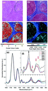
A protocol for rapid, label-free histochemical imaging of fibrotic liver
Mid-infrared microscopy is a non-destructive, quantitative and label-free spectroscopic imaging technique that, as a result of recent instrument advancements, is now at the point of enabling high-throughput automated biochemical screening of whole histology samples.
Analyst, 2017,142, 1179-1184
https://doi.org/10.1039/C6AN02080A
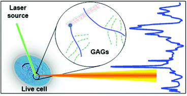
Probing glycosaminoglycan spectral signatures in live cells and their conditioned media by Raman microspectroscopy
GAG profiling in live cells by micro-Raman spectroscopy.
Analyst, 2017,142, 1333-1341
https://doi.org/10.1039/C6AN01951J
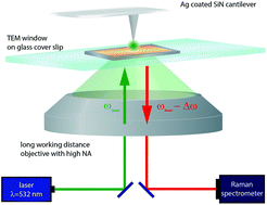
Towards a correlative approach for characterising single virus particles by transmission electron microscopy and nanoscale Raman spectroscopy
The morphology and structure of biological nanoparticles, such as viruses, can be efficiently analysed by transmission electron microscopy (TEM).
Analyst, 2017,142, 1342-1349
https://doi.org/10.1039/C6AN02151D
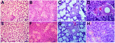
Monitoring the biochemical alterations in hypertension affected salivary gland tissues using Fourier transform infrared hyperspectral imaging
FTIR imaging shows biochemical differences between salivary glands from control and hypertensive rats.
Analyst, 2017,142, 1269-1275
https://doi.org/10.1039/C6AN02074G
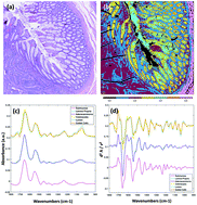
High definition infrared chemical imaging of colorectal tissue using a Spero QCL microscope
Mid-infrared microscopy has become a key technique in the field of biomedical science and spectroscopy. In this current study, we explore the use of a QCL infrared microscope to produce high definition, high throughput chemical images useful for the screening of biopsied colorectal tissue.
Analyst, 2017,142, 1381-1386
https://doi.org/10.1039/C6AN01916A
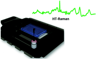
Development of a high throughput (HT) Raman spectroscopy method for rapid screening of liquid blood plasma from prostate cancer patients
High throughput Raman spectroscopy method for rapid and accurate diagnosis of prostate cancer using liquid plasma samples.
Analyst, 2017,142, 1216-1226
https://doi.org/10.1039/C6AN02100J
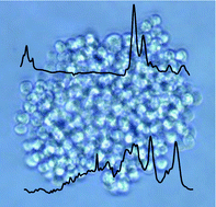
Effects of nilotinib on leukaemia cells using vibrational microspectroscopy and cell cloning
S-FTIR and Raman microspectroscopies identify spectral markers of sensitivity/resistance to nilotinib in leukaemia cell clones.
Analyst, 2017,142, 1299-1307
https://doi.org/10.1039/C6AN01914E
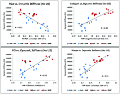
Near infrared spectroscopic assessment of developing engineered tissues: correlations with compositional and mechanical properties
Non-destructive near infrared spectroscopic data can be utilized for assessment of compositional and mechanical properties of engineered cartilage.
Analyst, 2017,142, 1320-1332
https://doi.org/10.1039/C6AN02167K
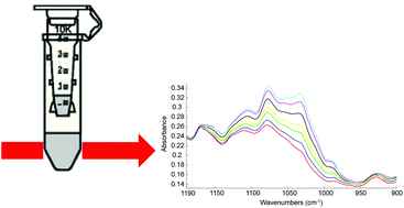
Ultra-filtration of human serum for improved quantitative analysis of low molecular weight biomarkers using ATR-IR spectroscopy
Monitoring of changes in the concentrations of the low molecular weight constituents enhanced by abundant proteins depletion.
Analyst, 2017,142, 1285-1298
https://doi.org/10.1039/C6AN01888B
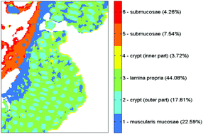
Digital de-waxing on FTIR images
This paper presents a procedure that digitally neutralizes the contribution of paraffin to FTIR hyperspectral images.
Analyst, 2017,142, 1358-1370
https://doi.org/10.1039/C6AN01975G
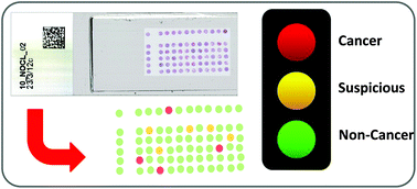
Infrared spectral histopathology using haematoxylin and eosin (H&E) stained glass slides: a major step forward towards clinical translation
Infrared spectral histopathology has shown great promise as an important diagnostic tool, with the potential to complement current pathological methods.
Analyst, 2017,142, 1258-1268
https://doi.org/10.1039/C6AN02224C
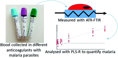
The effect of common anticoagulants in detection and quantification of malaria parasitemia in human red blood cells by ATR-FTIR spectroscopy
Total Reflectance Infrared Spectroscopy (ATR-FTIR) has the potential to become a new diagnostic tool for malaria and other diseases.
Analyst, 2017,142, 1192-1199
https://doi.org/10.1039/C6AN02075E
Infrared imaging of high density protein arrays
We propose in this paper that protein microarrays could be analysed by infrared imaging in place of enzymatic or fluorescence labelling.
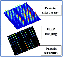
Analyst, 2017,142, 1371-1380
https://doi.org/10.1039/C6AN02048H
HTS-FTIR spectroscopy allows the classification of polyphenols according to their differential effects on the MDA-MB-231 breast cancer cell line
FTIR-based classification of the effect of polyphenols on a breast cancer cell line.
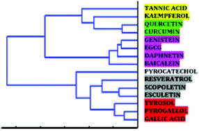
Analyst, 2017,142, 1244-1257
https://doi.org/10.1039/C6AN02135B
SIproc: an open-source biomedical data processing platform for large hyperspectral images
There has recently been significant interest within the vibrational spectroscopy community to apply quantitative spectroscopic imaging techniques to histology and clinical diagnosis.
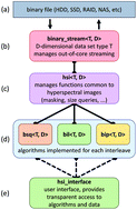
Analyst, 2017,142, 1350-1357
https://doi.org/10.1039/C6AN02082H
Optical properties of porcine dermis in the mid-infrared absorption band of glucose
Mid-infrared absorption and scattering properties of porcine dermis are quantified using quantum cascade laser-based goniometry.
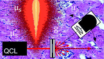
Analyst, 2017,142, 1235-1243
https://doi.org/10.1039/C6AN01757F
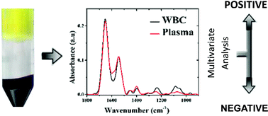
Early diagnosis of Alzheimer's disease using infrared spectroscopy of isolated blood samples followed by multivariate analyses
A simple blood test for the diagnosis of Alzheimer's disease using FTIR microscopy.
Analyst, 2017,142, 1276-1284
https://doi.org/10.1039/C6AN01580H
Virtual staining of colon cancer tissue by label-free Raman micro-spectroscopy
The great capability of virtual staining for label-free classification of colon cancer tissue has been demonstrated via Raman spectral imaging.

Analyst, 2017,142, 1207-1215
https://doi.org/10.1039/C6AN02072K
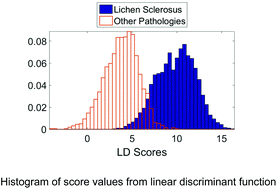
Raman spectroscopy and multivariate analysis for the non invasive diagnosis of clinically inconclusive vulval lichen sclerosus
The diagnostic performance of Raman spectroscopy for differentiating lichen sclerosus from other vulval conditions in fresh vulval biopsies is demonstrated.
Analyst, 2017,142, 1200-1206
https://doi.org/10.1039/C6AN02009G
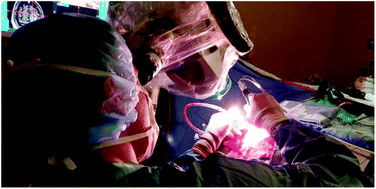
Raman spectroscopy in microsurgery: impact of operating microscope illumination sources on data quality and tissue classification
A filter system to perform in vivo Raman spectroscopy measurements under microscope lighting for seamless integration into the surgical workflow.
Analyst, 2017,142, 1185-1191
https://doi.org/10.1039/C6AN02061E
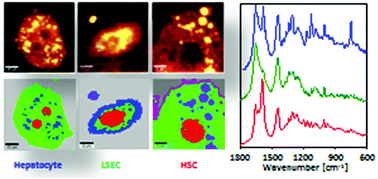
Label-free spectroscopic characterization of live liver sinusoidal endothelial cells (LSECs) isolated from the murine liver
Imaging with the use of Raman spectroscopy enables the characterization and distinction of live cells that were freshly isolated from murine livers.
Analyst, 2017,142, 1308-1319
https://doi.org/10.1039/C6AN02063A
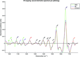
Rapid infrared mapping for highly accurate automated histology in Barrett's oesophagus
Barrett's oesophagus (BE) is a premalignant condition that can progress to oesophageal adenocarcinoma. Fourier transform infrared (FTIR) mapping can be used to identify neoplastic Barrett's with high accuracy in a clinically applicable timeframe.
Analyst, 2017,142, 1227-1234
https://doi.org/10.1039/C6AN01871H
About this collection
This collection of papers highlights work from the SPEC 2016 conference on Optical Diagnosis held in Montreal, Canada, 26-30 June 2016. Guest Edited by Fred Leblond and Kathy Gough.