
High spatial-resolution imaging of label-free in vivo protein aggregates by VISTA†

Abstract
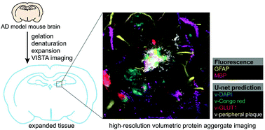
Amyloid aggregation, formed by aberrant proteins, is a pathological hallmark for neurodegenerative diseases, including Alzheimer's disease and Huntington's disease. High-resolution holistic mapping of the fine structures from these aggregates should facilitate our understanding of their pathological roles. Here, we achieved label-free high-resolution imaging of the polyQ and the amyloid-beta (Aβ) aggregates in cells and tissues utilizing a sample-expansion stimulated Raman strategy. We further focused on characterizing the Aβ plaques in 5XFAD mouse brain tissues. 3D volumetric imaging enabled visualization of the whole plaques, resolving both the fine protein filaments and the surrounding components. Coupling our expanded label-free Raman imaging with machine learning, we obtained specific segmentation of aggregate cores, peripheral filaments together with cell nuclei and blood vessels by pre-trained convolutional neural network models. Combining with 2-channel fluorescence imaging, we achieved a 6-color holistic view of the same sample. This ability for precise and multiplex high-resolution imaging of the protein aggregates and their micro-environment without the requirement of labeling would open new biomedical applications.
- This article is part of the themed collection: Biomedical Raman Imaging
 Please wait while we load your content...

