
Biomechanics as driver of aggregation of tethers in adherent membranes†

Abstract
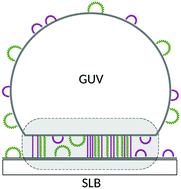
Cell adhesion is an important cellular process and is mediated by adhesion proteins residing on the cell membrane. Sometimes, two types of linker proteins are involved in adhesion, and they can segregate to form domains through a poorly understood size-exclusion process. We present an experimental and theoretical study of adhesion via linkers of two different sizes, realised in a mimetic model-system, based on giant unilamellar vesicles interacting with supported lipid bilayers. Here, adhesion is mediated by DNA linkers with two different lengths, but with the same binding enthalpy. We study the organisation of these linkers into domains as a function of relative fraction of long and short DNA constructs. Experimentally, we find that, irrespective of the composition, the adhesion domains are uniform with coexisting DNA bridge types, despite their relative difference in length of 9 nm. However, simulations suggest formation of nanodomains of the minority fraction at short length scales, which is below the optical resolution of the microscope. The nano-aggregation is more significant for long bridges, which are also more stable.
 Please wait while we load your content...

