Single X-ray crystal structure, DFT studies and topoisomerase I inhibition activity of a tailored ionic Ag(i) nalidixic acid–piperazinium drug entity specific for pancreatic cancer cells†
 a
and
Farukh
Arjmand
*a
a
and
Farukh
Arjmand
*a
Abstract
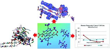
Novel ionic Ag(I)−piperazinium nalidixic acid conjugate 1 was synthesized as a potential antitumor agent and was thoroughly characterized by elemental analysis, FT-IR, 1H and 13C NMR and single X-ray crystal diffraction studies. Complex 1 crystallized in the triclinic space group P1 and comprises a dipiperazinium–Ag(I) cationic unit, two nalidixate (naI−) anionic moieties and a nitrate ion. The Ag(I) ion adopted a linear configuration upon coordination with two nitrogen atoms of piperazinium cations (pipzH+) arranged in a trans fashion. The density functional theory (DFT) studies of 1 revealed the HOMO and LUMO to be localized on the metal center viz., the dx2−y2 orbital and partially localized on the C27, C28, C29, C30, C31, C32, N7 and N6 atoms of the piperazinium moiety. Non covalent interaction (NCI) calculations were carried out to identify the weak non-covalent interactions from the topological analysis and reduced gradient of the electron density of complex 1. Our results revealed significant inter- and intramolecular non-covalent interactions between the naI− and [Ag(pip)2]2+ units. Furthermore, an analysis of Hirshfeld surfaces and fingerprint plots were carried out to ascertain a comparison between intermolecular interactions which provide interesting supramolecular architectures involving combinations of N–H⋯O, O–H⋯O and C–H⋯O linkages into a two-dimensional framework. In vitro binding studies of 1 with ct-DNA and tRNA revealed higher binding propensity for tRNA which was evidenced from its higher intrinsic binding constant, Kb and binding constant, K values and the mode of binding was found to be groove binding in nature. The catalytic activity of topoisomerase I enzyme in the presence of complex 1 was ascertained by gel electrophoretic assay which demonstrated significant inhibitory effects at a low concentration of 25 μM. The cytotoxicity activity of 1 was determined by SRB assay on MIA-PA-CA-2, HepG2, HeLa and MCF7 human cancer cell lines; these results exhibited specific and selective antitumor activity for the MIA-PA-CA-2 cancer cell line with a GI50 value <10.
 Please wait while we load your content...

