Preparation of fluorescent Au–SiO2 core–shell nanoparticles and nanorods with tunable silica shell thickness and surface modification for immunotargeting†
Abstract
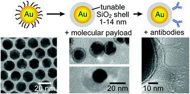
Gold–silica (Au–SiO2) core–shell nanoparticles (NPs) enable multifunctional properties for in vivo biomedical applications. However, scalable synthesis methods are lacking for the preparation of Au–SiO2 core–shell NPs less than 30 nm in overall diameter with a tunable silica shell less than 10 nm in thickness. Therefore, we prepared monodispersed Au–SiO2 core–shell NPs less than 30 nm in overall diameter with a uniform, tunable silica shell ∼1 to 14 nm in thickness using either citrate reduction followed by a modified Stöber method or oleylamine reduction followed by a reverse microemulsion method. Oleylamine reduction enabled up to 80-fold greater concentration yield compared to the citrate reduction method currently used for synthesizing Au core NPs. The formation of a tunable silica shell less than 10 nm in thickness was facilitated by controlling the molecular weight of the priming polymer (modified Stöber) or surfactant (reverse microemulsion) in addition to the concentration of the silane precursor, and was robust for encapsulating non-spherical morphologies such as Au nanorods. The reverse microemulsion method enabled several distinct advantages over the modified Stöber method, including greater control over the silica shell thickness, ∼16-fold greater yield in core–shell NP concentrations for scalable synthesis, and the ability to encapsulate controlled concentrations of a molecular payload (e.g., fluorophores with four different emission profiles) in the silica shell. Au–SiO2 core–shell NPs were also bioconjugated with immunoglobulin-G (IgG) as a model antibody to demonstrate immunotargeting. Bioactivity of Au–SiO2–IgG core–shell NPs was confirmed by agglomeration in the presence of protein A. The presence and proper orientation of IgG on NP surfaces was verified by direct observation in electron microscopy after negative staining. Therefore, the methods in this study for preparing and modifying Au–SiO2 core–shell NPs provide a platform for engineering core–shell NPs with size-dependent functional properties for multispectral/multimodal imaging, drug delivery, and combined theranostics.
- This article is part of the themed collection: JMC B Editor’s choice web collection: ‘‘seeing the unseen updated: advances in bioimaging’’
 Please wait while we load your content...

