
Morphology and characterization of Dematiaceous fungi on a cellulose paper substrate using synchrotron X-ray microtomography, scanning electron microscopy and confocal laser scanning microscopy in the context of cultural heritage
Abstract
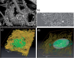
Dematiaceous (black pigmented) fungi interact with a cellular paper matrix growing on the surface and in paper bulk. Fungi-induced stains are one form of such interactions and are referred to as biodeterioration when applied to cultural heritage materials. The complexity of both paper and living systems, such as fungi, requires multi-scale analysis. The surface topography and spatial distribution of fungal monilioid hyphae, branched and un-branched chains and morphology of isodiametric enlargements of spores and yeast-like cells on the surface of paper were imaged by correlative microscopy, combining environmental scanning electron microscopy and imaging in backscattered electron mode (SEM-BSE) with confocal laser scanning microscopy (CLSM). The spherical fruiting bodies (perithecia) embedded in paper were not detected by CLSM, and ESEM provided data collected near the surface. Their interaction with paper was analyzed by 3D visualization using X-ray microtomography (XμCT). Until now the fungi and paper interfaces in the matrix (bulk) of paper have not been analyzed using XμCT. We present a novel method for investigating the interaction of fungal pigmented mycelia, spores and perithecia in the paper matrix using XμCT X and 3D visualization based on the XμCT data. The tomographs were generated on the designated ID19 beam at ESRF, Grenoble, France. The ultimate purpose of this investigation was to understand the mechanisms of fungi and paper interactions in order to develop preservation strategies for cultural heritage, such as historic and artistic works on paper infested by fungi.
- This article is part of the themed collection: Synchrotron radiation and neutrons in art and archaeology
 Please wait while we load your content...

