
Spectrally resolved optical microscopy using a transmission grating spectrograph: importance of spatial selection
Abstract
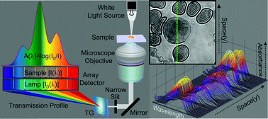
Spectral imaging using transmission grating (TG) based spectrographs has not been effectively utilized for spatially resolved absorption measurements of solid samples, and the role of spatial selection in spectral characteristics remains unexplored. We describe a simple yet efficient method to obtain reliable absorption spectra at sub micrometer spatial resolution for both non-fluorescent and fluorescent samples using a combination of a slit, TG and CCD detector coupled to an optical microscope. In this spectrally resolved optical microscopy (SROM) setup, the adjustable slit located before the TG enabled us to demonstrate that spatial selection greater than ∼1 μm is associated with a loss of spectral features, line broadening, and in certain situations, a shift in peak positions. We show that the use of near diffraction-limited slit-width is imperative for determination of reliable absorption profiles when a TG–CCD based spectrograph is used for spatially resolved spectroscopy measurements. The importance of high spatial selection in SROM becomes more apparent for heterogeneous samples with multiple absorbing species present in microscopically phase-separated regions, allowing for the identification of spectral signatures of individual components which cannot be resolved using ensemble measurements. This method can even be used to estimate the relative concentrations of absorbing species within sub-cellular regions, and therefore has the potential to map marker distribution in cellular environments.
 Please wait while we load your content...

