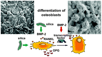
Three-dimensional printed (3D printed) bone material is needed to close the shortage and to avoid the potential health risks associated with autografts and allografts, in the treatment of bone fractures/nonunions or bone trauma. Here we describe the fabrication of 3D printed scaffold, initially prepared form Ca-sulfate that has been impregnated/biologized with Ca-phosphate or with silica. The 3D printed grids had a size mesh of 200 μm; the chemical composition was determined by energy dispersive X-ray spectroscopy or conventional chemical analysis. Using human SaOS-2 cells (human osteogenic cells) it is shown that both the Ca-sulfate, and the Ca-phosphate or the silica impregnated Ca-sulfate scaffold materials are biocompatible. Furthermore, they provide the cells with suitable matrices for hydroxyapatite synthesis in vitro, after stimulation with mineralization cocktail. By application of the quantitative real-time RT-PCR analysis technique it is shown that silica-impregnated scaffold induces SaOS-2 cells to express osteoprotegerin [OPG] and bone morphogenetic protein 2 [BMP-2]. Since the expression level of the receptor activator for NF-κB ligand [RANKL] is not influenced it is postulated that silica has the potential to neutralize the function of RANKL during osteoclast differentiation. It is concluded that 3D printed matrices, processed with silica, might function also as a morphogenetic material in vivo. Animal experiments are in progress to prove this assumption.

You have access to this article
 Please wait while we load your content...
Something went wrong. Try again?


 Please wait while we load your content...