Rapid diagnostics of liver steatosis by Raman spectroscopy via fiber optic probe: a pilot study†
Abstract
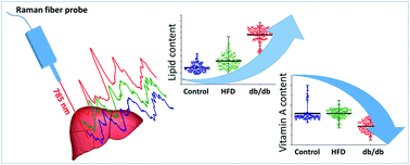
Raman spectroscopy via fiber optic probes omits some of the major limitations related to ex vivo preparation of tissue samples (e.g. fixation, freezing, cutting) and enables rapid registration of spectra, therefore, this technique has the potential to become a useful diagnostic tool. In this work, we evaluated the applicability of Raman spectroscopy via fiber optic probe for rapid assessment of lipid content in the liver in the context of its potential application as a tool to verify the degree of liver steatosis. Non-alcoholic fatty liver disease (NAFLD) is a common liver disorder that is characterized by excessive lipid accumulation within hepatic tissue and is associated with insulin resistance and metabolic syndrome. Raman spectroscopy via fiber optic probe was applied to investigate the biochemical status of the liver in mild (mice fed a high-fat diet) and severe (db/db mice) models of NAFLD. A considerable increase in lipid content without substantial alterations in composition was observed in mild liver steatosis. In contrast, more severe liver steatosis caused not only a significant lipid content increase, but also hepatic cholesterol accumulation accompanied by significant loss of hepatic vitamin A content. Chemometric analysis based on average Raman spectra recorded via fiber optic probe provided discrimination of mild and severe liver steatosis and control livers with high sensitivity and specificity. In conclusion, our work demonstrates that a relatively simple Raman setup equipped with a commercial fiber optic probe combined with basic chemometric analysis enables rapid quantification of liver steatosis.


 Please wait while we load your content...