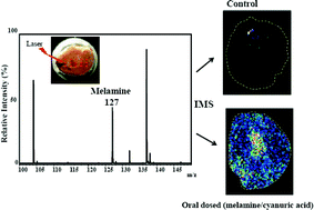
The study of the distribution of melamine in rat renal tissues by imaging mass spectrometry
Abstract
The recent tainting events of milk and pet

* Corresponding authors
a
Department of Chemistry, National Sun Yat-Sen University, Kaohsiung, Taiwan
E-mail:
jetea@mail.nsysu.edu.tw
Fax: +886-7-5253933
Tel: +886-7-5253933
b Department of Medical Imaging and Radiological Sciences, Kaohsiung Medical University, Kaohsiung, Taiwan
c National Sun Yat-Sen University—Kaohsiung Medical University Joint Research Center, Taiwan
d Department of Renal Care, Kaohsiung Medical University, Kaohsiung, Taiwan
e
Department of Biological Sciences, National Sun Yat-Sen University, Kaohsiung, Taiwan
E-mail:
hyjwang@gmail.com
Fax: +886-7-5252000-3611
Tel: +886-7-5252000-3611
f Cancer Center, Kaohsiung Medical University Hospital, Taiwan
The recent tainting events of milk and pet

 Please wait while we load your content...
Something went wrong. Try again?
H. Wang, L. Lo, Y. Tyan, H. Chen, M. Yang, T. Y. Chen, H. J. Wang and J. Shiea, Anal. Methods, 2010, 2, 1974 DOI: 10.1039/C0AY00266F
To request permission to reproduce material from this article, please go to the Copyright Clearance Center request page.
If you are an author contributing to an RSC publication, you do not need to request permission provided correct acknowledgement is given.
If you are the author of this article, you do not need to request permission to reproduce figures and diagrams provided correct acknowledgement is given. If you want to reproduce the whole article in a third-party publication (excluding your thesis/dissertation for which permission is not required) please go to the Copyright Clearance Center request page.
Read more about how to correctly acknowledge RSC content.
 Fetching data from CrossRef.
This may take some time to load.
Loading related content
