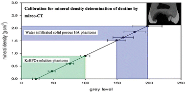
Laboratory micro-CT systems, although limited by beam hardening effect and instability of the source, have been utilized to measure mineral density in combination with specific image processing methods. However, few attempts have been made to accurately determine mineral density profiles in dentine due to the lack of suitable calibration standards. The aim of this study was to develop a calibration method to evaluate mineral density profiles in dentine including changes associated with dentinal caries. A series of K2HPO4 solution phantoms in a concentration range between 0 and 0.9 g cm−3-coupled to a set of water infiltrated porous solid hydroxyapatite (HA) phantoms, with mineral densities ranging from 1.52 to 2.08 g cm−3, was used in this investigation. First we evaluated the micrometer-scale homogeneity and noise in the HA phantoms using a commercial laboratory micro-CT system. Then an experimental validation was performed of the linearity over the entire density range of these two different calibration materials. The results show the HA phantoms extended the calibration curve obtained from K2HPO4 solution phantoms to densites as high as 2.08 g cm−3; the linearity remains stable at different energy levels. Finally, compared to the reference micro-CT calibration methods, the advantages of this new method are discussed. We conclude that this calibration method allows a more rational assessment of mineral density of dentine by micro-CT and has a promising potential for future studies.
You have access to this article
 Please wait while we load your content...
Something went wrong. Try again?
Please wait while we load your content...
Something went wrong. Try again?


