Fabrication of magnetic nanoprobes for ultrahigh-field magnetic resonance imaging
Abstract
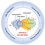
Ultrahigh-field magnetic resonance imaging (UHF-MRI) has been attracting tremendous attention in biomedical imaging owing to its high signal-to-noise ratio, superior spatial resolution, and fast imaging speed. However, at UHF-MRI, there is a lack of proper imaging probes that can impart superior imaging sensitivity of disease lesions because conventional contrast agents generally produce pronounced susceptibility artifacts and induce very strong T2 decay effects, thus hindering satisfactory imaging performance. This review focused on the recent development of high-performance nanoprobes that can improve the sensitivity and specificity of UHF-MRI. Firstly, the contrast enhancement mechanism of nanoprobes at UHF-MRI has been elucidated. In particular, the strategies for modulating nanoprobe performance, including size effects, metal alloying and magnetic-dopant effects, surface effects, and stimuli-response regulation, have been comprehensively discussed. Furthermore, we illustrate the remarkable advances in the design of UHF-MRI nanoprobes for medical diagnosis, such as early-stage primary tumor and metastasis imaging, angiography, and dynamic monitoring of biosignaling factors in vivo. Finally, we provide a summary and outlook on the development of cutting-edge UHF-MRI nanoprobes for advanced biomedical imaging.

- This article is part of the themed collections: Nanoscale 2023 Emerging Investigators and Recent Review Articles


 Please wait while we load your content...