The peculiarities of charge distribution and spatiotemporal changes in electronic and atomic structure of bone tissue
Abstract
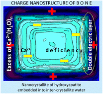
X-ray diffraction and inner-shell photoemission studies of rat cortical bone were performed. The distinct and systematic changes in the nanostructure of young, adult and mature bone are revealed. We observe the concurrent changes in core electrons binding energies, degrees of crystallinity, sizes of crystallites, Ca2+ deficiency and hydroxyapatite lattice constant c in bone mineral. These spatiotemporal changes are examined and assigned with the hierarchical organization of bone tissue, its ternary apatite–collagen–water composition as well as with the superperiodic charge alterations at the nanoscale. Using the extracted spectroscopic and structural parameters, the mineral matrix of bone as a function of age is reconstructed in the charge – space coordinates. We show that mineralized bone is a kind of electric battery composed from nanometric cells formed from negatively charged nanocrystallites embedded into positively charged intercrystallite water. The cells are strongly charged in a newborn bone and discharge with age. The charge dissipation is due to the counter Ca2+ and OH− ionic currents. Relationships between the spatiotemporal and charge regulation processes and their effect on the mechanical properties of bone are discussed in detail.


 Please wait while we load your content...