
All-in-one superparamagnetic and SERS-active niosomes for dual-targeted in vitro detection of breast cancer cells†

Abstract
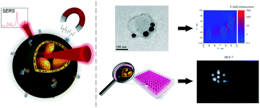
In vitro and in vivo biosensing through surface-enhanced Raman scattering often suffer from signal contamination diminishing both the limit of detection and quantification. However, overcoming the lack of specificity requires excessive nanoparticle concentrations, which may lead to adverse side effects if applied to patients. In this study we propose encapsulation of iron oxide (FexOy) and gold (Au) nanoparticles (NPs) into the bilayer structure of transferrin-modified niosomes. This approach enables achieving greatly enhanced and contamination-free SERS-signals in vitro as well as a dual-targeting functionality towards MCF-7 breast cancer cells. An in-depth characterization of FexOyNPs- and AuNPs-loaded niosomes (AuNPs/FexOyNPs/NIO) after magnetic downstream processing reveals defined hybrid niosome structures, which show a long-term SERS-signal stability in various media such as MCF-7 cell culture medium. In vitro 2D-SERS imaging unveil a successful incorporation of a non-toxic dose of hybrid NPs into MCF-7 cells, which leads to strong and almost contamination-free SERS-signals. The measured signal-to-noise ratio of the in vitro signal exceeds the values required by DIN 32645 for the successful validation of a detection method. The hybrid niosomes can thus be considered a promising and efficient agent for the establishment and commercialization of a highly sensitive detection kit for monitoring cancerous tissue.
- This article is part of the themed collection: Breast Cancer Awareness Month 2025
 Please wait while we load your content...

