
In situ analysis of the nucleation of O- and Zn-polar ZnO nanowires using synchrotron-based X-ray diffraction†

Abstract
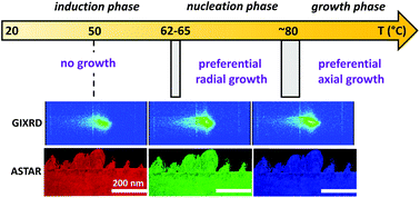
The selection of the polarity of ZnO nanowires grown by chemical bath deposition offers a great advantage for their integration into a wide variety of engineering devices. However, the nucleation process of ZnO nanowires and its dependence on their polarity is still unknown despite its importance for optimizing their morphology and properties and thus to enhance the related device performances. To tackle this major issue, we combine an in situ analysis of the nucleation process of O- and Zn-polar ZnO nanowires on O- and Zn-polar ZnO single crystals, respectively, using synchrotron radiation-based grazing incidence X-ray diffraction with ex situ transmission and scanning electron microscopy. We show that the formation of ZnO nanowires obeys three successive phases from the induction, through nucleation to growth phases. The characteristics of each phase, including the nucleation temperature, the shape and dimension of nuclei, as well as their radial and axial development are found to depend on the polarity of ZnO nanowires. A comprehensive description reporting the dominant physicochemical processes in each phase and their dependence on the polarity of ZnO nanowires is presented, revisiting their formation process step-by-step. These findings provide a deeper understanding of the phenomena at work during the growth of ZnO nanowires by chemical bath deposition and open the perspective to develop a more accurate control of their properties at each step of the formation process.
 Please wait while we load your content...

