
Temporal imaging of drug dynamics in live cells using stimulated Raman scattering microscopy and a perfusion cell culture system†
 a
Andrew S.
Merchant,
a
Rebecca
Fearon,
a
Nicholas C. O.
Tomkinson,
*b
Karen
Faulds
*a
and
Duncan
Graham
*a
a
Andrew S.
Merchant,
a
Rebecca
Fearon,
a
Nicholas C. O.
Tomkinson,
*b
Karen
Faulds
*a
and
Duncan
Graham
*a
Abstract
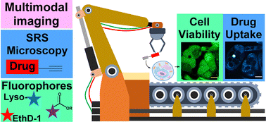
Stimulated Raman scattering (SRS) microscopy is a powerful technique for visualising the cellular uptake and distribution of drugs and small molecules in live cells under biocompatible imaging conditions. The use of bio-orthogonal groups within the drug molecule, including alkynes and nitriles, has enabled the direct detection of a plethora of bioactive molecules in a minimally perturbative fashion. Limited progress has been made towards real-time detection of drug uptake and distribution into live cells under physiological conditions, despite the accordant potential it presents for preclinical drug development. SRS microscopy has been applied to the study of cellular dynamics of the drug 7RH, which is a potent inhibitor of dicoidin domain receptor 1 (DDR1) and prevents cellular adhesion, proliferation and migration in vitro. The uptake of 7RH into a variety of mammalian cell models was shown to be independent of DDR1 expression. Using a perfusion chamber, the recurrent treatment of live cancer cells was achieved, enabling 7RH uptake to be visualised in real-time using SRS microscopy, after which the viability of the same cellular population was assessed using commercially available fluorescent markers in a multimodal imaging experiment. The effect of 7RH treatment in combination with the chemotherapeutic, cisplatin was investigated using sequential perfusion and time-lapse imaging in the same live cell population, to demonstrate the application of the approach. SRS microscopy also identified potent inhibition of cellular adhesion and migration in breast cancer cell models with increasing 7RH treatment concentrations, thus representing a novel read-out methodology for phenotypic assays of this kind. The direct assessment of drug–cell interactions under physiological conditions offers significant potential for the preclinical drug development process.
 Please wait while we load your content...

