3D imaging of single bacterial cells using surface-enhanced Raman spectroscopy with a multivariate curve resolution model†
 b
Jingjing
Du
*a
b
Jingjing
Du
*a
Abstract
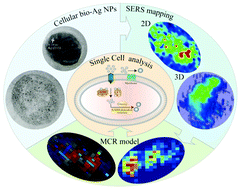
Imaging biomolecules within a single bacterial cell is crucial for understanding cellular genetic mechanisms. Herein, we exploited a surface-enhanced Raman spectroscopy (SERS) imaging strategy for single cell analysis. Cellular biosynthesized Ag nanoparticles (NPs) provided the necessary enhancement for SERS imaging. Multiple complementary techniques, including high-resolution transmission electron microscopy (HR-TEM), high-angle annular dark-field (HAADF)-scanning transmission electron microscopy (STEM), and energy-dispersive X-ray spectroscopy (EDX), were used to characterize the biogenic Ag NPs in cells. Three-dimensional SERS imaging maps displayed spectral information of biomolecules within the single cell. The multivariate curve resolution (MCR) model and principal component analysis (PCA) model were used to analyze the cellular SERS imaging maps. The MCR model, with a specific constraint of non-negativity, resulted in meaningful identification of biomolecules associated with Ag reduction. Focusing on the molecular level reveals that Pantoea sp. IMH utilizes several mechanisms to synthesize Ag NPs, including cytoplasm reduction by glucose or nicotinamide adenine dinucleotide (NADH)-dependent reductase, and extracellular reduction by an electron transfer chain containing quinone and cytochrome C. Our results shed new light on the Ag NP biosynthesis mechanism and single cell Raman analysis.
- This article is part of the themed collection: Analyst HOT Articles 2021
 Please wait while we load your content...

