Direct observation of protein structural transitions through entire amyloid aggregation processes in water using 2D-IR spectroscopy†
Abstract
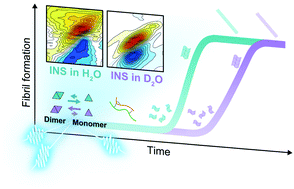
Amyloid proteins that undergo self-assembly to form insoluble fibrillar aggregates have attracted much attention due to their role in biological and pathological significance in amyloidosis. This study aims to understand the amyloid aggregation dynamics of insulin (INS) in H2O using two-dimensional infrared (2D-IR) spectroscopy. Conventional IR studies have been performed in D2O to avoid spectral congestion despite distinct H–D isotope effects. We observed a slowdown of the INS fibrillation process in D2O compared to that in H2O. The 2D-IR results reveal that different quaternary structures of INS at the onset of the nucleation phase caused the distinct fibrillation pathways of INS in H2O and D2O. A few different biophysical analysis, including solution-phase small-angle X-ray scattering combined with molecular dynamics simulations and other spectroscopic techniques, support our 2D-IR investigation results, providing insight into mechanistic details of distinct structural transition dynamics of INS in water. We found the delayed structural transition in D2O is due to the kinetic isotope effect at an early stage of fibrillation of INS in D2O, i.e., enhanced dimer formation of INS in D2O. Our 2D-IR and biophysical analysis provide insight into mechanistic details of structural transition dynamics of INS in water. This study demonstrates an innovative 2D-IR approach for studying protein dynamics in H2O, which will open the way for observing protein dynamics under biological conditions without IR spectroscopic interference by water vibrations.


 Please wait while we load your content...