
Mapping blood biochemistry by Raman spectroscopy at the cellular level†

Abstract
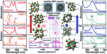
We report how Raman difference imaging provides insight on cellular biochemistry in vivo as a function of sub-cellular dimensions and the cellular environment. We show that this approach offers a sensitive diagnostic to address blood biochemistry at the cellular level. We examine Raman microscopic images of the distribution of the different hemoglobins in both healthy (discocyte) and unhealthy (echinocyte) blood cells and interpret these images using pre-calculated, accurate pre-resonant Raman tensors for scattering intensities specific to hemoglobins. These tensors are developed from theoretical calculations of models of the oxy, deoxy and met forms of heme benchmarked against the experimental visible spectra of the corresponding hemoglobins. The calculations also enable assignments of the electronic transitions responsible for the colour of blood: these are mainly ligand to metal charge transfer transitions.
 Please wait while we load your content...

