Alignment of Au nanorods along de novo designed protein nanofibers studied with automated image analysis†
Abstract
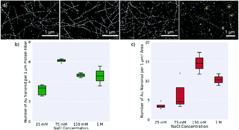
In this study, we focus on exploring the directional assembly of anisotropic Au nanorods along de novo designed 1D protein nanofiber templates. Using machine learning and automated image processing, we analyze scanning electron microscopy (SEM) images to study how the attachment density and alignment fidelity are influenced by variables such as the aspect ratio of the Au nanorods, and the salt concentration of the solution. We find that the Au nanorods prefer to align parallel to the protein nanofibers. This preference decreases with increasing salt concentration, but is only weakly sensitive to the nanorod aspect ratio. While the overall specific Au nanorod attachment density to the protein fibers increases with increasing solution ionic strength, this increase is dominated primarily by non-specific binding to the substrate background, and we find that greater specific attachment (nanorods attached to the nanofiber template as compared to the substrates) occurs at the lower studied salt concentrations, with the maximum ratio of specific to non-specific binding occurring when the protein fiber solutions are prepared in 75 mM NaCl concentration.


 Please wait while we load your content...