Dual contrast agents for fluorescence and photoacoustic imaging: evaluation in a murine model of prostate cancer†
Abstract
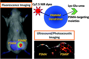
Prostate-specific membrane antigen (PSMA) is a promising diagnostic and therapeutic target for prostate cancer (PC). Poly(amidoamine) [PAMAM] dendrimers serve as versatile scaffolds for imaging agents and drug delivery that can be tailored to different sizes and compositions depending upon the application. We have developed PSMA-targeted PAMAM dendrimers for real-time detection of PC using fluorescence (FL) and photoacoustic (PA) imaging. A generation-4, ethylenediamine core, amine-terminated dendrimer was consecutively conjugated with on average 10 lysine-glutamate-urea PSMA targeting moieties and a different number of sulfo-cyanine7.5 (Cy7.5) near-infrared dyes (2, 4, 6 and 8 denoted as conjugates II, III, IV and V, respectively). The remaining terminal primary amines were capped with butane-1,2-diol functionalities. We also prepared a conjugate composed of Cy7.5-lysine-suberic acid-lysine glutamate-urea (I) and control dendrimer conjugate (VI). Among all conjugates, IV showed superior in vivo target specificity in male NOD-SCID mice bearing isogenic PSMA+ PC3 PIP and PSMA− PC3 flu xenografts and suitable physicochemical properties for FL and PA imaging. Such agents may prove useful in PC cancer detection and subsequent surgical guidance during excision of PSMA-expressing lesions.


 Please wait while we load your content...