Quantitative proteomic analysis of MDCK cell adhesion†
 *ab
*ab
Abstract
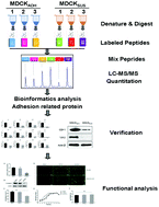
MDCK cells are a key reagent in modern vaccine production. As MDCK cells are normally adherent, creation of suspension cells for vaccine production using genetic engineering approaches is highly desirable. However, little is known regarding the mechanisms and effectors underlying MDCK cell adhesion. In this study, we performed a comparative analysis of whole protein levels between MDCK adhesion and suspension cells using an iTRAQ-based (isobaric tags for relative and absolute quantitation) proteomics approach. We found that expression of several proteins involved in cell adhesion exhibit reduced expression in suspension cells, including at the mRNA level. Proteins whose expression was reduced in suspension cells include cadherin 1 (CDH1), catenin beta-1 (CTNNB1), and catenin alpha-1 (CTNNA1), which are involved in intercellular adhesion; junction plakoglobin (JUP), desmoplakin (DSP), and desmoglein 3 (DSG3), which are desmosome components; and transglutaminase 2 (TGM2) and alpha-actinin-1 (ACTN1), which regulate the adhesion between cells and the extracellular matrix. A functional verification experiment showed that inhibition of E-cadherin significantly reduced intercellular adhesion of MDCK cells. E-Cadherin did not significantly affect the proliferation of MDCK cells and the replication of influenza virus. These findings reveal possible mechanisms underlying adhesion of MDCK cells and will guide the creation of MDCK suspension cells by genetic engineering.
 Please wait while we load your content...