
Microfluidic model with air-walls reveals fibroblasts and keratinocytes modulate melanoma cell phenotype, migration, and metabolism†
 *a
Shujah
Rehman,
cde
*a
Shujah
Rehman,
cde
Abstract
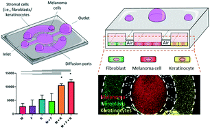
Melanoma evolution is a complex process. The role epidermal keratinocytes and dermal fibroblasts play in this process and the mechanisms involved in tumor–stroma interactions remain poorly understood. Here, we used a microfluidic platform to evaluate the cross-talk between human primary melanoma cells, keratinocytes and dermal fibroblasts. The microfluidic device included multiple circular chambers separated by a series of narrow connection channels. The microdevice design allowed us to develop a new cell patterning method based on air-walls, removing the need for hydrogel barriers, porous membranes, or external equipment. Using this method, we co-cultured melanoma cells in the presence of keratinocytes and/or dermal fibroblasts. The results demonstrated that the presence of dermal fibroblasts and keratinocytes led to changes in melanoma cell morphology and growth pattern. Molecular analysis revealed changes in the chemokine secretion pattern, identifying multiple secreted factors involved in tumor progression. Finally, optical metabolic imaging showed that melanoma cells, fibroblasts, and keratinocytes exhibited different metabolic features. Additionally, the presence of stromal cells led to a metabolic shift in melanoma cells, highlighting the role the skin microenvironment on melanoma evolution.
 Please wait while we load your content...

